


Acute Sinusitis (Acute Rhinosinusitis): Classification, Clinical Manifestations, Diagnosis, and Treatment
A detailed review of rhinosinusitis, including classification, symptoms, diagnostic approaches, and current treatment strategies.
Anesthesia
Pain management and sedation techniques
Angiology
Arterial and venous pathologies
Cardiology
Acquired and congenital heart diseases
Dentistry
Diseases of teeth, gums, and the oral cavity
Dermatology
Disorders of the skin and subcutaneous tissue
Endocrinology
Disorders of the glands and hormonal imbalance
Gastroenterology
Stomach, intestinal, and digestive diseases
Gynecology
Diseases of female reproductive organs
Hepatology
Liver, gallbladder, and biliary tract diseases
Neurology
Brain, spinal cord, and peripheral nerve disorders
Obstetrics
Pregnancy complications and abnormal fetal positions
Oncology
Cancer types, benign and malignant tumors
Ophthalmology
Conditions affecting the eyes and vision
Otorhinolaryngology
Ear, nose, and throat diseases
Pediatrics
Child health, development, and clinical conditions
Pulmonology
Lung and respiratory tract diseases
Traumatology
Acute injuries and musculoskeletal trauma
Urology
Urinary tract and male reproductive disorders

This article is for informational purposes only
The content on this website, including text, graphics, and other materials, is provided for informational purposes only. It is not intended as advice or guidance. Regarding your specific medical condition or treatment, please consult your healthcare provider.
Acute inflammatory diseases of the pharynx – a pathological condition caused by the activation of pathogenic microflora on the surface of the pharyngeal mucosa and manifested by local and general symptoms.
These include:
Acute tonsillitis (sore throat) in most cases is caused by pathogenic bacteria, in particular about 80% beta-hemolytic group A streptococcus (BHSA). In addition, the source of infection can be streptococci, staphylococci, pneumococci, enterococci, hemophilus bacillus, a large number of viruses, especially adenoviruses, herpesviruses. When local immunity is weakened, opportunistic microflora living in the oral cavity (Streptococcus viridans, Leptothrix, fusobacteria, fungi) can also cause inflammation.
In acute inflammation of the palatine tonsils is determined by their swelling and hyperemia, in the same way change and nearby structures (uvula, soft palate, back wall of the pharynx), the tongue covered with white plaque, these signs are characteristic of catarrhal tonsillitis.
In the follicular form, in addition to the above picture, small millet dots of white-yellow color appear in the follicles.
In lacunar tonsillitis on hyperemic and edematous tonsils spread
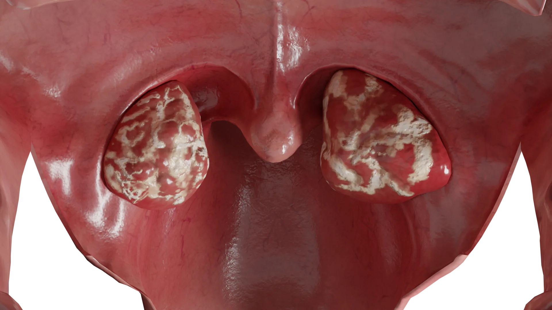
Acute tonsillitis is characterized by a sudden acute onset. Patients notice a pronounced pain in the throat, intensified by swallowing, which may irradiate to the ear or neck. Because of the swelling and painful palatine tonsils there is difficulty in opening the mouth, nasality, bad breath. Plaque on the tonsils is easily removed with a spatula, the surface does not bleed. General symptoms appear: fever to febrile values (38-40°C), chills, headache, regional lymph nodes are enlarged and sharply painful. The disease lasts 5-7 days.
Frequent complications are characteristic, local complications include: paratonsillitis, paratonsillar abscess, cervical lymphadenitis; general complications include: rheumatism, glomerulonephritis, arthritis, sepsis, endocarditis.
Diagnosis is established on the basis of a characteristic clinical picture, the results of pharyngoscopy. Determination of leukocytes and C-reactive protein, rheumatoid factor in blood, urinalysis, bacteriological examination of the detachment or strepto-test (for diagnostics of BHSA) is performed.
If it is not possible to perform a bacterial examination or strepto-test, the Centor-McAzek scale is used. If the sum of scores is 3 or higher, a bacterial pathogen is suspected and antibiotic therapy is prescribed.
The Centor-McAzek Scale:
| Criterion | Score |
|---|---|
| Body temperature above 38°C | 1 |
| No cough | 1 |
| Enlargement and soreness of the cervical LUs | 1 |
| Swelling and plaque on the tonsils | 1 |
| Age (3-14 years) | 1 |
| Age (15-44 years) | 0 |
| Age (≥45 years) | -1 |
For the treatment of acute bacterial tonsillitis, antibacterial drugs of the penicillin series are used for 10 days, in case of allergy – cephalosporins or macrolides. In addition to antibiotics, non-steroidal anti-inflammatory drugs are prescribed. For a speedy recovery prescribe a sparing high-calorie diet, it is also recommended to observe bed rest.
Leukemia is a malignant blood disease in which immature or atypical blood cells are formed.
In leukemia, the formation of immune system cells in the bone marrow is impaired, and immature and undifferentiated cells – blasts – are released into the blood. These forms are not able to fully repel the infection. For the development of an infectious process in the oropharynx, a slight violation of the integrity of the mucosa, through which the pathogenic microflora enters, is sufficient.
Due to a sharp decline in local and general immunity develop pathological processes in the tonsils. Pathogenic microflora begins to spread uncontrollably in the oropharynx, causing inflammation of the tonsils. Tonsillitis quickly passes all stages (catarrhal, lacunar, follicular) and eventually necrotic changes in the tonsils occur. The oropharyngeal mucosa is infiltrated, palatine tonsils hypertrophied, covered with fibrinous hard to separate plaque. Subsequent necrosis may spread to the mucosa of gums, soft palate.
The first signs of leukemia include general weakness, subfebrile temperature, body aches. Hemorrhagic syndrome is characteristic: there is a widespread small hemorrhagic rash on the skin and mucous membranes, epistaxis, from a small wound there is prolonged bleeding, which can end fatally. Local changes in the oropharynx develop in 3-5 days from the onset of the disease. There is a sequential development of all stages of tonsillitis, which ends with the development of ulcerative-necrotic sore throat. There is a pronounced pain in the throat, intensified by swallowing, irradiating to the ear. Against the background of pathological changes in the amygdalae is characterized by pyretic fever (increase in body temperature to 39-40°C), sweating, chills. Regional lymph nodes are enlarged, but sometimes generalized lymphadenopathy is observed, the liver and spleen are also enlarged. With the development of the necrotic form, there is a putrid odor from the mouth, the general condition rapidly deteriorates, the course becomes malignant. With the progression of the process may develop sepsis and multi-organ failure with lethal outcome.
Initially, a general examination and otorhinopharyngoscopy are performed. However, it is difficult to clinically suspect leukemia. It is obligatory to perform a general blood test, which reveals a decrease in all formational elements, in addition to leukocytes, which increase several times due to immature cells (blasts). In some cases, a pronounced decrease in leukocytes is observed in the leukopenic form. To definitively establish the diagnosis, a bone marrow puncture is performed. Leukemia is characterized by suppression of all sprouts of hematopoiesis and the presence of a large number of blasts. Consultation with an oncohematologist is recommended.
Initially, broad-spectrum antibiotic therapy is initially prescribed to prevent a severe course of leukemia. Specific therapy is carried out by doctors of oncohematology profile, which includes
Agranulocytosis is not an independent diagnosis, but is a clinical and hematologic syndrome. In this pathology, the number of neutrophilic granulocytes (< 0.5*109/L) is sharply reduced, up to their complete absence. More often occurs in women over 40 years old. Depending on the causative factors distinguish myelotoxic, immune and autoimmune agranulocytosis:
Against the background of any of the above forms of the disease, infectious processes occur in places of traumatization or contact with a large number of pathogens, including the development of acute tonsillitis. Lesions of tonsils with agranulocytosis is a characteristic symptom.
Myelotoxic form of agranulocytosis occurs due to suppression of myelopoiesis progenitor cells production in the bone marrow, while the level of lymphocytes, reticulocytes, and platelets also decreases.
Immune agranulocytosis is characterized by the formation of antibodies (a complex composed of drugs and blood proteins) that are directed against white blood cells.
Autoimmune agranulocytosis triggers the independent production of antineutrophil antibodies, which cause their death.
With a decrease in leukocytes, the penetration of any microorganism through the damaged mucosa of the tonsils causes a pathological process that leads to the development of acute tonsillitis. In mild forms, the process is limited to catarrhal sore throat, which is characterized by infiltration and hyperemia on the palatine tonsils. However, with a severe course develops ulcerative-necrotic sore throat, affecting the mucosa and underlying structures, spreading down to the bone tissue. After rejection of necrotic masses, deep ulcers remain. Microscopically, there are no neutrophils in places of necrosis.
The disease begins with pyretic fever (a rise in body temperature to 40°C), chills, sweating, and body aches.
Simultaneously with the general changes there are pathological processes in the oropharynx. Characterized by severe
In myelotoxic agranulocytosis is characterized by the presence of hemorrhagic syndrome, manifested by bleeding gums, nosebleeds, the formation of bruises and hematomas, hematuria.
Among the most frequent complications are perforation of the soft palate, sepsis, mediastinitis with fatal outcome.
It is necessary to carefully collect an anamnesis, clarify what drugs the patient takes, the presence of concomitant diseases.
General examination and pharyngoscopy are performed. It is obligatory to perform a general blood test, which reveals leukopenia due to a decrease in neutrophil granulocytes (total leukocyte count less than 1.0×10⁹ / L, neutropenia below 0.5×10⁹ / L, relative lymphocytosis and monocytosis). In myelotoxic agranulocytosis, the number of erythrocytes and platelets is reduced. Then a bone marrow study is performed, where a violation of leukopoiesis is noted. If autoimmune agranulocytosis is suspected, a blood test for antineutrophil antibodies is performed. In all cases, it is necessary to consult a hematologist.
Initially, it is necessary to discontinue all drugs that could lead to the development of agranulocytosis. Patients are treated in the hematology department, where hemotransfusions are performed, drugs that stimulate leukopoiesis, antibacterial and antifungal therapy are prescribed.
It is necessary to regularly perform sanitation of oropharynx with removal of necrotic masses, treatment with antiseptic solutions. In the immune nature of the disease glucocorticoids are administered in high dosage, in the presence of a large number of circulating immune complexes plasmapheresis is indicated. A sparing high-calorie diet is prescribed, if necessary, the transition to parenteral nutrition. It is necessary to prevent infectious complications. The prognosis for this disease is severe.
Infectious inflammatory changes in the pharynx are most commonly caused by viruses (about 70%), less commonly by bacteria (30%).
Among the viruses are:
Among bacterial pathogens, b-hemolytic streptococcus group A is predominant, pneumococci, mycoplasmas, hemophilus bacillus, Staphylococcus aureus are found less frequently.
Separately, mechanical factors should be singled out as a cause of acute pharyngitis, such as trauma, burns, irritation by caustic substances, allergens.
There is a
Acute pharyngitis is characterized by the prevalence of local complaints over the general symptomatology. Patients are bothered by
When the patient goes to the doctor
Find more scientifically accurate content on our social media


With viral etiology (more often), only
If a bacterial infection is suspected, broad-spectrum antibacterial drugs are prescribed systemically.
In both cases, the use of anti-inflammatory drugs is indicated . Absorbent preparations with lidocaine, menthol, essential oils have proved to be good for pain symptom relief. It is recommended to moisturize the air in the room, drink plenty of fluids.
1. What does sore throat look like and how does it manifest itself?
2. Is sore throat transmissible and how is it contracted?
3. What causes sore throat?
4. How does tonsillitis differ from sore throat and pharyngitis?
5. What is pharyngitis and how does it manifest itself?
6. How can viral tonsillitis be distinguished from bacterial tonsillitis?
7. What is chronic tonsillitis and can it be cured?
8. What are the dangers of untreated tonsillitis?
9. Is pharyngitis contagious and what are its symptoms?
List of Sources
1.
VOKA Catalog.
https://catalog.voka.io/
2.
Total Otolaryngology—Head and Neck Surgery, Anthony P. Sclafani, Robin A. Dyleski, Michael J. Pitman, Stimson P. Schantz. Thieme Medical Publishers, Inc., 2015. ISBN 978-1-60406-646-3.
3.
Beerbohm H. Diseases of the ear, throat and nose / Hans Berbom, Oliver Kaschke, Thadeus Navka, Andrew Swift; transl. from English. – 2nd ed. – Moscow : MEDpress-Inform, 2016. – 776 с. : ill. ISBN 978-5-00030-322-1
4.
Swidsinski A, Göktas O, Bessler C, Loening-Baucke V, Hale LP, Andree H, Weizenegger M, Hölzl M, Scherer H, Lochs H. Spatial organization of microbiota in quiescent adenoiditis and tonsillitis. J Clin Pathol. 2007 Mar;60(3):253-60. doi: 10.1136/jcp.2006.037309. Epub 2006 May 12. PMID: 16698947; PMCID: PMC1860565.
5.
Anderson J, Paterek E. Tonsillitis. [Updated 2023 Aug 8]. In: StatPearls [Internet]. Treasure Island (FL): StatPearls Publishing; 2025 Jan-.
Available from: https://www.ncbi.nlm.nih.gov/books/NBK544342/
6.
Nguyen VTN, Ngo L, Stratton E, Arriola D. Tonsillitis. Prim Care. 2025 Mar;52(1):27-35. doi: 10.1016/j.pop.2024.09.005. Epub 2024 Dec 30. PMID: 39939088.
7.
Bird JH, Biggs TC, King EV. Controversies in the management of acute tonsillitis: an evidence-based review. Clin Otolaryngol. 2014 Dec;39(6):368-74. doi: 10.1111/coa.12299. PMID: 25418818; PMCID: PMC7162355.
8.
Thakur JS, Mohindroo NK, Sharma DR, Mohindroo S, Thakur A. Tonsillitis with acute myeloid leukemia: a case series for caution. Ear Nose Throat J. 2013 Apr-May;92(4-5):E22-3. doi: 10.1177/014556131309200425. PMID: 23599112.
9.
Andrès E, Mourot-Cottet R, Maloisel F, Séverac F, Keller O, Vogel T, Tebacher M, Weber JC, Kaltenbach G, Gottenberg JE, Goichot B, Sibilia J, Korganow AS, Herbrecht R. Idiosyncratic drug-induced neutropenia & agranulocytosis. QJM. 2017 May;110(5):299-305. doi: 10.1093/qjmed/hcw220. Epub 2017 Jan 9. PMID: 28069912.
Loading test 6 questions
Table of Contents


Summarize article with AI
Choose your preferable AI assistant:
Link successfully copied to clipboard
Thank you!
Your message is sent!
Our experts will contact you shortly. If you have any additional questions, please contact us at info@voka.io


