Odontogenic Sinusitis: Classification, Etiology, Clinical Presentation, Diagnosis, and Treatment
A detailed overview of odontogenic maxillary sinusitis: causes, symptoms of acute and chronic inflammation, diagnosis, and modern treatment approaches.


Anesthesia
Pain management and sedation techniques

Angiology
Arterial and venous pathologies

Cardiology
Acquired and congenital heart diseases

Dentistry
Diseases of teeth, gums, and the oral cavity

Dermatology
Disorders of the skin and subcutaneous tissue

Embryology
Stages of embryogenesis and congenital anomalies

Endocrinology
Disorders of the glands and hormonal imbalance

Gastroenterology
Stomach, intestinal, and digestive diseases

Gynecology
Diseases of female reproductive organs

Hematology
Hematopoiesis and blood-related disorders

Hepatology
Liver, gallbladder, and biliary tract diseases

Histology
Microscopic tissue and cell structures

Infectious diseases
Bacterial, viral, and parasitic infections

Neurology
Brain, spinal cord, and peripheral nerve disorders

Obstetrics
Pregnancy complications and abnormal fetal positions

Oncology
Cancer types, benign and malignant tumors

Ophthalmology
Conditions affecting the eyes and vision

Orthopedics
Bone, joint, and soft tissue disorders

Otorhinolaryngology
Ear, nose, and throat diseases

Pediatrics
Child health, development, and clinical conditions

Physiology
Biological processes within organs and systems

Pulmonology
Lung and respiratory tract diseases

Traumatology
Acute injuries and musculoskeletal trauma

Urology
Urinary tract and male reproductive disorders
Anesthesia
Pain management and sedation techniques
Angiology
Arterial and venous pathologies
Cardiology
Acquired and congenital heart diseases
Dentistry
Diseases of teeth, gums, and the oral cavity
Dermatology
Disorders of the skin and subcutaneous tissue
Embryology
Stages of embryogenesis and congenital anomalies
Endocrinology
Disorders of the glands and hormonal imbalance
Gastroenterology
Stomach, intestinal, and digestive diseases
Gynecology
Diseases of female reproductive organs
Hematology
Hematopoiesis and blood-related disorders
Hepatology
Liver, gallbladder, and biliary tract diseases
Histology
Microscopic tissue and cell structures
Infectious diseases
Bacterial, viral, and parasitic infections
Neurology
Brain, spinal cord, and peripheral nerve disorders
Obstetrics
Pregnancy complications and abnormal fetal positions
Oncology
Cancer types, benign and malignant tumors
Ophthalmology
Conditions affecting the eyes and vision
Orthopedics
Bone, joint, and soft tissue disorders
Otorhinolaryngology
Ear, nose, and throat diseases
Pediatrics
Child health, development, and clinical conditions
Physiology
Biological processes within organs and systems
Pulmonology
Lung and respiratory tract diseases
Traumatology
Acute injuries and musculoskeletal trauma
Urology
Urinary tract and male reproductive disorders
Specialties
AnesthesiaAngiologyCardiologyDentistryDermatologyEmbryologyEndocrinologyGastroenterologyGynecologyHematologyHepatologyHistologyInfectious diseasesNeurologyObstetricsOncologyOphthalmologyOrthopedicsOtorhinolaryngologyPediatricsPhysiologyPulmonologyTraumatologyUrology
This article is for informational purposes only
The content on this website, including text, graphics, and other materials, is provided for informational purposes only. It is not intended as advice or guidance. Regarding your specific medical condition or treatment, please consult your healthcare provider.
Alterations in the shape of the external nose may be associated with a congenital or acquired defect of the external nose. These changes may cause aesthetic dissatisfaction or even impair an individual’s breathing function.
The condition typically affects the cartilaginous and/or bony parts of the pyramid.
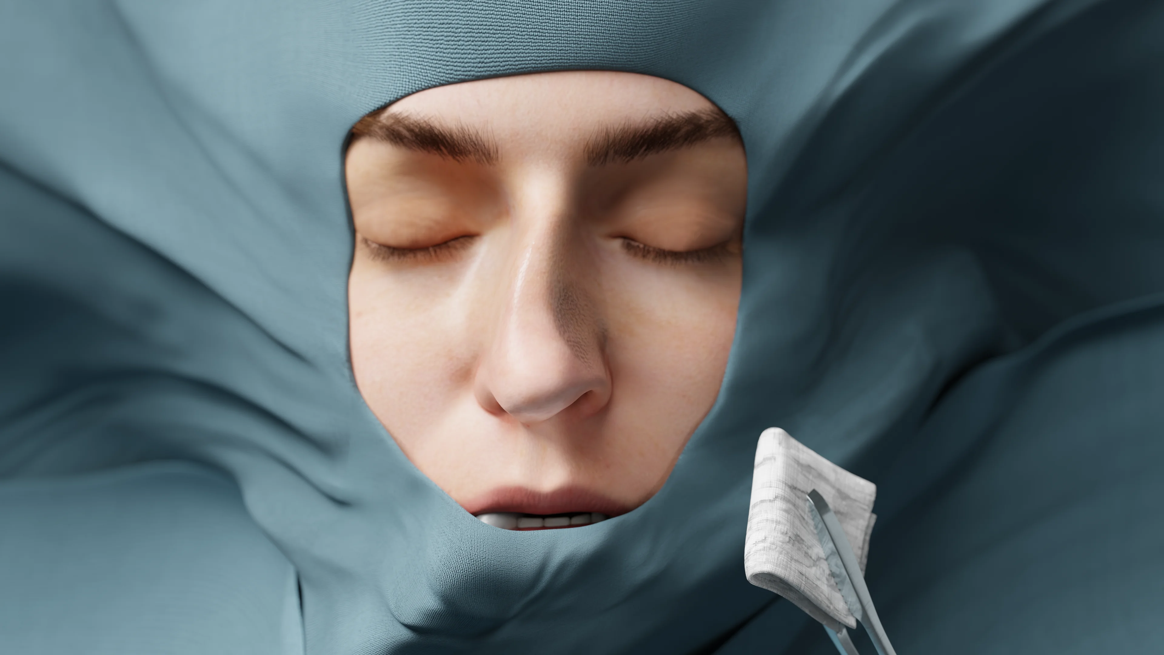
Changes in nose shape may be caused by various factors:
Leonardo da Vinci is thought to have devised the theory of ideal facial symmetry, also known as the golden ratio, which is now used to determine nasal irregularities. According to his theory, the left and right halves of the face should mirror each other.
Da Vinci also introduced the golden ratio (1 : 1.618) concept, which helps identify the right facial proportions. The intervals between facial features (eyes, nose, mouth) should correspond to this ratio.
To analyze the facial profile, modern plastic surgery utilizes geometric lines and points, such as the nasion, rhinion, pronasale, and subnasale, and determines the nasofrontal and nasolabial angles. When the proportions are distorted, the external nose shape is considered to be altered.
Note that this article does not deal with the aesthetic analysis of the face. Below, you can find the most common types of nasal deformities.
Currently, there is no established classification system for external nasal deformities. Our goal is to describe the most common types.
When the cartilaginous part of the nose fails to develop properly, the columella, apex, and alae of the nose change their shape, position and orientation. Nasal cartilages are also responsible for the stiffness of the distal third of the nasal dorsum. The bony part of the nose is responsible for the shape of the bridge of the nose, the profile of the nose, and the presence of a hump or saddle-shaped deformity.
3D models – external nasal deformities:
 Rhinokyphosis (hawk nose)
 Rhinolordosis (saddle nose)
 Platyrhinia (flat nose)
 Brachyrhinia (wide short nose)
 Leptorhinia (narrow nose)
 Rhinoskoliosis (ace nose)
These conditions affect the appearance of the nose and may impair its functions, such as breathing or smelling.
A healthcare professional should perform a visual examination to evaluate the external nose shape and ask the patient about complaints.
The external nose shape may be corrected using minimally invasive techniques. These include fillers injected into specific areas of the nose. However, this method is not suitable for more severe defects and the result is temporary.
Surgical therapy aims to correct the external and/or internal nose, its appearance, and breathing function. Depending on the deformity type, rhinoplasty may be either closed or open.
Rhinophyma results from persistent chronic inflammation of the skin secondary to rosacea. It is very common among middle-aged and senior males and is rarely seen in females. Genetic predisposition is also one of the key factors. Prolonged UV exposure and unhealthy lifestyles (smoking, alcohol abuse, excessive consumption of spices) tend to exacerbate the condition. Hormonal disorders may also promote and aggravate rhinophyma.
Connective tissue, blood vessels, and sebaceous glands start to proliferate secondary to chronic skin inflammation. Gradually, the nasal skin develops dense red nodules covered with superficial vessels. Then the nodules transform into cyanopurpuric tuberous build-ups, and telangiectasia may develop. Sebaceous glands become significantly enlarged and produce excessive amounts of discharge.
Patients are primarily concerned about their disfigured appearance. Their nose becomes cyanotic, tuberous, and significantly enlarged. The pores are also bigger, and the skin is oily and shiny. In extreme cases, the condition may cause pain and discomfort. Symptoms tend to worsen over time.
Diagnosis is based on medical history and a physical examination. Dermatologist/cosmetologist advice is generally recommended.
Find more scientifically accurate content on our social media



Medical treatment may halt the disease.
Patients are advised to break their bad habits and limit exposure to high temperatures and UV.
Antibacterial ointments, azelaic acid, and retinoids have proven to be effective.
Laser treatment of pathologically altered tissues may be an option. This method targets hypertrophied vessels and reduces inflammation.
Significant overgrowth of the external nasal tissues typically necessitates surgery. In this case, specialized tools and electric impulses are used to excise excessive tissues.
1. What are the main types of external nasal deformities?
2. What are the causes of rhinophyma and how quickly does it develop?
3. How is rhinophyma treated?
4. What are the common causes of external nasal deformities?
5. How are structural deformities of the nose, such as rhinokifosis or rhinoscoliosis, treated?
References
1.
VOKA 3D Anatomy & Pathology – Complete Anatomy and Pathology 3D Atlas. VOKA 3D Anatomy & Pathology.
Available from: https://catalog.voka.io/
2.
Sclafani AP, Dyleski RA, Pitman MJ, Schantz SP. Total otolaryngology—head and neck surgery. New York: Thieme Medical Publishers; 2015. ISBN: 978-1-60406-646-3.
3.
Behrbohm H, Kaschke O, Nawka T, Swift A. Bolezni ukha, gorla i nosa [Ear, nose, and throat diseases]. 2nd ed. Moscow: MEDpress-inform; 2016. 776 p. [In Russian.] ISBN 978-5-00030-322-1.
4.
Suh MK, Jeong E. Correction of deviated nose. Arch Craniofac Surg. 2018 Jun;19(2):85-93. doi: 10.7181/acfs.2018.01914. PMID: 29996638; PMCID: PMC6057124.
5.
Sam A, Deshmukh PT, Patil C, Jain S, Patil R. Nasal septal deviation and external nasal deformity: a correlative study of 100 cases. Indian J Otolaryngol Head Neck Surg. 2012 Dec;64(4):312-318. doi: 10.1007/s12070-011-0311-x. PMID: 24294569; PMCID: PMC3477430.
6.
Rhinoplasty Archive. Rhinoplasty dissection manual. Chapter 1 [Internet].
Available from: https://www.rhinoplastyarchive.com/articles/rhinoplasty-fundamentals/rhinoplasty-dissection-manual-chapter-1
7.
Kao WK, Ho T. The management of posttraumatic nasal deformities. Facial Plast Surg. 2023 Dec;39(6):630-637. doi: 10.1055/a-2152-8670. PMID: 37567568.
8.
Huseynov J, Mozet C. Übersicht und aktuelle Empfehlung zur Therapie des Rhinophyms [A systematic review and current recommendation for treatment of rhinophyma]. Laryngorhinootologie. 2020 Nov;99(11):772-778. German. doi: 10.1055/a-1208-5284. PMID: 33111293.
9.
Chauhan R, Loewenstein SN, Hassanein AH. Rhinophyma: prevalence, severity, impact and management. Clin Cosmet Investig Dermatol. 2020 Aug 11;13:537-551. doi: 10.2147/CCID.S201290. PMID: 32848439; PMCID: PMC7429105.
Make VOKA your preferred source
See more VOKA articles in Google Search
Table of Contents









Summarize article with AI
Choose your preferable AI assistant:
Link successfully copied to clipboard
Thank you!
Your message is sent!
Our experts will contact you shortly. If you have any additional questions, please contact us at info@voka.io