
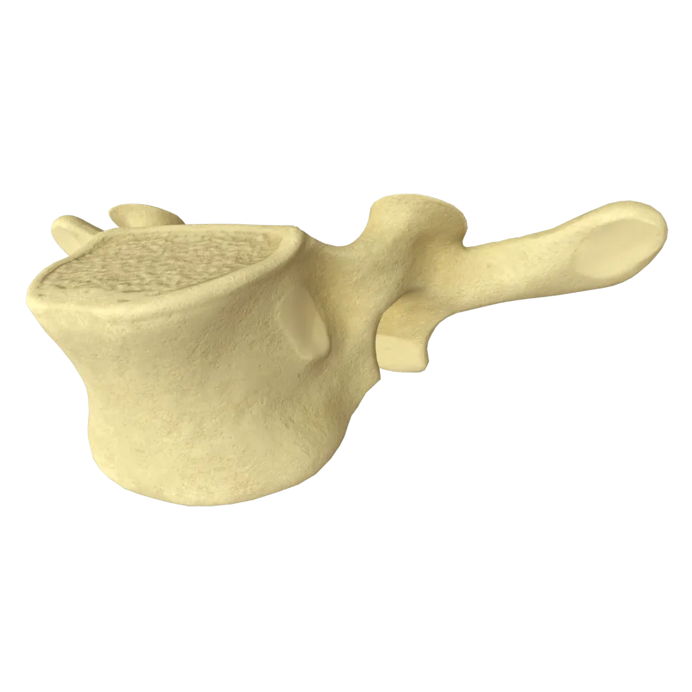
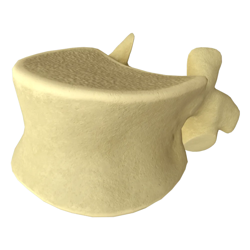
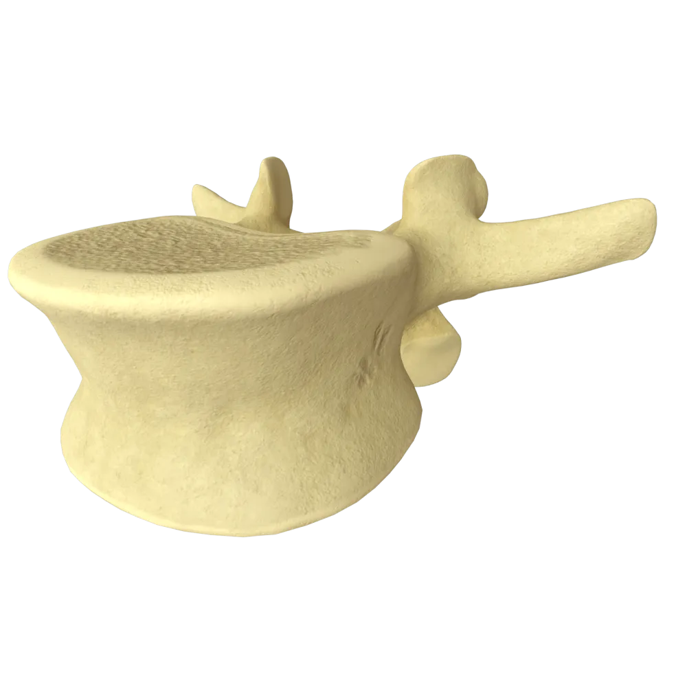
Axis vertebra anatomy quiz
Quiz on the normal anatomy of the axis vertebra (CII): structure, processes, joints, ligaments, and functional relationships.
1/20
bold
text
1. Which structure is morphologically unique to the axis vertebra (CII) and distinguishes it from all other cervical vertebrae?
-
Bifid spinous process
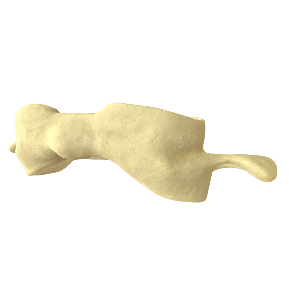
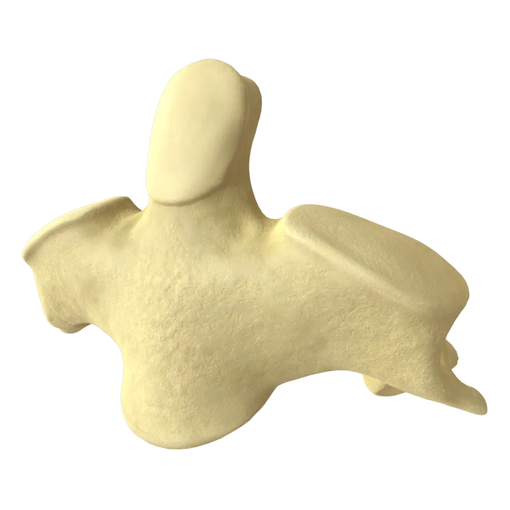
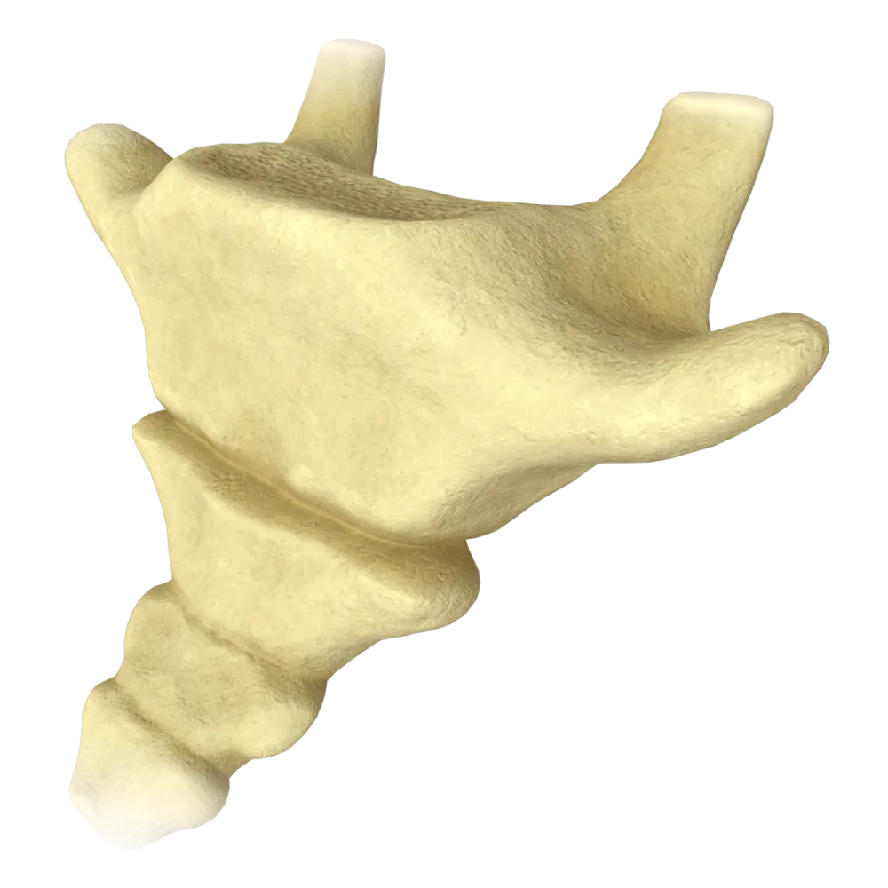
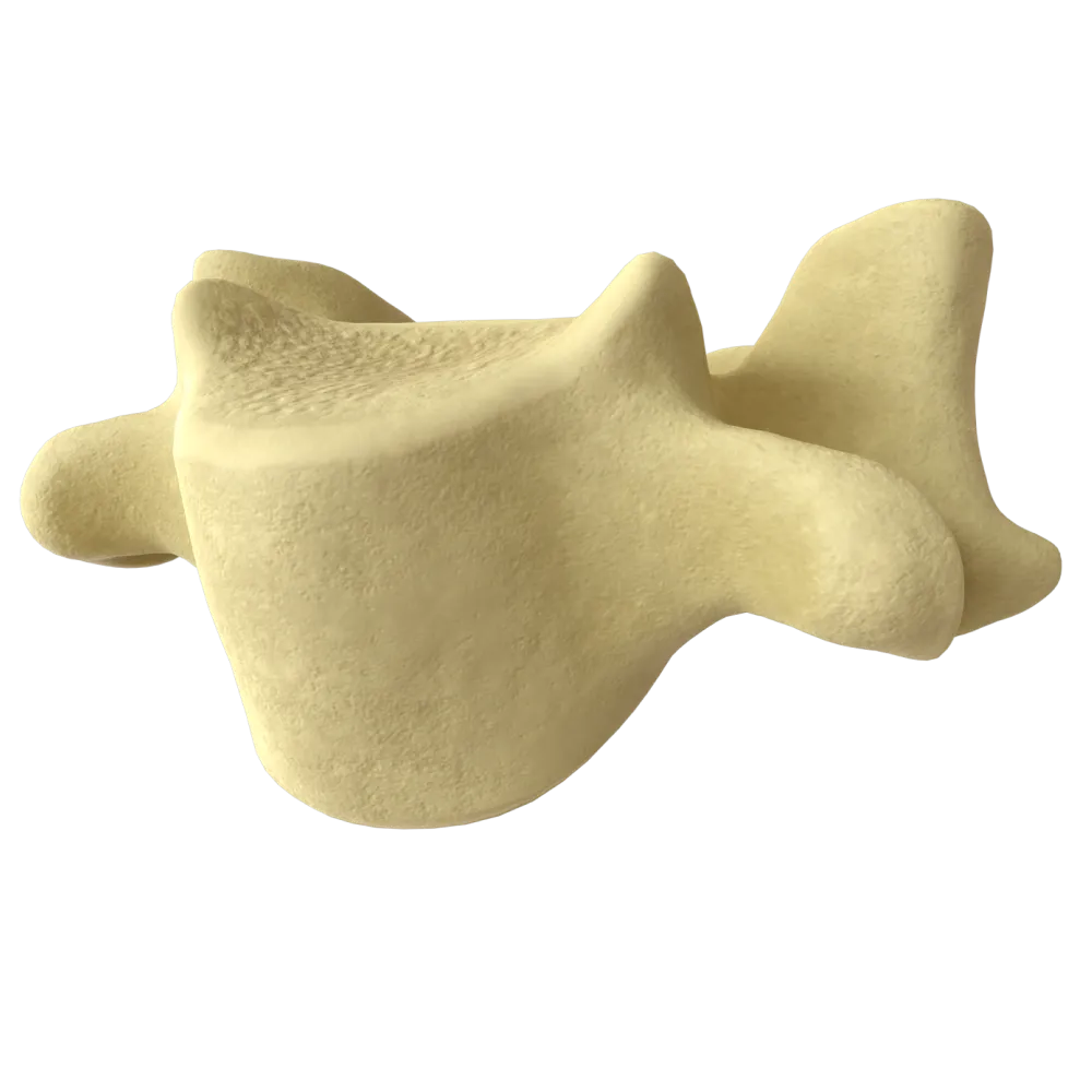
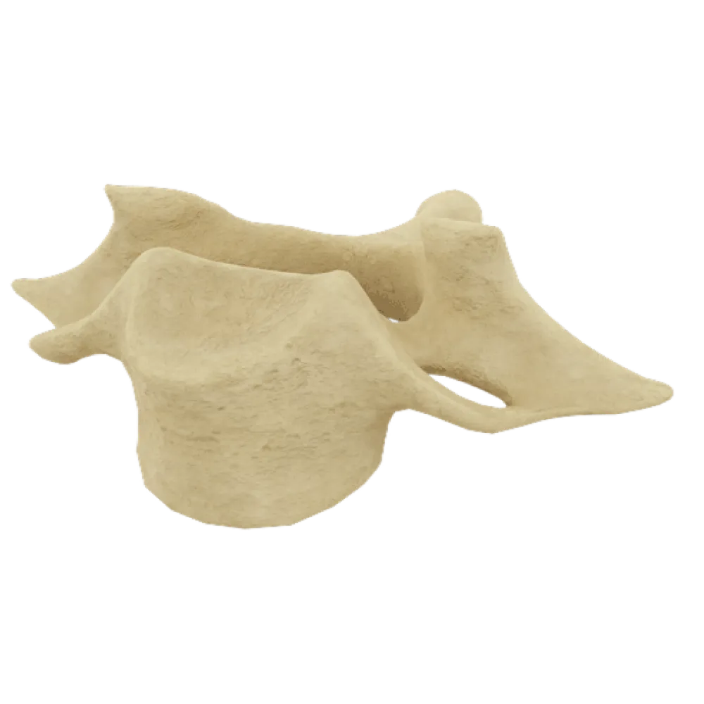
The dens of the axis (dens axis) is a bony process extending vertically from the body of CII. This structure is unique to the axis and is not found in any other vertebra.
-
Dens of the axis (dens axis)
The dens of the axis (dens axis) is a bony process extending vertically from the body of CII. This structure is unique to the axis and is not found in any other vertebra.
-
Transverse foramen (foramen transversarium)
The dens of the axis (dens axis) is a bony process extending vertically from the body of CII. This structure is unique to the axis and is not found in any other vertebra.
-
Articular surfaces on the vertebral body
The dens of the axis (dens axis) is a bony process extending vertically from the body of CII. This structure is unique to the axis and is not found in any other vertebra.
-
I find it difficult to answer
The dens of the axis (dens axis) is a bony process extending vertically from the body of CII. This structure is unique to the axis and is not found in any other vertebra.
2. What is the embryological origin of the dens of the axis (dens axis)?
-
A derivative of the body of the axis itself
Embryologically, the dens of the axis corresponds to the body of the atlas (CI). During development, the body of the first cervical vertebra “migrates” cranially and fuses with the body of CII, forming the dens axis.
-
A derivative of the reduced body of the atlas (CI)
Embryologically, the dens of the axis corresponds to the body of the atlas (CI). During development, the body of the first cervical vertebra “migrates” cranially and fuses with the body of CII, forming the dens axis.
-
Hypertrophied transverse process of CI
Embryologically, the dens of the axis corresponds to the body of the atlas (CI). During development, the body of the first cervical vertebra “migrates” cranially and fuses with the body of CII, forming the dens axis.
-
A remnant of the C0–CI intervertebral disc
Embryologically, the dens of the axis corresponds to the body of the atlas (CI). During development, the body of the first cervical vertebra “migrates” cranially and fuses with the body of CII, forming the dens axis.
-
I find it difficult to answer
Embryologically, the dens of the axis corresponds to the body of the atlas (CI). During development, the body of the first cervical vertebra “migrates” cranially and fuses with the body of CII, forming the dens axis.
3. What is the apex of the dens of the axis called?
-
Apex of the dens
The apex of the dens is designated as apex dentis. The apical ligament of the dens (ligamentum apicis dentis), which extends to the anterior margin of the foramen magnum, attaches to it.
-
Anterior articular surface of the dens
The apex of the dens is designated as apex dentis. The apical ligament of the dens (ligamentum apicis dentis), which extends to the anterior margin of the foramen magnum, attaches to it.
-
Neck of the dens
The apex of the dens is designated as apex dentis. The apical ligament of the dens (ligamentum apicis dentis), which extends to the anterior margin of the foramen magnum, attaches to it.
-
Base of the dens
The apex of the dens is designated as apex dentis. The apical ligament of the dens (ligamentum apicis dentis), which extends to the anterior margin of the foramen magnum, attaches to it.
-
I find it difficult to answer
The apex of the dens is designated as apex dentis. The apical ligament of the dens (ligamentum apicis dentis), which extends to the anterior margin of the foramen magnum, attaches to it.
4. Which articular surface of the dens of the axis articulates with the anterior arch of the atlas?
-
Posterior articular surface of the dens
The anterior articular surface of the dens (facies articularis anterior dentis) articulates with the facet for the dens (fovea dentis) on the inner surface of the anterior arch of the atlas, forming the median atlantoaxial joint (articulatio atlantoaxialis mediana).
-
Apex of the dens
The anterior articular surface of the dens (facies articularis anterior dentis) articulates with the facet for the dens (fovea dentis) on the inner surface of the anterior arch of the atlas, forming the median atlantoaxial joint (articulatio atlantoaxialis mediana).
-
Anterior articular surface of the dens
The anterior articular surface of the dens (facies articularis anterior dentis) articulates with the facet for the dens (fovea dentis) on the inner surface of the anterior arch of the atlas, forming the median atlantoaxial joint (articulatio atlantoaxialis mediana).
-
Base of the dens
The anterior articular surface of the dens (facies articularis anterior dentis) articulates with the facet for the dens (fovea dentis) on the inner surface of the anterior arch of the atlas, forming the median atlantoaxial joint (articulatio atlantoaxialis mediana).
-
I find it difficult to answer
The anterior articular surface of the dens (facies articularis anterior dentis) articulates with the facet for the dens (fovea dentis) on the inner surface of the anterior arch of the atlas, forming the median atlantoaxial joint (articulatio atlantoaxialis mediana).
5. With which structure does the posterior articular surface of the dens of the axis (facies articularis posterior dentis) articulate?
-
With the posterior arch of the atlas
The posterior articular surface of the dens (facies articularis posterior dentis) articulates with the transverse ligament of the atlas (ligamentum transversum atlantis), which holds the dens against the anterior arch of the atlas and is the principal stabilizer of the median atlantoaxial joint.
-
With the transverse ligament of the atlas (ligamentum transversum atlantis)
The posterior articular surface of the dens (facies articularis posterior dentis) articulates with the transverse ligament of the atlas (ligamentum transversum atlantis), which holds the dens against the anterior arch of the atlas and is the principal stabilizer of the median atlantoaxial joint.
-
With the occipital bone.
The posterior articular surface of the dens (facies articularis posterior dentis) articulates with the transverse ligament of the atlas (ligamentum transversum atlantis), which holds the dens against the anterior arch of the atlas and is the principal stabilizer of the median atlantoaxial joint.
-
With the body of the third cervical vertebra (CIII)
The posterior articular surface of the dens (facies articularis posterior dentis) articulates with the transverse ligament of the atlas (ligamentum transversum atlantis), which holds the dens against the anterior arch of the atlas and is the principal stabilizer of the median atlantoaxial joint.
-
I find it difficult to answer
The posterior articular surface of the dens (facies articularis posterior dentis) articulates with the transverse ligament of the atlas (ligamentum transversum atlantis), which holds the dens against the anterior arch of the atlas and is the principal stabilizer of the median atlantoaxial joint.
6. What are the superior articular surfaces of the axis that participate in the formation of the lateral atlantoaxial joints called?
-
Inferior articular surfaces
The superior articular surfaces of CII (facies articulares superiores) are located on the superior articular processes (processus articulares superiores) and articulate with the inferior articular surfaces of the lateral masses of the atlas, forming the paired lateral atlantoaxial joints (articulationes atlantoaxiales laterales).
-
Superior articular surfaces
The superior articular surfaces of CII (facies articulares superiores) are located on the superior articular processes (processus articulares superiores) and articulate with the inferior articular surfaces of the lateral masses of the atlas, forming the paired lateral atlantoaxial joints (articulationes atlantoaxiales laterales).
-
Uncinate processes
The superior articular surfaces of CII (facies articulares superiores) are located on the superior articular processes (processus articulares superiores) and articulate with the inferior articular surfaces of the lateral masses of the atlas, forming the paired lateral atlantoaxial joints (articulationes atlantoaxiales laterales).
-
Costal articular surfaces
The superior articular surfaces of CII (facies articulares superiores) are located on the superior articular processes (processus articulares superiores) and articulate with the inferior articular surfaces of the lateral masses of the atlas, forming the paired lateral atlantoaxial joints (articulationes atlantoaxiales laterales).
-
I find it difficult to answer
The superior articular surfaces of CII (facies articulares superiores) are located on the superior articular processes (processus articulares superiores) and articulate with the inferior articular surfaces of the lateral masses of the atlas, forming the paired lateral atlantoaxial joints (articulationes atlantoaxiales laterales).
7. What is the joint formed by the dens of the axis and the anterior arch of the atlas called?
-
Lateral atlanto-axial joint
The median atlantoaxial joint (articulatio atlantoaxialis mediana) is formed by the dens of the axis: anteriorly, with the facet for the dens on the anterior arch of the atlas, and posteriorly, with the transverse ligament of the atlas.
-
Atlanto-occipital joint
The median atlantoaxial joint (articulatio atlantoaxialis mediana) is formed by the dens of the axis: anteriorly, with the facet for the dens on the anterior arch of the atlas, and posteriorly, with the transverse ligament of the atlas.
-
Median atlanto-axial joint
The median atlantoaxial joint (articulatio atlantoaxialis mediana) is formed by the dens of the axis: anteriorly, with the facet for the dens on the anterior arch of the atlas, and posteriorly, with the transverse ligament of the atlas.
-
C1–C2 zygapophyseal joint
The median atlantoaxial joint (articulatio atlantoaxialis mediana) is formed by the dens of the axis: anteriorly, with the facet for the dens on the anterior arch of the atlas, and posteriorly, with the transverse ligament of the atlas.
-
I find it difficult to answer
The median atlantoaxial joint (articulatio atlantoaxialis mediana) is formed by the dens of the axis: anteriorly, with the facet for the dens on the anterior arch of the atlas, and posteriorly, with the transverse ligament of the atlas.
8. What type of movement is the primary movement in the median atlantoaxial joint?
-
Flexion and extension
The median atlanto-axial joint is cylindrical in shape, which permits its primary movement—rotation (rotational movement) around the vertical axis of the dens. Approximately 50% of all axial rotation of the neck occurs here.
-
Rotation (rotational movement) around the vertical axis
The median atlanto-axial joint is cylindrical in shape, which permits its primary movement—rotation (rotational movement) around the vertical axis of the dens. Approximately 50% of all axial rotation of the neck occurs here.
-
Lateral flexion
The median atlanto-axial joint is cylindrical in shape, which permits its primary movement—rotation (rotational movement) around the vertical axis of the dens. Approximately 50% of all axial rotation of the neck occurs here.
-
Circumductio (circumduction)
The median atlanto-axial joint is cylindrical in shape, which permits its primary movement—rotation (rotational movement) around the vertical axis of the dens. Approximately 50% of all axial rotation of the neck occurs here.
-
I find it difficult to answer
The median atlanto-axial joint is cylindrical in shape, which permits its primary movement—rotation (rotational movement) around the vertical axis of the dens. Approximately 50% of all axial rotation of the neck occurs here.
9. Which ligament attaches to the apex of the dens of the axis (apex dentis) and extends to the anterior margin of the foramen magnum?
-
Transverse ligament of the atlas
The apical ligament of the dens (ligamentum apicis dentis) is a median structure extending from the apex dentis to the anterior margin of the foramen magnum.
-
Ligamentum nuchae
The apical ligament of the dens (ligamentum apicis dentis) is a median structure extending from the apex dentis to the anterior margin of the foramen magnum.
-
Apical ligament of the dens
The apical ligament of the dens (ligamentum apicis dentis) is a median structure extending from the apex dentis to the anterior margin of the foramen magnum.
-
Tectorial membrane
The apical ligament of the dens (ligamentum apicis dentis) is a median structure extending from the apex dentis to the anterior margin of the foramen magnum.
-
I find it difficult to answer
The apical ligament of the dens (ligamentum apicis dentis) is a median structure extending from the apex dentis to the anterior margin of the foramen magnum.
10. To which bony structures do the alar ligaments (ligamenta alaria), which originate from the dens of the axis, attach?
-
To the anterior arch of the atlas and the occipital condyles
The alar ligaments (ligamenta alaria) arise from the lateral surfaces of the dens of CII and attach to the occipital condyles (pars condylaris ossis occipitalis) and the lateral masses of the atlas. They are the principal structures limiting excessive rotation and lateral flexion.
-
To the occipital condyles and the lateral masses of the atlas
The alar ligaments (ligamenta alaria) arise from the lateral surfaces of the dens of CII and attach to the occipital condyles (pars condylaris ossis occipitalis) and the lateral masses of the atlas. They are the principal structures limiting excessive rotation and lateral flexion.
-
Only to the basilar part of the occipital bone
The alar ligaments (ligamenta alaria) arise from the lateral surfaces of the dens of CII and attach to the occipital condyles (pars condylaris ossis occipitalis) and the lateral masses of the atlas. They are the principal structures limiting excessive rotation and lateral flexion.
-
To the posterior arch of the atlas
The alar ligaments (ligamenta alaria) arise from the lateral surfaces of the dens of CII and attach to the occipital condyles (pars condylaris ossis occipitalis) and the lateral masses of the atlas. They are the principal structures limiting excessive rotation and lateral flexion.
-
I find it difficult to answer
The alar ligaments (ligamenta alaria) arise from the lateral surfaces of the dens of CII and attach to the occipital condyles (pars condylaris ossis occipitalis) and the lateral masses of the atlas. They are the principal structures limiting excessive rotation and lateral flexion.
11. What is the transverse ligament of the atlas (ligamentum transversum atlantis) in the context of the ligamentous apparatus of the axis?
-
A ligament connecting the body of CII to the body of CIII
The transverse ligament of the atlas (ligamentum transversum atlantis) is the horizontal part of the cruciform ligament of the atlas (ligamentum cruciforme atlantis). It is located posterior to the dens of CII and holds it against the anterior arch of CI, preventing dorsal displacement of the dens into the vertebral canal.
-
The horizontal part of the cruciform ligament of the atlas, which holds the dens of CII against the anterior arch of CI
The transverse ligament of the atlas (ligamentum transversum atlantis) is the horizontal part of the cruciform ligament of the atlas (ligamentum cruciforme atlantis). It is located posterior to the dens of CII and holds it against the anterior arch of CI, preventing dorsal displacement of the dens into the vertebral canal.
-
A ligament attached to the apex dentis
The transverse ligament of the atlas (ligamentum transversum atlantis) is the horizontal part of the cruciform ligament of the atlas (ligamentum cruciforme atlantis). It is located posterior to the dens of CII and holds it against the anterior arch of CI, preventing dorsal displacement of the dens into the vertebral canal.
-
An external ligament covering the posterior arch of the atlas
The transverse ligament of the atlas (ligamentum transversum atlantis) is the horizontal part of the cruciform ligament of the atlas (ligamentum cruciforme atlantis). It is located posterior to the dens of CII and holds it against the anterior arch of CI, preventing dorsal displacement of the dens into the vertebral canal.
-
I find it difficult to answer
The transverse ligament of the atlas (ligamentum transversum atlantis) is the horizontal part of the cruciform ligament of the atlas (ligamentum cruciforme atlantis). It is located posterior to the dens of CII and holds it against the anterior arch of CI, preventing dorsal displacement of the dens into the vertebral canal.
12. What is the composition of the cruciform ligament of the atlas (ligamentum cruciforme atlantis)?
-
Only the transverse ligament of the atlas
The cruciform ligament of the atlas (ligamentum cruciforme atlantis) consists of the horizontally oriented transverse ligament of the atlas (ligamentum transversum atlantis) and vertical longitudinal bands (fasciculi longitudinales) extending from the body of CII to the basilar part of the occipital bone, thereby forming a cruciform configuration.
-
The transverse ligament of the atlas and vertical longitudinal bands (fasciculi longitudinales)
The cruciform ligament of the atlas (ligamentum cruciforme atlantis) consists of the horizontally oriented transverse ligament of the atlas (ligamentum transversum atlantis) and vertical longitudinal bands (fasciculi longitudinales) extending from the body of CII to the basilar part of the occipital bone, thereby forming a cruciform configuration.
-
The transverse ligament of the atlas and the alar ligaments
The cruciform ligament of the atlas (ligamentum cruciforme atlantis) consists of the horizontally oriented transverse ligament of the atlas (ligamentum transversum atlantis) and vertical longitudinal bands (fasciculi longitudinales) extending from the body of CII to the basilar part of the occipital bone, thereby forming a cruciform configuration.
-
The apical ligament of the dens and the alar ligaments
The cruciform ligament of the atlas (ligamentum cruciforme atlantis) consists of the horizontally oriented transverse ligament of the atlas (ligamentum transversum atlantis) and vertical longitudinal bands (fasciculi longitudinales) extending from the body of CII to the basilar part of the occipital bone, thereby forming a cruciform configuration.
-
I find it difficult to answer
The cruciform ligament of the atlas (ligamentum cruciforme atlantis) consists of the horizontally oriented transverse ligament of the atlas (ligamentum transversum atlantis) and vertical longitudinal bands (fasciculi longitudinales) extending from the body of CII to the basilar part of the occipital bone, thereby forming a cruciform configuration.
13. Which strong ligament covers the cruciform ligament of the atlas posteriorly and is the cranial continuation of the posterior longitudinal ligament of the vertebral column?
-
Ligamentum flavum
The tectorial membrane (membrana tectoria) is a broad ligament that constitutes the cranial continuation of the posterior longitudinal ligament. It covers the cruciform ligament of the atlas and the alar ligaments posteriorly and attaches to the clivus of the occipital bone (clivus).
-
Tectorial membrane
The tectorial membrane (membrana tectoria) is a broad ligament that constitutes the cranial continuation of the posterior longitudinal ligament. It covers the cruciform ligament of the atlas and the alar ligaments posteriorly and attaches to the clivus of the occipital bone (clivus).
-
Ligamentum nuchae
The tectorial membrane (membrana tectoria) is a broad ligament that constitutes the cranial continuation of the posterior longitudinal ligament. It covers the cruciform ligament of the atlas and the alar ligaments posteriorly and attaches to the clivus of the occipital bone (clivus).
-
Posterior atlanto-occipital membrane
The tectorial membrane (membrana tectoria) is a broad ligament that constitutes the cranial continuation of the posterior longitudinal ligament. It covers the cruciform ligament of the atlas and the alar ligaments posteriorly and attaches to the clivus of the occipital bone (clivus).
-
I find it difficult to answer
The tectorial membrane (membrana tectoria) is a broad ligament that constitutes the cranial continuation of the posterior longitudinal ligament. It covers the cruciform ligament of the atlas and the alar ligaments posteriorly and attaches to the clivus of the occipital bone (clivus).
14. What is the foramen in the transverse process of the axis called, and which vascular structure passes through it?
-
Foramen transversarium; vertebral artery (a. vertebralis)
The transverse foramen (foramen transversarium) of the transverse process of CII transmits the vertebral artery (a. vertebralis).
-
Foramen intervertebrale; spinal nerve
The transverse foramen (foramen transversarium) of the transverse process of CII transmits the vertebral artery (a. vertebralis).
-
Foramen nutricium; nutrient artery of the vertebra
The transverse foramen (foramen transversarium) of the transverse process of CII transmits the vertebral artery (a. vertebralis).
-
Canalis vertebralis; spinal cord
The transverse foramen (foramen transversarium) of the transverse process of CII transmits the vertebral artery (a. vertebralis).
-
I find it difficult to answer
The transverse foramen (foramen transversarium) of the transverse process of CII transmits the vertebral artery (a. vertebralis).
15. Several muscles attach to the spinous process of the axis. Which of the following muscles does NOT attach to the processus spinosus axis?
-
M. semispinalis cervicis
M. rectus capitis posterior minor originates from the tuberculum posterius of the arch of the atlas (CI), not from the axis. The following muscles attach to the processus spinosus axis (CII): m. rectus capitis posterior major muscle semispinalis cervicis, and m. multifidus muscle.
-
Rectus capitis posterior major muscle
M. rectus capitis posterior minor originates from the tuberculum posterius of the arch of the atlas (CI), not from the axis. The following muscles attach to the processus spinosus axis (CII): m. rectus capitis posterior major muscle semispinalis cervicis, and m. multifidus muscle.
-
Rectus capitis posterior minor muscle
M. rectus capitis posterior minor originates from the tuberculum posterius of the arch of the atlas (CI), not from the axis. The following muscles attach to the processus spinosus axis (CII): m. rectus capitis posterior major muscle semispinalis cervicis, and m. multifidus muscle.
-
Multifidus muscle
M. rectus capitis posterior minor originates from the tuberculum posterius of the arch of the atlas (CI), not from the axis. The following muscles attach to the processus spinosus axis (CII): m. rectus capitis posterior major muscle semispinalis cervicis, and m. multifidus muscle.
-
I find it difficult to answer
M. rectus capitis posterior minor originates from the tuberculum posterius of the arch of the atlas (CI), not from the axis. The following muscles attach to the processus spinosus axis (CII): m. rectus capitis posterior major muscle semispinalis cervicis, and m. multifidus muscle.
16. Does the second cervical vertebra have superior articular processes?
-
Yes, they are well developed and project upward from the vertebral arch, as in all other vertebrae
Unlike typical vertebrae, the axis (C2) lacks classical, discrete superior articular processes (processus articulares superiores). Instead, large superior articular surfaces (facies articulares superiores) are located on either side of the dens (on the lateral parts of the vertebral body). They face superiorly and laterally and articulate with the inferior articular surfaces of the atlas (C1), forming the lateral atlantoaxial joints. The inferior articular processes (processus articulares inferiores) of C2, however, are developed in the typical manner.
-
No, instead of processes, there are superior articular surfaces (facies articulares superiores) located directly on the vertebral body
Unlike typical vertebrae, the axis (C2) lacks classical, discrete superior articular processes (processus articulares superiores). Instead, large superior articular surfaces (facies articulares superiores) are located on either side of the dens (on the lateral parts of the vertebral body). They face superiorly and laterally and articulate with the inferior articular surfaces of the atlas (C1), forming the lateral atlantoaxial joints. The inferior articular processes (processus articulares inferiores) of C2, however, are developed in the typical manner.
-
No, the axis lacks all articular processes
Unlike typical vertebrae, the axis (C2) lacks classical, discrete superior articular processes (processus articulares superiores). Instead, large superior articular surfaces (facies articulares superiores) are located on either side of the dens (on the lateral parts of the vertebral body). They face superiorly and laterally and articulate with the inferior articular surfaces of the atlas (C1), forming the lateral atlantoaxial joints. The inferior articular processes (processus articulares inferiores) of C2, however, are developed in the typical manner.
-
Yes, they fuse with the anterior arch of the atlas to form a single immobile block
Unlike typical vertebrae, the axis (C2) lacks classical, discrete superior articular processes (processus articulares superiores). Instead, large superior articular surfaces (facies articulares superiores) are located on either side of the dens (on the lateral parts of the vertebral body). They face superiorly and laterally and articulate with the inferior articular surfaces of the atlas (C1), forming the lateral atlantoaxial joints. The inferior articular processes (processus articulares inferiores) of C2, however, are developed in the typical manner.
-
I find it difficult to answer
Unlike typical vertebrae, the axis (C2) lacks classical, discrete superior articular processes (processus articulares superiores). Instead, large superior articular surfaces (facies articulares superiores) are located on either side of the dens (on the lateral parts of the vertebral body). They face superiorly and laterally and articulate with the inferior articular surfaces of the atlas (C1), forming the lateral atlantoaxial joints. The inferior articular processes (processus articulares inferiores) of C2, however, are developed in the typical manner.
17. Does the second cervical vertebra have a superior vertebral notch (incisura vertebralis superior)?
-
Yes, it is as well developed as in the other cervical vertebrae
C2 has no discrete superior articular process; instead, a broad articular facet lies directly on the vertebral body. Consequently, the typical superior notch is absent (or represented by a barely perceptible groove posterior to the joint).
-
It is absent or extremely poorly developed
C2 has no discrete superior articular process; instead, a broad articular facet lies directly on the vertebral body. Consequently, the typical superior notch is absent (or represented by a barely perceptible groove posterior to the joint).
-
Yes, it forms a deep bony canal for the exit of the C1 nerve
C2 has no discrete superior articular process; instead, a broad articular facet lies directly on the vertebral body. Consequently, the typical superior notch is absent (or represented by a barely perceptible groove posterior to the joint).
-
No, the spinous process is located in its place
C2 has no discrete superior articular process; instead, a broad articular facet lies directly on the vertebral body. Consequently, the typical superior notch is absent (or represented by a barely perceptible groove posterior to the joint).
-
I find it difficult to answer
C2 has no discrete superior articular process; instead, a broad articular facet lies directly on the vertebral body. Consequently, the typical superior notch is absent (or represented by a barely perceptible groove posterior to the joint).
18. The inferior articular surfaces of the axis (facies articulares inferiores) form joints with the articular surfaces of which vertebra?
-
With the superior articular processes of the atlas (CI)
The inferior articular processes of the axis (processus articulares inferiores) bear inferior articular surfaces (facies articulares inferiores), which articulate with the superior articular surfaces of the third cervical vertebra (CIII), forming typical zygapophyseal joints (articulationes zygapophysiales).
-
With the superior articular processes of the third cervical vertebra (CIII)
The inferior articular processes of the axis (processus articulares inferiores) bear inferior articular surfaces (facies articulares inferiores), which articulate with the superior articular surfaces of the third cervical vertebra (CIII), forming typical zygapophyseal joints (articulationes zygapophysiales).
-
With the occipital condyles
The inferior articular processes of the axis (processus articulares inferiores) bear inferior articular surfaces (facies articulares inferiores), which articulate with the superior articular surfaces of the third cervical vertebra (CIII), forming typical zygapophyseal joints (articulationes zygapophysiales).
-
With the inferior articular processes of CIII
The inferior articular processes of the axis (processus articulares inferiores) bear inferior articular surfaces (facies articulares inferiores), which articulate with the superior articular surfaces of the third cervical vertebra (CIII), forming typical zygapophyseal joints (articulationes zygapophysiales).
-
I find it difficult to answer
The inferior articular processes of the axis (processus articulares inferiores) bear inferior articular surfaces (facies articulares inferiores), which articulate with the superior articular surfaces of the third cervical vertebra (CIII), forming typical zygapophyseal joints (articulationes zygapophysiales).
19. Which of the following anatomical structures is NOT characteristic of the second cervical vertebra (the axis)?
-
Dens
Anterior and posterior arches instead of a large body are characteristic of the first cervical vertebra—the atlas (C1).
-
Anterior arch (arcus anterior)
Anterior and posterior arches instead of a large body are characteristic of the first cervical vertebra—the atlas (C1).
-
Transverse foramen (foramen transversarium)
Anterior and posterior arches instead of a large body are characteristic of the first cervical vertebra—the atlas (C1).
-
Bifid spinous process
Anterior and posterior arches instead of a large body are characteristic of the first cervical vertebra—the atlas (C1).
-
I find it difficult to answer
Anterior and posterior arches instead of a large body are characteristic of the first cervical vertebra—the atlas (C1).
20. Which of the following names is NOT applied to the human second cervical vertebra (C2)?
-
Axis
Atlas (atlas) is the name of the first cervical vertebra (C1).
-
Atlas
Atlas (atlas) is the name of the first cervical vertebra (C1).
-
Axis
Atlas (atlas) is the name of the first cervical vertebra (C1).
-
Epistropheus
Atlas (atlas) is the name of the first cervical vertebra (C1).
-
I find it difficult to answer
Atlas (atlas) is the name of the first cervical vertebra (C1).
Retake this quiz?
Your current progress will be reset.
