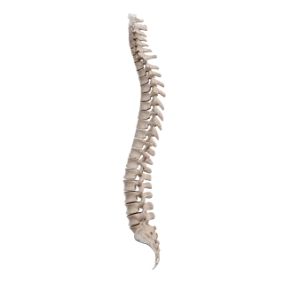
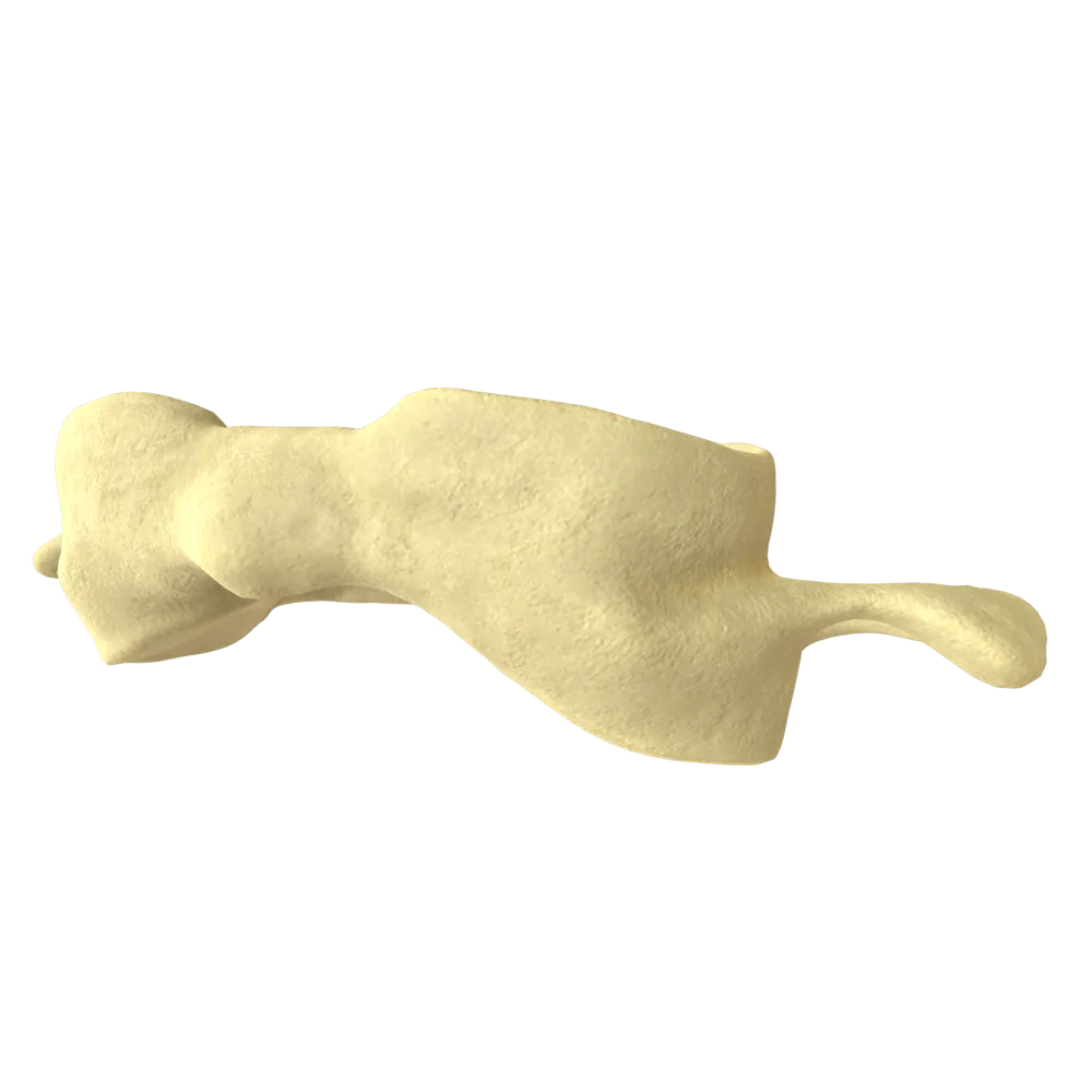
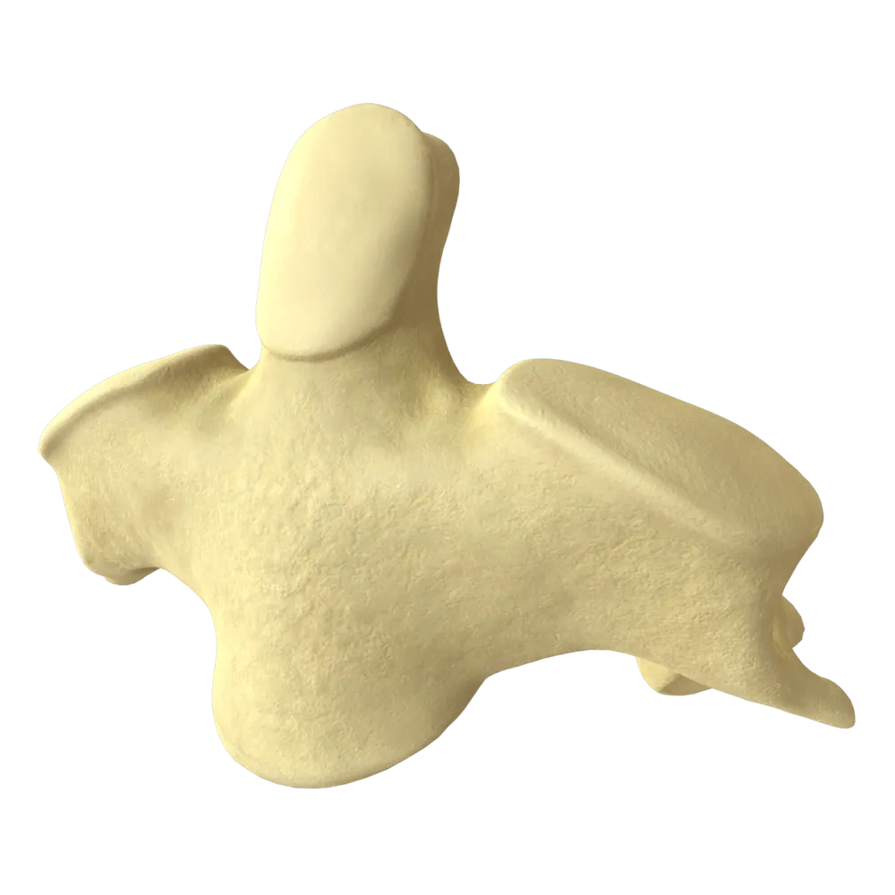

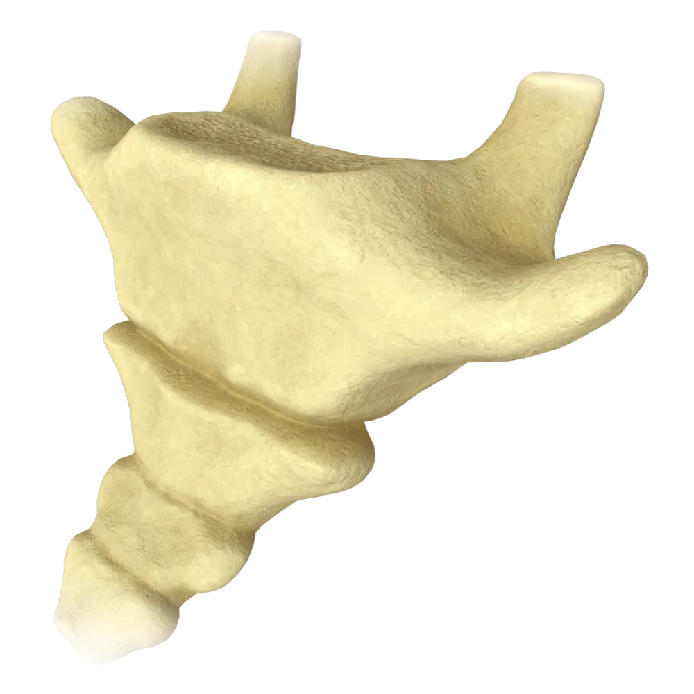
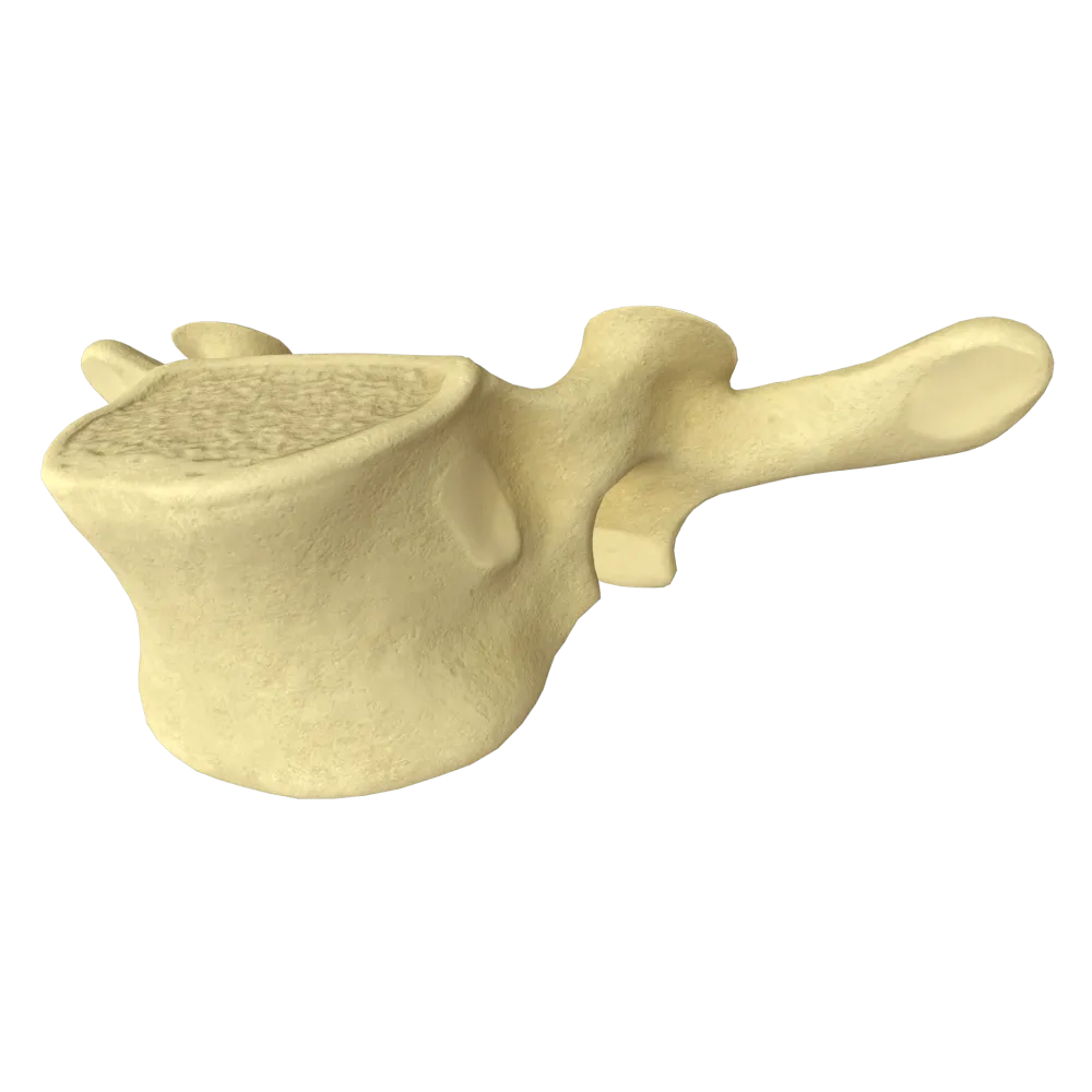
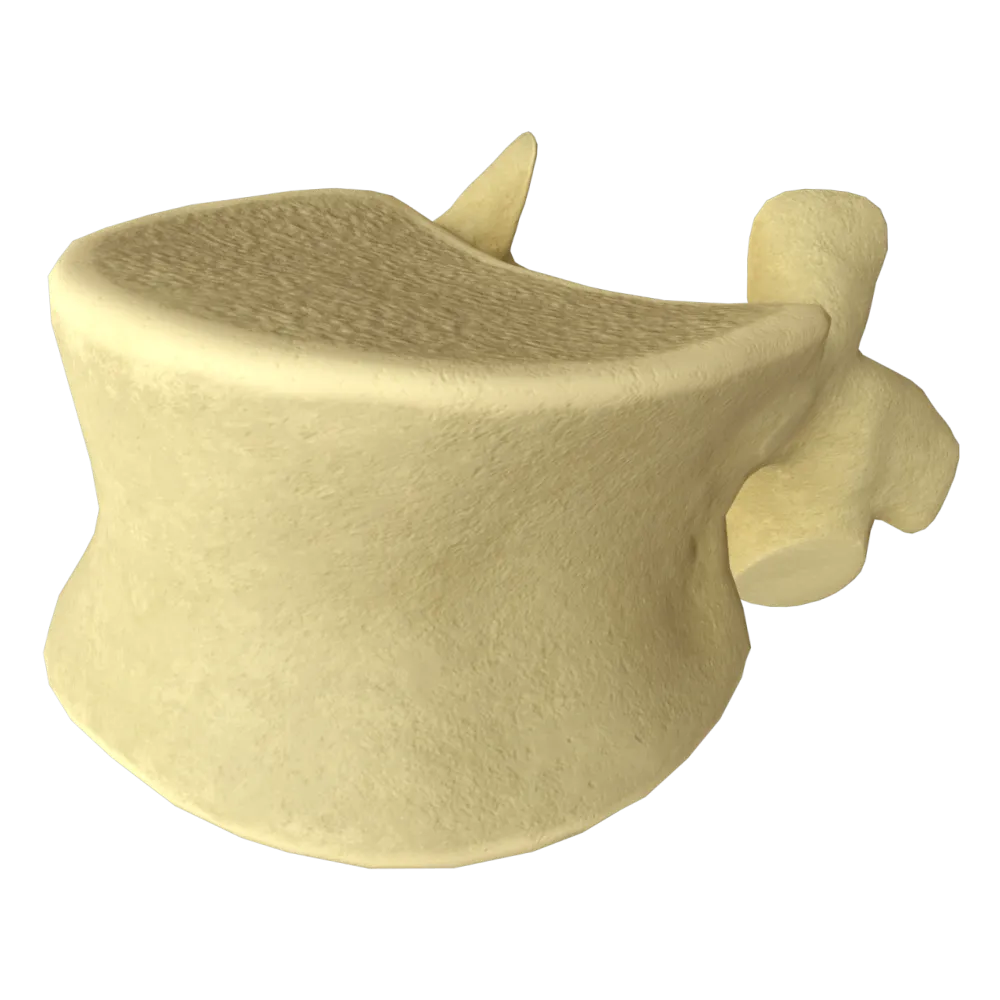
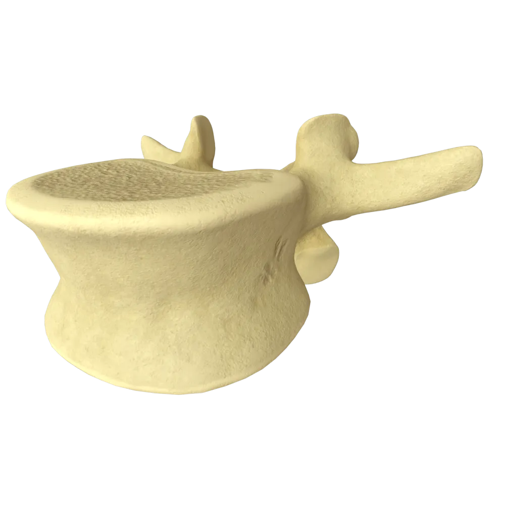
Sixth cervical vertebra anatomy quiz
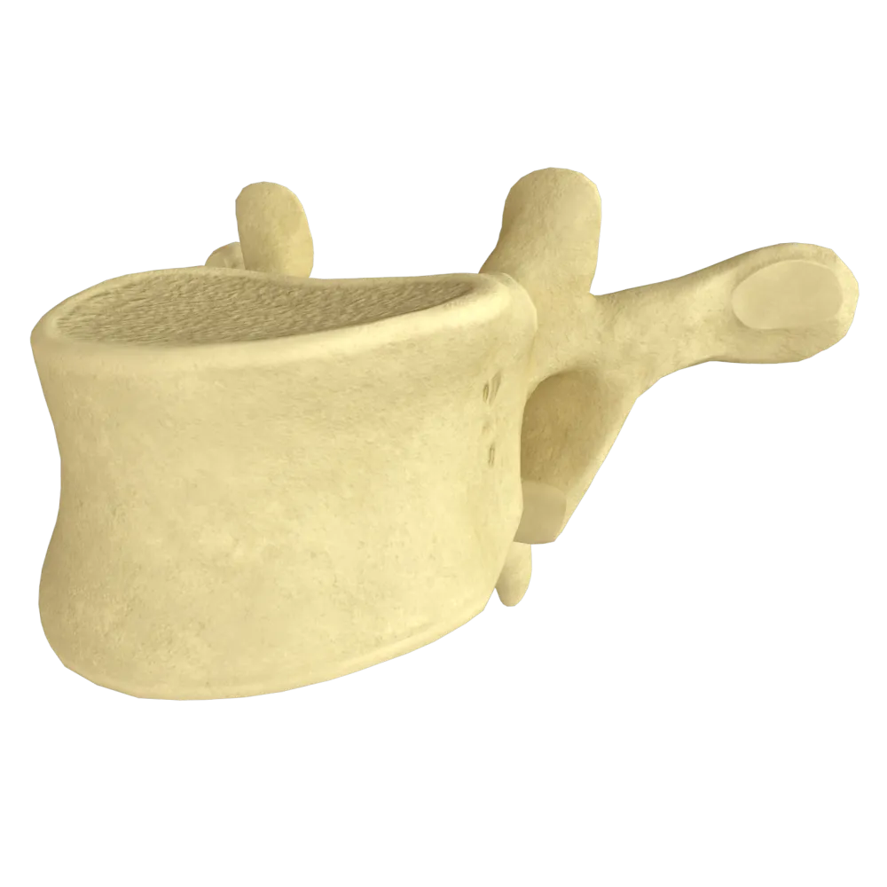
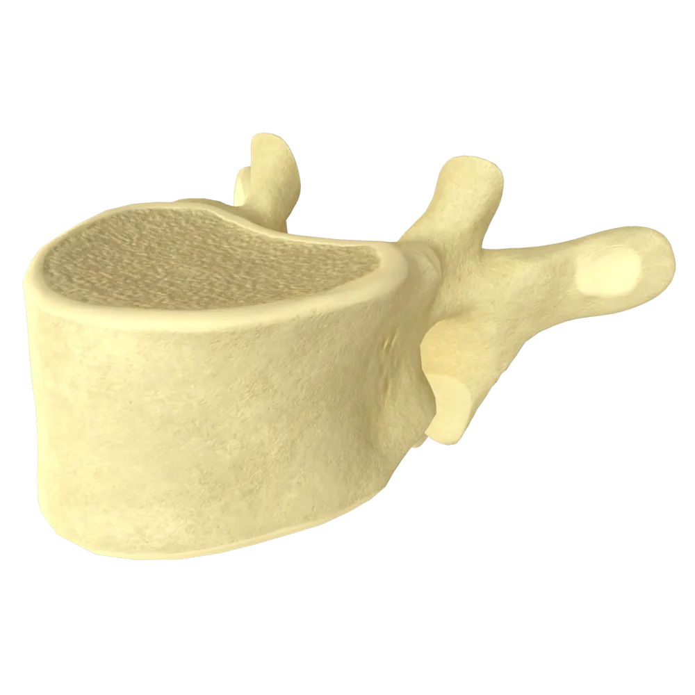
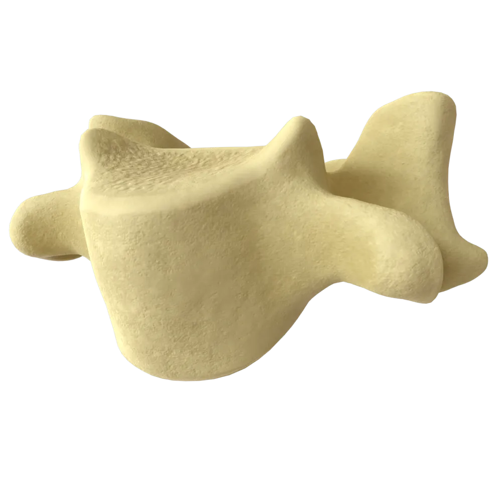
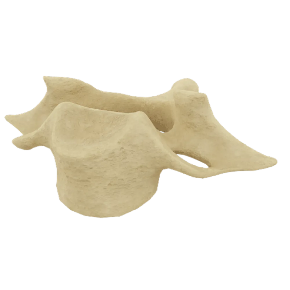
Normal anatomy quiz of the sixth cervical vertebra: structure, processes, foramina, articular surfaces, and associated structures.
1/20
bold
text
1. Which anatomical feature of the body of the sixth cervical vertebra distinguishes it from the thoracic vertebrae?
-
Presence of costal facets on the lateral surfaces of the body
Cervical vertebrae, including C6, have uncinate processes (processus uncinatus) on the superolateral margins of the body, which form the uncovertebral joints (joints of Luschka). This is a characteristic feature of the cervical region that is absent in the thoracic vertebrae.
-
Presence of uncinate processes (processus uncinatus) on the superolateral margins of the body
Cervical vertebrae, including C6, have uncinate processes (processus uncinatus) on the superolateral margins of the body, which form the uncovertebral joints (joints of Luschka). This is a characteristic feature of the cervical region that is absent in the thoracic vertebrae.
-
Absence of foramina in the transverse processes
Cervical vertebrae, including C6, have uncinate processes (processus uncinatus) on the superolateral margins of the body, which form the uncovertebral joints (joints of Luschka). This is a characteristic feature of the cervical region that is absent in the thoracic vertebrae.
-
Presence of two superior and two inferior costal demifacets
Cervical vertebrae, including C6, have uncinate processes (processus uncinatus) on the superolateral margins of the body, which form the uncovertebral joints (joints of Luschka). This is a characteristic feature of the cervical region that is absent in the thoracic vertebrae.
-
I find it difficult to answer
Cervical vertebrae, including C6, have uncinate processes (processus uncinatus) on the superolateral margins of the body, which form the uncovertebral joints (joints of Luschka). This is a characteristic feature of the cervical region that is absent in the thoracic vertebrae.
2. Through which structure of the sixth cervical vertebra does the vertebral artery (arteria vertebralis) pass?
-
Through the intervertebral foramen (foramen intervertebrale)
The vertebral artery passes through the transverse foramina (foramina transversaria) of the cervical vertebrae from C6 to C1. It generally enters the canal formed by the transverse foramina at the C6 level.
-
Through the vertebral foramen (foramen vertebrale)
The vertebral artery passes through the transverse foramina (foramina transversaria) of the cervical vertebrae from C6 to C1. It generally enters the canal formed by the transverse foramina at the C6 level.
-
Through the transverse foramen (foramen transversarium)
The vertebral artery passes through the transverse foramina (foramina transversaria) of the cervical vertebrae from C6 to C1. It generally enters the canal formed by the transverse foramina at the C6 level.
-
Through the nutrient foramen (foramen nutricium) of the vertebral body
The vertebral artery passes through the transverse foramina (foramina transversaria) of the cervical vertebrae from C6 to C1. It generally enters the canal formed by the transverse foramina at the C6 level.
-
I find it difficult to answer
The vertebral artery passes through the transverse foramina (foramina transversaria) of the cervical vertebrae from C6 to C1. It generally enters the canal formed by the transverse foramina at the C6 level.
3. What is the name of the anterior tubercle of the transverse process of the sixth cervical vertebra that has clinical topographic anatomical significance?
-
Lesgaft's tubercle
The anterior tubercle of the transverse process of C6 is called the carotid tubercle (tuberculum caroticum, Chassaignac's tubercle). The common carotid artery lies adjacent to it, allowing the artery to be compressed against it to stop bleeding.
-
Chassaignac's tubercle (tuberculum caroticum)
The anterior tubercle of the transverse process of C6 is called the carotid tubercle (tuberculum caroticum, Chassaignac's tubercle). The common carotid artery lies adjacent to it, allowing the artery to be compressed against it to stop bleeding.
-
Lisfranc's tubercle
The anterior tubercle of the transverse process of C6 is called the carotid tubercle (tuberculum caroticum, Chassaignac's tubercle). The common carotid artery lies adjacent to it, allowing the artery to be compressed against it to stop bleeding.
-
Gérard-Marchant's tubercle
The anterior tubercle of the transverse process of C6 is called the carotid tubercle (tuberculum caroticum, Chassaignac's tubercle). The common carotid artery lies adjacent to it, allowing the artery to be compressed against it to stop bleeding.
-
I find it difficult to answer
The anterior tubercle of the transverse process of C6 is called the carotid tubercle (tuberculum caroticum, Chassaignac's tubercle). The common carotid artery lies adjacent to it, allowing the artery to be compressed against it to stop bleeding.
4. What is the shape of the vertebral foramen (foramen vertebrale) of the sixth cervical vertebra?
-
Round
The vertebral foramen of the cervical vertebrae, including C6, is triangular with rounded angles and is relatively large compared with those of the thoracic and lumbar vertebrae, providing a capacious vertebral canal in the cervical region.
-
Triangular with rounded angles
The vertebral foramen of the cervical vertebrae, including C6, is triangular with rounded angles and is relatively large compared with those of the thoracic and lumbar vertebrae, providing a capacious vertebral canal in the cervical region.
-
Oval, elongated in the sagittal direction
The vertebral foramen of the cervical vertebrae, including C6, is triangular with rounded angles and is relatively large compared with those of the thoracic and lumbar vertebrae, providing a capacious vertebral canal in the cervical region.
-
Reniform (bean-shaped)
The vertebral foramen of the cervical vertebrae, including C6, is triangular with rounded angles and is relatively large compared with those of the thoracic and lumbar vertebrae, providing a capacious vertebral canal in the cervical region.
-
I find it difficult to answer
The vertebral foramen of the cervical vertebrae, including C6, is triangular with rounded angles and is relatively large compared with those of the thoracic and lumbar vertebrae, providing a capacious vertebral canal in the cervical region.
5. How are the articular surfaces of the inferior articular processes of the sixth cervical vertebra oriented?
-
Directed anteriorly and inferiorly, at an angle of approximately 45° to the horizontal plane
The articular surfaces of the cervical vertebrae are positioned almost horizontally (at an angle of approximately 45° to the horizontal, with the inferior processes inclined anteriorly and inferiorly), allowing a large range of motion in the cervical region, including flexion, extension, and rotation.
-
Directed strictly vertically, as in the lumbar vertebrae
The articular surfaces of the cervical vertebrae are positioned almost horizontally (at an angle of approximately 45° to the horizontal, with the inferior processes inclined anteriorly and inferiorly), allowing a large range of motion in the cervical region, including flexion, extension, and rotation.
-
Directed posteriorly and medially, as in the thoracic vertebrae
The articular surfaces of the cervical vertebrae are positioned almost horizontally (at an angle of approximately 45° to the horizontal, with the inferior processes inclined anteriorly and inferiorly), allowing a large range of motion in the cervical region, including flexion, extension, and rotation.
-
Directed strictly horizontally, parallel to the plane of the vertebral body
The articular surfaces of the cervical vertebrae are positioned almost horizontally (at an angle of approximately 45° to the horizontal, with the inferior processes inclined anteriorly and inferiorly), allowing a large range of motion in the cervical region, including flexion, extension, and rotation.
-
I find it difficult to answer
The articular surfaces of the cervical vertebrae are positioned almost horizontally (at an angle of approximately 45° to the horizontal, with the inferior processes inclined anteriorly and inferiorly), allowing a large range of motion in the cervical region, including flexion, extension, and rotation.
6. Which spinal nerve exits through the intervertebral foramen between C6 and C7?
-
Fifth cervical spinal nerve (C5)
In the cervical region, spinal nerves are numbered according to the vertebra below: the C7 nerve exits through the intervertebral foramen between C6 and C7. This is the standard numbering rule for the cervical nerves (the exception is C8, which exits between C7 and Th1).
-
Sixth cervical spinal nerve (C6)
In the cervical region, spinal nerves are numbered according to the vertebra below: the C7 nerve exits through the intervertebral foramen between C6 and C7. This is the standard numbering rule for the cervical nerves (the exception is C8, which exits between C7 and Th1).
-
Seventh cervical spinal nerve (C7)
In the cervical region, spinal nerves are numbered according to the vertebra below: the C7 nerve exits through the intervertebral foramen between C6 and C7. This is the standard numbering rule for the cervical nerves (the exception is C8, which exits between C7 and Th1).
-
Eighth cervical spinal nerve (C8)
In the cervical region, spinal nerves are numbered according to the vertebra below: the C7 nerve exits through the intervertebral foramen between C6 and C7. This is the standard numbering rule for the cervical nerves (the exception is C8, which exits between C7 and Th1).
-
I find it difficult to answer
In the cervical region, spinal nerves are numbered according to the vertebra below: the C7 nerve exits through the intervertebral foramen between C6 and C7. This is the standard numbering rule for the cervical nerves (the exception is C8, which exits between C7 and Th1).
7. What is the inferior notch (incisura vertebralis inferior) of the arch of the sixth cervical vertebra called, and which structure does it form together with the vertebra below?
-
Forms the vertebral foramen together with the body of C7
The inferior vertebral notch of C6 and the superior vertebral notch of C7 together form the intervertebral foramen (foramen intervertebrale), through which the C7 spinal nerve passes with its roots and vessels.
-
Forms the intervertebral foramen (foramen intervertebrale) together with the superior notch of C7
The inferior vertebral notch of C6 and the superior vertebral notch of C7 together form the intervertebral foramen (foramen intervertebrale), through which the C7 spinal nerve passes with its roots and vessels.
-
Forms the transverse foramen together with the costotransverse joint
The inferior vertebral notch of C6 and the superior vertebral notch of C7 together form the intervertebral foramen (foramen intervertebrale), through which the C7 spinal nerve passes with its roots and vessels.
-
Forms the carotid triangle together with the anterior scalene muscle
The inferior vertebral notch of C6 and the superior vertebral notch of C7 together form the intervertebral foramen (foramen intervertebrale), through which the C7 spinal nerve passes with its roots and vessels.
-
I find it difficult to answer
The inferior vertebral notch of C6 and the superior vertebral notch of C7 together form the intervertebral foramen (foramen intervertebrale), through which the C7 spinal nerve passes with its roots and vessels.
8. To what does the anterior longitudinal ligament attach at the sixth cervical vertebra (ligamentum longitudinale anterius)?
-
To the transverse processes
The anterior longitudinal ligament runs along the anterior surfaces of the vertebral bodies from the occipital bone to the sacrum, firmly blending with the intervertebral discs and margins of the vertebral bodies, including C6.
-
To the spinous process
The anterior longitudinal ligament runs along the anterior surfaces of the vertebral bodies from the occipital bone to the sacrum, firmly blending with the intervertebral discs and margins of the vertebral bodies, including C6.
-
To the vertebral body (anterior surface)
The anterior longitudinal ligament runs along the anterior surfaces of the vertebral bodies from the occipital bone to the sacrum, firmly blending with the intervertebral discs and margins of the vertebral bodies, including C6.
-
To the articular processes
The anterior longitudinal ligament runs along the anterior surfaces of the vertebral bodies from the occipital bone to the sacrum, firmly blending with the intervertebral discs and margins of the vertebral bodies, including C6.
-
I find it difficult to answer
The anterior longitudinal ligament runs along the anterior surfaces of the vertebral bodies from the occipital bone to the sacrum, firmly blending with the intervertebral discs and margins of the vertebral bodies, including C6.
9. What is the characteristic feature of the spinous process of the sixth cervical vertebra compared with the spinous process of C7?
-
The spinous process of C6 is longer than that of C7 and is readily palpable
The spinous processes of C3–C6 are bifid at their tips (bifidi), whereas C7 has a long, non-bifid spinous process that projects beneath the skin (vertebra prominens). The spinous process of C6 is shorter and less prominent than that of C7.
-
The spinous process of C6 is bifid at its tip and shorter than the invisible (prominent) process of C7
The spinous processes of C3–C6 are bifid at their tips (bifidi), whereas C7 has a long, non-bifid spinous process that projects beneath the skin (vertebra prominens). The spinous process of C6 is shorter and less prominent than that of C7.
-
The spinous process of C6 is not bifid and is directed strictly horizontally
The spinous processes of C3–C6 are bifid at their tips (bifidi), whereas C7 has a long, non-bifid spinous process that projects beneath the skin (vertebra prominens). The spinous process of C6 is shorter and less prominent than that of C7.
-
The spinous process of C6 is absent, as in the atlas
The spinous processes of C3–C6 are bifid at their tips (bifidi), whereas C7 has a long, non-bifid spinous process that projects beneath the skin (vertebra prominens). The spinous process of C6 is shorter and less prominent than that of C7.
-
I find it difficult to answer
The spinous processes of C3–C6 are bifid at their tips (bifidi), whereas C7 has a long, non-bifid spinous process that projects beneath the skin (vertebra prominens). The spinous process of C6 is shorter and less prominent than that of C7.
10. Which ligament attaches to the tips of the spinous processes of the cervical vertebrae, including C6, forming a midline structure on the posterior aspect of the neck?
-
Nuchal ligament (ligamentum nuchae)
The nuchal ligament (ligamentum nuchae) is a strong, midline elastic ligament that attaches to the tips of the spinous processes of the C1–C7 cervical vertebrae and to the external occipital crest. It is the cervical analogue of the supraspinous ligament and limits flexion.
-
Posterior longitudinal ligament (ligamentum longitudinale posterius)
The nuchal ligament (ligamentum nuchae) is a strong, midline elastic ligament that attaches to the tips of the spinous processes of the C1–C7 cervical vertebrae and to the external occipital crest. It is the cervical analogue of the supraspinous ligament and limits flexion.
-
Interspinous ligament (ligamentum interspinale)
The nuchal ligament (ligamentum nuchae) is a strong, midline elastic ligament that attaches to the tips of the spinous processes of the C1–C7 cervical vertebrae and to the external occipital crest. It is the cervical analogue of the supraspinous ligament and limits flexion.
-
Supraspinous ligament (ligamentum supraspinale)
The nuchal ligament (ligamentum nuchae) is a strong, midline elastic ligament that attaches to the tips of the spinous processes of the C1–C7 cervical vertebrae and to the external occipital crest. It is the cervical analogue of the supraspinous ligament and limits flexion.
-
I find it difficult to answer
The nuchal ligament (ligamentum nuchae) is a strong, midline elastic ligament that attaches to the tips of the spinous processes of the C1–C7 cervical vertebrae and to the external occipital crest. It is the cervical analogue of the supraspinous ligament and limits flexion.
11. What type of joint is formed by the superior and inferior articular processes of adjacent cervical vertebrae, including C6?
-
Saddle joint (articulatio sellaris)
The zygapophyseal (facet) joints of the cervical region—the intervertebral joints (articulationes zygapophysiales)—are plane joints (articulationes planae). Their flat articular surfaces permit gliding movements.
-
Plane joint (articulatio plana)
The zygapophyseal (facet) joints of the cervical region—the intervertebral joints (articulationes zygapophysiales)—are plane joints (articulationes planae). Their flat articular surfaces permit gliding movements.
-
Spheroidal joint (articulatio spheroidea)
The zygapophyseal (facet) joints of the cervical region—the intervertebral joints (articulationes zygapophysiales)—are plane joints (articulationes planae). Their flat articular surfaces permit gliding movements.
-
Ellipsoid joint (articulatio ellipsoidea)
The zygapophyseal (facet) joints of the cervical region—the intervertebral joints (articulationes zygapophysiales)—are plane joints (articulationes planae). Their flat articular surfaces permit gliding movements.
-
I find it difficult to answer
The zygapophyseal (facet) joints of the cervical region—the intervertebral joints (articulationes zygapophysiales)—are plane joints (articulationes planae). Their flat articular surfaces permit gliding movements.
12. Which of the following structures is part of the transverse process of the sixth cervical vertebra?
-
Costal facet (fovea costalis)
The superior surface of the transverse process of the cervical vertebrae (C3–C6) has a groove for the spinal nerve (sulcus nervi spinalis), in which the anterior ramus of the spinal nerve lies. The process is bounded by the anterior and posterior tubercles.
-
Groove for the spinal nerve (sulcus nervi spinalis)
The superior surface of the transverse process of the cervical vertebrae (C3–C6) has a groove for the spinal nerve (sulcus nervi spinalis), in which the anterior ramus of the spinal nerve lies. The process is bounded by the anterior and posterior tubercles.
-
Mastoid process (processus mastoideus)
The superior surface of the transverse process of the cervical vertebrae (C3–C6) has a groove for the spinal nerve (sulcus nervi spinalis), in which the anterior ramus of the spinal nerve lies. The process is bounded by the anterior and posterior tubercles.
-
Pterygoid process (processus pterygoideus)
The superior surface of the transverse process of the cervical vertebrae (C3–C6) has a groove for the spinal nerve (sulcus nervi spinalis), in which the anterior ramus of the spinal nerve lies. The process is bounded by the anterior and posterior tubercles.
-
I find it difficult to answer
The superior surface of the transverse process of the cervical vertebrae (C3–C6) has a groove for the spinal nerve (sulcus nervi spinalis), in which the anterior ramus of the spinal nerve lies. The process is bounded by the anterior and posterior tubercles.
13. Which spinal nerve exits through the intervertebral foramen between C5 and C6?
-
Fifth cervical spinal nerve (C5)
In the cervical region, spinal nerves are numbered according to the vertebra below: the C6 nerve exits through the intervertebral foramen between C6 and C7. This is the standard numbering rule for the cervical nerves (the exception is C8, which exits between C7 and Th1).
-
Sixth cervical spinal nerve (C6)
In the cervical region, spinal nerves are numbered according to the vertebra below: the C6 nerve exits through the intervertebral foramen between C6 and C7. This is the standard numbering rule for the cervical nerves (the exception is C8, which exits between C7 and Th1).
-
Seventh cervical spinal nerve (C7)
In the cervical region, spinal nerves are numbered according to the vertebra below: the C6 nerve exits through the intervertebral foramen between C6 and C7. This is the standard numbering rule for the cervical nerves (the exception is C8, which exits between C7 and Th1).
-
Eighth cervical spinal nerve (C8)
In the cervical region, spinal nerves are numbered according to the vertebra below: the C6 nerve exits through the intervertebral foramen between C6 and C7. This is the standard numbering rule for the cervical nerves (the exception is C8, which exits between C7 and Th1).
-
I find it difficult to answer
In the cervical region, spinal nerves are numbered according to the vertebra below: the C6 nerve exits through the intervertebral foramen between C6 and C7. This is the standard numbering rule for the cervical nerves (the exception is C8, which exits between C7 and Th1).
14. Which muscle attaches to the anterior tubercles of the transverse processes of the cervical vertebrae, including C6?
-
Scalenus medius muscle (musculus scalenus medius)
The anterior scalene muscle (m. scalenus anterior) attaches to the anterior tubercles of the transverse processes of C3–C6. The scalenus medius muscle attaches to the posterior tubercles of C2–C7, and the posterior scalene muscle attaches to the posterior tubercles of C5–C7.
-
Anterior scalene muscle (musculus scalenus anterior)
The anterior scalene muscle (m. scalenus anterior) attaches to the anterior tubercles of the transverse processes of C3–C6. The scalenus medius muscle attaches to the posterior tubercles of C2–C7, and the posterior scalene muscle attaches to the posterior tubercles of C5–C7.
-
Longissimus cervicis muscle (musculus longissimus cervicis)
The anterior scalene muscle (m. scalenus anterior) attaches to the anterior tubercles of the transverse processes of C3–C6. The scalenus medius muscle attaches to the posterior tubercles of C2–C7, and the posterior scalene muscle attaches to the posterior tubercles of C5–C7.
-
Semispinalis capitis muscle (musculus semispinalis capitis)
The anterior scalene muscle (m. scalenus anterior) attaches to the anterior tubercles of the transverse processes of C3–C6. The scalenus medius muscle attaches to the posterior tubercles of C2–C7, and the posterior scalene muscle attaches to the posterior tubercles of C5–C7.
-
I find it difficult to answer
The anterior scalene muscle (m. scalenus anterior) attaches to the anterior tubercles of the transverse processes of C3–C6. The scalenus medius muscle attaches to the posterior tubercles of C2–C7, and the posterior scalene muscle attaches to the posterior tubercles of C5–C7.
15. In relation to the body of the sixth cervical vertebra, the posterior longitudinal ligament (ligamentum longitudinale posterius) is located:
-
On the anterior surface of the vertebral body, external to the anterior longitudinal ligament
The posterior longitudinal ligament (lig. longitudinale posterius) runs along the posterior surfaces of the vertebral bodies within the vertebral canal, from the body of C2 to the sacrum. It limits extension and reinforces the posterior aspect of the vertebral bodies.
-
On the posterior surface of the vertebral body, within the vertebral canal
The posterior longitudinal ligament (lig. longitudinale posterius) runs along the posterior surfaces of the vertebral bodies within the vertebral canal, from the body of C2 to the sacrum. It limits extension and reinforces the posterior aspect of the vertebral bodies.
-
Between the vertebral bodies, in place of the intervertebral disc
The posterior longitudinal ligament (lig. longitudinale posterius) runs along the posterior surfaces of the vertebral bodies within the vertebral canal, from the body of C2 to the sacrum. It limits extension and reinforces the posterior aspect of the vertebral bodies.
-
External to the vertebral arch, beneath the nuchal ligament
The posterior longitudinal ligament (lig. longitudinale posterius) runs along the posterior surfaces of the vertebral bodies within the vertebral canal, from the body of C2 to the sacrum. It limits extension and reinforces the posterior aspect of the vertebral bodies.
-
I find it difficult to answer
The posterior longitudinal ligament (lig. longitudinale posterius) runs along the posterior surfaces of the vertebral bodies within the vertebral canal, from the body of C2 to the sacrum. It limits extension and reinforces the posterior aspect of the vertebral bodies.
16. How many foramina are present in the transverse processes of a typical cervical vertebra, including C6?
-
One foramen on each side (two in total)
Each transverse process of a typical cervical vertebra (C3–C6) has one transverse foramen (foramen transversarium)—one on each side, for a total of two. The vertebral artery, the corresponding vein, and the sympathetic plexus pass through them.
-
Two foramina on each side (four in total)
Each transverse process of a typical cervical vertebra (C3–C6) has one transverse foramen (foramen transversarium)—one on each side, for a total of two. The vertebral artery, the corresponding vein, and the sympathetic plexus pass through them.
-
Three foramina on each side (six in total)
Each transverse process of a typical cervical vertebra (C3–C6) has one transverse foramen (foramen transversarium)—one on each side, for a total of two. The vertebral artery, the corresponding vein, and the sympathetic plexus pass through them.
-
The transverse processes of C6 have no foramina
Each transverse process of a typical cervical vertebra (C3–C6) has one transverse foramen (foramen transversarium)—one on each side, for a total of two. The vertebral artery, the corresponding vein, and the sympathetic plexus pass through them.
-
I find it difficult to answer
Each transverse process of a typical cervical vertebra (C3–C6) has one transverse foramen (foramen transversarium)—one on each side, for a total of two. The vertebral artery, the corresponding vein, and the sympathetic plexus pass through them.
17. Which of the following ligaments connects the arches of adjacent vertebrae, including the arch of C6, and consists predominantly of elastic fibers?
-
Intertransverse ligaments (ligamenta intertransversaria)
The ligamenta flava (ligamenta flava) connect the arches of adjacent vertebrae and consist predominantly of elastic fibers (up to 80%), which gives them their yellow color and enables the arches to return to their original position after flexion of the vertebral column.
-
Ligamenta flava (ligamenta flava)
The ligamenta flava (ligamenta flava) connect the arches of adjacent vertebrae and consist predominantly of elastic fibers (up to 80%), which gives them their yellow color and enables the arches to return to their original position after flexion of the vertebral column.
-
Interspinous ligaments (ligamenta interspinalia)
The ligamenta flava (ligamenta flava) connect the arches of adjacent vertebrae and consist predominantly of elastic fibers (up to 80%), which gives them their yellow color and enables the arches to return to their original position after flexion of the vertebral column.
-
Anterior longitudinal ligament (ligamentum longitudinale anterius)
The ligamenta flava (ligamenta flava) connect the arches of adjacent vertebrae and consist predominantly of elastic fibers (up to 80%), which gives them their yellow color and enables the arches to return to their original position after flexion of the vertebral column.
-
I find it difficult to answer
The ligamenta flava (ligamenta flava) connect the arches of adjacent vertebrae and consist predominantly of elastic fibers (up to 80%), which gives them their yellow color and enables the arches to return to their original position after flexion of the vertebral column.
18. Why is the anterior tubercle of the transverse process of the sixth cervical vertebra (C6) called the carotid tubercle (tuberculum caroticum)?
-
A muscle responsible for relaxing the neck during sleep attaches to it
The anterior tubercle of the transverse process of C6 (also known as Chassaignac's tubercle) is more prominent than those of the other cervical vertebrae. It has key clinical significance: the common carotid artery (arteria carotis communis) passes immediately anterior to it. In severe severe arterial bleeding in the head or upper neck, this artery can be effectively compressed with a finger against the carotid tubercle of C6 to achieve emergency hemostasis.
-
The common carotid artery is compressed against it to stop bleeding
The anterior tubercle of the transverse process of C6 (also known as Chassaignac's tubercle) is more prominent than those of the other cervical vertebrae. It has key clinical significance: the common carotid artery (arteria carotis communis) passes immediately anterior to it. In severe severe arterial bleeding in the head or upper neck, this artery can be effectively compressed with a finger against the carotid tubercle of C6 to achieve emergency hemostasis.
-
The carotid nerve, which regulates sleep phases, passes through its canal
The anterior tubercle of the transverse process of C6 (also known as Chassaignac's tubercle) is more prominent than those of the other cervical vertebrae. It has key clinical significance: the common carotid artery (arteria carotis communis) passes immediately anterior to it. In severe severe arterial bleeding in the head or upper neck, this artery can be effectively compressed with a finger against the carotid tubercle of C6 to achieve emergency hemostasis.
-
When it is damaged, a person immediately loses consciousness ("falls asleep")
The anterior tubercle of the transverse process of C6 (also known as Chassaignac's tubercle) is more prominent than those of the other cervical vertebrae. It has key clinical significance: the common carotid artery (arteria carotis communis) passes immediately anterior to it. In severe severe arterial bleeding in the head or upper neck, this artery can be effectively compressed with a finger against the carotid tubercle of C6 to achieve emergency hemostasis.
-
I find it difficult to answer
The anterior tubercle of the transverse process of C6 (also known as Chassaignac's tubercle) is more prominent than those of the other cervical vertebrae. It has key clinical significance: the common carotid artery (arteria carotis communis) passes immediately anterior to it. In severe severe arterial bleeding in the head or upper neck, this artery can be effectively compressed with a finger against the carotid tubercle of C6 to achieve emergency hemostasis.
19. Which spinal nerve exits through the intervertebral foramen at the C6–C7 level and contributes to the formation of the brachial plexus?
-
spinal nerve C5
The C7 spinal nerve exits through the intervertebral foramen between C6 and C7 and is formed by the anterior (motor) and posterior (sensory) roots of C7. The C7 nerve is part of the brachial plexus and forms its middle trunk.
-
spinal nerve C6
The C7 spinal nerve exits through the intervertebral foramen between C6 and C7 and is formed by the anterior (motor) and posterior (sensory) roots of C7. The C7 nerve is part of the brachial plexus and forms its middle trunk.
-
spinal nerve C7
The C7 spinal nerve exits through the intervertebral foramen between C6 and C7 and is formed by the anterior (motor) and posterior (sensory) roots of C7. The C7 nerve is part of the brachial plexus and forms its middle trunk.
-
spinal nerve C8
The C7 spinal nerve exits through the intervertebral foramen between C6 and C7 and is formed by the anterior (motor) and posterior (sensory) roots of C7. The C7 nerve is part of the brachial plexus and forms its middle trunk.
-
I find it difficult to answer
The C7 spinal nerve exits through the intervertebral foramen between C6 and C7 and is formed by the anterior (motor) and posterior (sensory) roots of C7. The C7 nerve is part of the brachial plexus and forms its middle trunk.
20. Which of the following muscles attaches to the posterior tubercles of the transverse processes of the C2–C7 cervical vertebrae, including C6?
-
Anterior scalene muscle (musculus scalenus anterior)
The scalenus medius muscle (m. scalenus medius) is the largest of the scalene muscles and attaches to the posterior tubercles of the transverse processes of C2–C7, including C6. The anterior scalene muscle attaches to the anterior tubercles of C3–C6.
-
Longus colli muscle (musculus longus colli)
The scalenus medius muscle (m. scalenus medius) is the largest of the scalene muscles and attaches to the posterior tubercles of the transverse processes of C2–C7, including C6. The anterior scalene muscle attaches to the anterior tubercles of C3–C6.
-
Scalenus medius muscle (musculus scalenus medius)
The scalenus medius muscle (m. scalenus medius) is the largest of the scalene muscles and attaches to the posterior tubercles of the transverse processes of C2–C7, including C6. The anterior scalene muscle attaches to the anterior tubercles of C3–C6.
-
Sternocleidomastoid muscle (musculus sternocleidomastoideus)
The scalenus medius muscle (m. scalenus medius) is the largest of the scalene muscles and attaches to the posterior tubercles of the transverse processes of C2–C7, including C6. The anterior scalene muscle attaches to the anterior tubercles of C3–C6.
-
I find it difficult to answer
The scalenus medius muscle (m. scalenus medius) is the largest of the scalene muscles and attaches to the posterior tubercles of the transverse processes of C2–C7, including C6. The anterior scalene muscle attaches to the anterior tubercles of C3–C6.
Retake this quiz?
Your current progress will be reset.
