
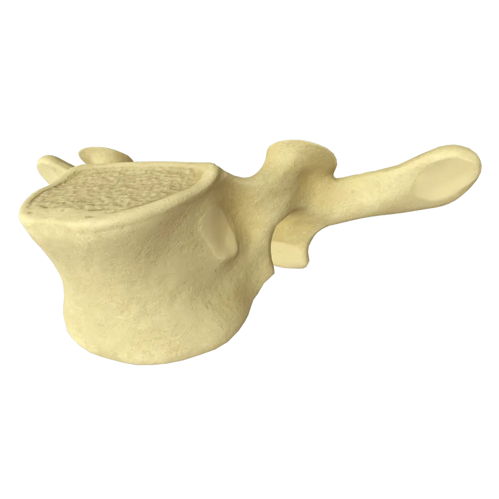
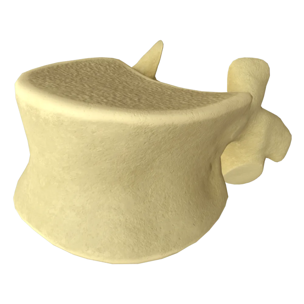
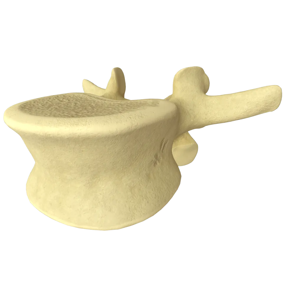
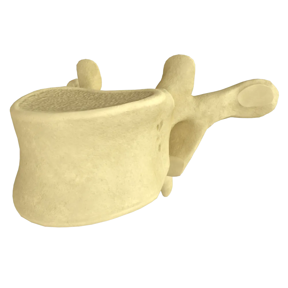
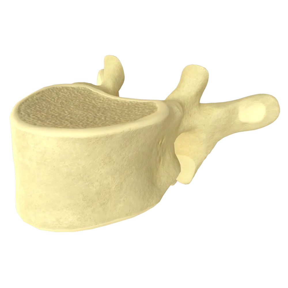
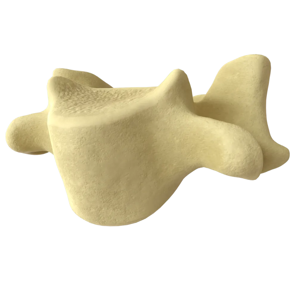
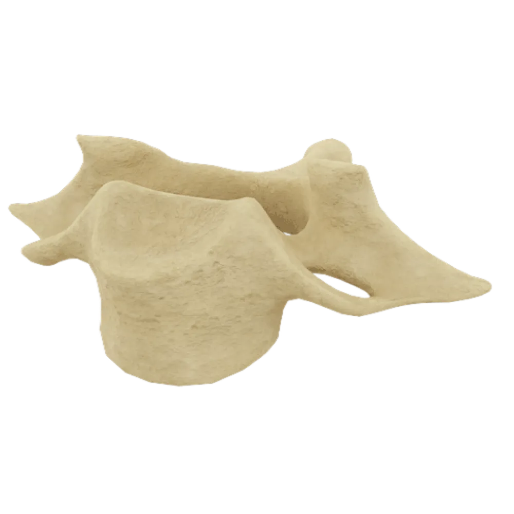
First thoracic vertebra anatomy quiz
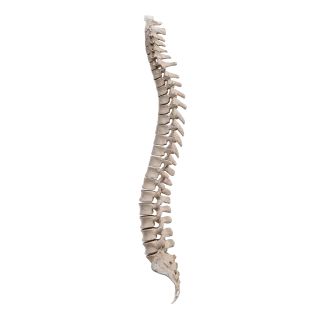
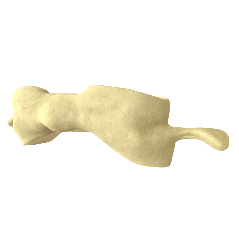
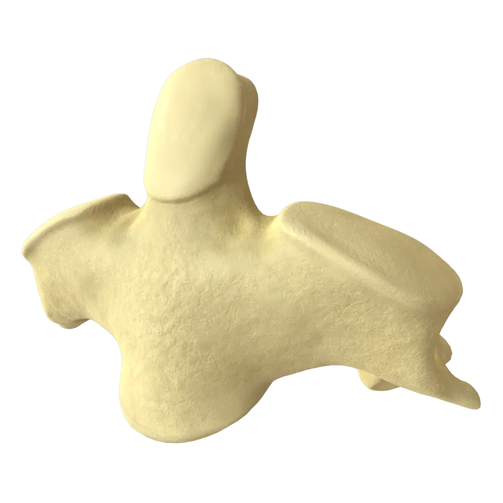
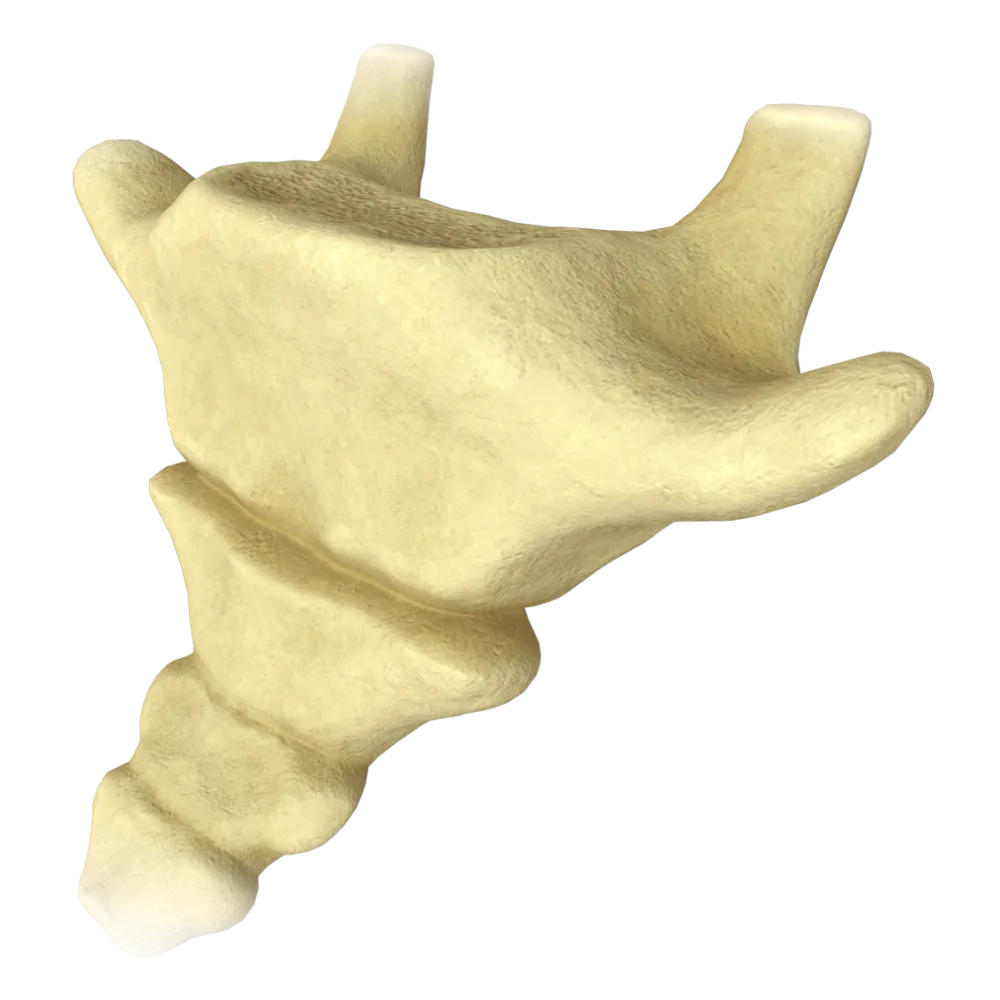
Normal anatomy quiz of the first thoracic vertebra: structure, processes, articular surfaces, connections, and functional features.
1/20
bold
text
1. What is the ordinal position of the first thoracic vertebra in the vertebral column, counting from the cranial base?
-
Eighth
The vertebral column comprises 7 cervical vertebrae, followed by the thoracic vertebrae. The first thoracic vertebra (Th1) is the eighth vertebra from the cranial base, immediately following the seventh cervical vertebra (C7).
-
Twelfth
The vertebral column comprises 7 cervical vertebrae, followed by the thoracic vertebrae. The first thoracic vertebra (Th1) is the eighth vertebra from the cranial base, immediately following the seventh cervical vertebra (C7).
-
Twenty-sixth
The vertebral column comprises 7 cervical vertebrae, followed by the thoracic vertebrae. The first thoracic vertebra (Th1) is the eighth vertebra from the cranial base, immediately following the seventh cervical vertebra (C7).
-
Eighth cervical
The vertebral column comprises 7 cervical vertebrae, followed by the thoracic vertebrae. The first thoracic vertebra (Th1) is the eighth vertebra from the cranial base, immediately following the seventh cervical vertebra (C7).
-
I find it difficult to answer
The vertebral column comprises 7 cervical vertebrae, followed by the thoracic vertebrae. The first thoracic vertebra (Th1) is the eighth vertebra from the cranial base, immediately following the seventh cervical vertebra (C7).
2. How does the body of the first thoracic vertebra (Th1) differ from the bodies of typical thoracic vertebrae (Th2–Th9)?
-
Presence of a complete costal facet on the superior part of the vertebral body
Typical thoracic vertebrae (Th2–Th9) have two demifacets (superior and inferior) on the lateral surfaces of the body because the head of one rib articulates with the bodies of two adjacent vertebrae. The first thoracic vertebra (Th1) is atypical: because the first rib articulates entirely with its superior part, a complete costal facet is located here.
-
Complete absence of costal facets on the vertebral body
Typical thoracic vertebrae (Th2–Th9) have two demifacets (superior and inferior) on the lateral surfaces of the body because the head of one rib articulates with the bodies of two adjacent vertebrae. The first thoracic vertebra (Th1) is atypical: because the first rib articulates entirely with its superior part, a complete costal facet is located here.
-
Presence of a transverse foramen in the vertebral body
Typical thoracic vertebrae (Th2–Th9) have two demifacets (superior and inferior) on the lateral surfaces of the body because the head of one rib articulates with the bodies of two adjacent vertebrae. The first thoracic vertebra (Th1) is atypical: because the first rib articulates entirely with its superior part, a complete costal facet is located here.
-
Presence of four complete costal facets on the vertebral body
Typical thoracic vertebrae (Th2–Th9) have two demifacets (superior and inferior) on the lateral surfaces of the body because the head of one rib articulates with the bodies of two adjacent vertebrae. The first thoracic vertebra (Th1) is atypical: because the first rib articulates entirely with its superior part, a complete costal facet is located here.
-
I find it difficult to answer
Typical thoracic vertebrae (Th2–Th9) have two demifacets (superior and inferior) on the lateral surfaces of the body because the head of one rib articulates with the bodies of two adjacent vertebrae. The first thoracic vertebra (Th1) is atypical: because the first rib articulates entirely with its superior part, a complete costal facet is located here.
3. What is the complete costal facet located on the lateral surface of the body of the first thoracic vertebra called?
-
Superior costal facet
The body of Th1 has a complete superior costal facet (fovea costalis superior) for articulation with the head of the first rib, rather than a demifacet as in typical thoracic vertebrae.
-
Inferior costal facet
The body of Th1 has a complete superior costal facet (fovea costalis superior) for articulation with the head of the first rib, rather than a demifacet as in typical thoracic vertebrae.
-
Transverse costal facet
The body of Th1 has a complete superior costal facet (fovea costalis superior) for articulation with the head of the first rib, rather than a demifacet as in typical thoracic vertebrae.
-
Articular facet of head of rib
The body of Th1 has a complete superior costal facet (fovea costalis superior) for articulation with the head of the first rib, rather than a demifacet as in typical thoracic vertebrae.
-
I find it difficult to answer
The body of Th1 has a complete superior costal facet (fovea costalis superior) for articulation with the head of the first rib, rather than a demifacet as in typical thoracic vertebrae.
4. With the head of which rib does the inferior demifacet (fovea costalis inferior) of the body of the first thoracic vertebra articulate?
-
With the head of the second rib, together with the superior demifacet of the body of Th2
The inferior demifacet of the body of Th1 forms a joint with the head of the second rib together with the superior demifacet of the body of Th2.
-
With the head of the first rib independently
The inferior demifacet of the body of Th1 forms a joint with the head of the second rib together with the superior demifacet of the body of Th2.
-
With the head of the third rib as part of an accessory joint
The inferior demifacet of the body of Th1 forms a joint with the head of the second rib together with the superior demifacet of the body of Th2.
-
With the tubercle of the second rib via the transverse costal facet
The inferior demifacet of the body of Th1 forms a joint with the head of the second rib together with the superior demifacet of the body of Th2.
-
I find it difficult to answer
The inferior demifacet of the body of Th1 forms a joint with the head of the second rib together with the superior demifacet of the body of Th2.
5. What is the structure of the spinous process of the first thoracic vertebra compared with typical thoracic vertebrae?
-
Long and directed horizontally
The spinous process of Th1 is long and nearly horizontal. This distinguishes it from typical middle thoracic vertebrae, whose processes are sharply inclined inferiorly.
-
Short and bifid at the end, as in cervical vertebrae
The spinous process of Th1 is long and nearly horizontal. This distinguishes it from typical middle thoracic vertebrae, whose processes are sharply inclined inferiorly.
-
Long and sharply inclined inferiorly, as in Th5–Th8
The spinous process of Th1 is long and nearly horizontal. This distinguishes it from typical middle thoracic vertebrae, whose processes are sharply inclined inferiorly.
-
Absent and replaced by two tubercles
The spinous process of Th1 is long and nearly horizontal. This distinguishes it from typical middle thoracic vertebrae, whose processes are sharply inclined inferiorly.
-
I find it difficult to answer
The spinous process of Th1 is long and nearly horizontal. This distinguishes it from typical middle thoracic vertebrae, whose processes are sharply inclined inferiorly.
6. Which statement is correct regarding the transverse process of the first thoracic vertebra?
-
Presence of a transverse costal facet (fovea costalis transversalis)
The transverse processes of Th1 bear a costal facet typical of thoracic vertebrae (fovea costalis transversalis) for articulation with the tubercle of the first rib in the costotransverse joint.
-
Absence of a costal facet due to its atypical structure
The transverse processes of Th1 bear a costal facet typical of thoracic vertebrae (fovea costalis transversalis) for articulation with the tubercle of the first rib in the costotransverse joint.
-
Presence of a transverse foramen (foramen transversarium)
The transverse processes of Th1 bear a costal facet typical of thoracic vertebrae (fovea costalis transversalis) for articulation with the tubercle of the first rib in the costotransverse joint.
-
Bifurcation at the end
The transverse processes of Th1 bear a costal facet typical of thoracic vertebrae (fovea costalis transversalis) for articulation with the tubercle of the first rib in the costotransverse joint.
-
I find it difficult to answer
The transverse processes of Th1 bear a costal facet typical of thoracic vertebrae (fovea costalis transversalis) for articulation with the tubercle of the first rib in the costotransverse joint.
7. Through which joint do the transverse processes of the first thoracic vertebra articulate with the tubercles of the first ribs?
-
Costotransverse joint
The costotransverse joint (articulatio costotransversaria) is formed by the transverse costal facet and the tubercle of the rib. This joint is present at Th1–Th10.
-
Joint of head of rib
The costotransverse joint (articulatio costotransversaria) is formed by the transverse costal facet and the tubercle of the rib. This joint is present at Th1–Th10.
-
Costovertebral joint
The costotransverse joint (articulatio costotransversaria) is formed by the transverse costal facet and the tubercle of the rib. This joint is present at Th1–Th10.
-
Sternocostal joint
The costotransverse joint (articulatio costotransversaria) is formed by the transverse costal facet and the tubercle of the rib. This joint is present at Th1–Th10.
-
I find it difficult to answer
The costotransverse joint (articulatio costotransversaria) is formed by the transverse costal facet and the tubercle of the rib. This joint is present at Th1–Th10.
8. How many processes does the first thoracic vertebra (Th1) have in total?
-
7
Although the first thoracic vertebra (Th1) has distinctive features of the body structure (the presence of a complete costal facet for the first rib), it has the same number of processes as most human vertebrae—7 processes.
-
3
Although the first thoracic vertebra (Th1) has distinctive features of the body structure (the presence of a complete costal facet for the first rib), it has the same number of processes as most human vertebrae—7 processes.
-
5
Although the first thoracic vertebra (Th1) has distinctive features of the body structure (the presence of a complete costal facet for the first rib), it has the same number of processes as most human vertebrae—7 processes.
-
12
Although the first thoracic vertebra (Th1) has distinctive features of the body structure (the presence of a complete costal facet for the first rib), it has the same number of processes as most human vertebrae—7 processes.
-
I find it difficult to answer
Although the first thoracic vertebra (Th1) has distinctive features of the body structure (the presence of a complete costal facet for the first rib), it has the same number of processes as most human vertebrae—7 processes.
9. How are the articular surfaces of the superior articular processes of the first thoracic vertebra oriented in the frontal plane?
-
Directed predominantly posteriorly and superiorly (dorsally)
In thoracic vertebrae, including Th1, the articular surfaces of the superior articular processes are oriented predominantly dorsally (posteriorly), resulting in the frontal orientation of the joints.
-
Directed predominantly laterally, as in lumbar vertebrae
In thoracic vertebrae, including Th1, the articular surfaces of the superior articular processes are oriented predominantly dorsally (posteriorly), resulting in the frontal orientation of the joints.
-
Directed strictly vertically and medially
In thoracic vertebrae, including Th1, the articular surfaces of the superior articular processes are oriented predominantly dorsally (posteriorly), resulting in the frontal orientation of the joints.
-
Directed anteriorly and inferiorly
In thoracic vertebrae, including Th1, the articular surfaces of the superior articular processes are oriented predominantly dorsally (posteriorly), resulting in the frontal orientation of the joints.
-
I find it difficult to answer
In thoracic vertebrae, including Th1, the articular surfaces of the superior articular processes are oriented predominantly dorsally (posteriorly), resulting in the frontal orientation of the joints.
10. Which vertebra articulates cranially with the first thoracic vertebra in the intervertebral joint?
-
Seventh cervical vertebra (C7)
Cranially, Th1 articulates with the inferior articular processes of C7 (vertebra prominens) in paired zygapophyseal joints.
-
Sixth cervical vertebra (C6)
Cranially, Th1 articulates with the inferior articular processes of C7 (vertebra prominens) in paired zygapophyseal joints.
-
Axis (C2)
Cranially, Th1 articulates with the inferior articular processes of C7 (vertebra prominens) in paired zygapophyseal joints.
-
Atlas (C1)
Cranially, Th1 articulates with the inferior articular processes of C7 (vertebra prominens) in paired zygapophyseal joints.
-
I find it difficult to answer
Cranially, Th1 articulates with the inferior articular processes of C7 (vertebra prominens) in paired zygapophyseal joints.
11. What accounts for the presence of a complete superior costal facet (rather than a demifacet) on the body of the first thoracic vertebra?
-
The first rib articulates exclusively with Th1, without involving the body of the adjacent vertebra
The first rib articulates only with the body of Th1, without forming a joint with C7. Therefore, Th1 bears a complete costal facet (rather than a demifacet) for the head of the first rib.
-
The first rib has a double head requiring two complete facets
The first rib articulates only with the body of Th1, without forming a joint with C7. Therefore, Th1 bears a complete costal facet (rather than a demifacet) for the head of the first rib.
-
C7 also participates in the joint with the first rib, leaving Th1 available for a complete facet
The first rib articulates only with the body of Th1, without forming a joint with C7. Therefore, Th1 bears a complete costal facet (rather than a demifacet) for the head of the first rib.
-
The first rib is rudimentary and forms only one joint
The first rib articulates only with the body of Th1, without forming a joint with C7. Therefore, Th1 bears a complete costal facet (rather than a demifacet) for the head of the first rib.
-
I find it difficult to answer
The first rib articulates only with the body of Th1, without forming a joint with C7. Therefore, Th1 bears a complete costal facet (rather than a demifacet) for the head of the first rib.
12. Which intervertebral disc is located between the first thoracic and seventh cervical vertebrae?
-
C7–Th1 intervertebral disc
The C7–Th1 intervertebral disc is located between the bodies of C7 and Th1 and is designated according to the adjacent vertebrae. It is the lowest cervical disc and forms part of the cervicothoracic junction.
-
Th1–Th2 intervertebral disc
The C7–Th1 intervertebral disc is located between the bodies of C7 and Th1 and is designated according to the adjacent vertebrae. It is the lowest cervical disc and forms part of the cervicothoracic junction.
-
C6–C7 intervertebral disc
The C7–Th1 intervertebral disc is located between the bodies of C7 and Th1 and is designated according to the adjacent vertebrae. It is the lowest cervical disc and forms part of the cervicothoracic junction.
-
C5–C6 intervertebral disc
The C7–Th1 intervertebral disc is located between the bodies of C7 and Th1 and is designated according to the adjacent vertebrae. It is the lowest cervical disc and forms part of the cervicothoracic junction.
-
I find it difficult to answer
The C7–Th1 intervertebral disc is located between the bodies of C7 and Th1 and is designated according to the adjacent vertebrae. It is the lowest cervical disc and forms part of the cervicothoracic junction.
13. Which ligament, attached to the anterior surfaces of the bodies of the thoracic vertebrae, also covers the body of the first thoracic vertebra?
-
Anterior longitudinal ligament
The anterior longitudinal ligament (lig. longitudinale anterius) runs along the anterior surfaces of the bodies of all vertebrae, including Th1, from the occipital bone to the sacrum.
-
Ligamentum longitudinale posterius
The anterior longitudinal ligament (lig. longitudinale anterius) runs along the anterior surfaces of the bodies of all vertebrae, including Th1, from the occipital bone to the sacrum.
-
Ligamentum flavum
The anterior longitudinal ligament (lig. longitudinale anterius) runs along the anterior surfaces of the bodies of all vertebrae, including Th1, from the occipital bone to the sacrum.
-
Ligamentum supraspinale
The anterior longitudinal ligament (lig. longitudinale anterius) runs along the anterior surfaces of the bodies of all vertebrae, including Th1, from the occipital bone to the sacrum.
-
I find it difficult to answer
The anterior longitudinal ligament (lig. longitudinale anterius) runs along the anterior surfaces of the bodies of all vertebrae, including Th1, from the occipital bone to the sacrum.
14. Which ligament connects the arches of adjacent vertebrae, including Th1, and is known for its high content of elastic fibers?
-
Ligamentum flavum
The ligamentum flavum (lig. flavum) connects the arches of adjacent vertebrae. It is rich in elastic fibers (up to 80%), which impart its yellow color and provide elastic resistance during flexion.
-
Ligamentum interspinale
The ligamentum flavum (lig. flavum) connects the arches of adjacent vertebrae. It is rich in elastic fibers (up to 80%), which impart its yellow color and provide elastic resistance during flexion.
-
Ligamentum longitudinale posterius
The ligamentum flavum (lig. flavum) connects the arches of adjacent vertebrae. It is rich in elastic fibers (up to 80%), which impart its yellow color and provide elastic resistance during flexion.
-
Intertransverse ligament
The ligamentum flavum (lig. flavum) connects the arches of adjacent vertebrae. It is rich in elastic fibers (up to 80%), which impart its yellow color and provide elastic resistance during flexion.
-
I find it difficult to answer
The ligamentum flavum (lig. flavum) connects the arches of adjacent vertebrae. It is rich in elastic fibers (up to 80%), which impart its yellow color and provide elastic resistance during flexion.
15. Which spinal nerve exits through the intervertebral foramen between the first and second thoracic vertebrae?
-
First thoracic spinal nerve (Th1)
The first thoracic spinal nerve (Th1) exits through the intervertebral foramen formed by the inferior vertebral notch of Th1 and the superior vertebral notch of Th2. This is the standard rule for numbering thoracic nerves.
-
Eighth cervical spinal nerve (C8)
The first thoracic spinal nerve (Th1) exits through the intervertebral foramen formed by the inferior vertebral notch of Th1 and the superior vertebral notch of Th2. This is the standard rule for numbering thoracic nerves.
-
Second thoracic spinal nerve (Th2)
The first thoracic spinal nerve (Th1) exits through the intervertebral foramen formed by the inferior vertebral notch of Th1 and the superior vertebral notch of Th2. This is the standard rule for numbering thoracic nerves.
-
First lumbar spinal nerve (L1)
The first thoracic spinal nerve (Th1) exits through the intervertebral foramen formed by the inferior vertebral notch of Th1 and the superior vertebral notch of Th2. This is the standard rule for numbering thoracic nerves.
-
I find it difficult to answer
The first thoracic spinal nerve (Th1) exits through the intervertebral foramen formed by the inferior vertebral notch of Th1 and the superior vertebral notch of Th2. This is the standard rule for numbering thoracic nerves.
16. Which region of the vertebral column is directly adjacent cranially to the first thoracic vertebra?
-
Cervical part (pars cervicalis)
Th1 is the first vertebra of the thoracic region and is directly adjacent cranially to the cervical region of the vertebral column, forming the cervicothoracic junction at the C7–Th1 level.
-
Lumbar part (pars lumbalis)
Th1 is the first vertebra of the thoracic region and is directly adjacent cranially to the cervical region of the vertebral column, forming the cervicothoracic junction at the C7–Th1 level.
-
Sacral part (pars sacralis)
Th1 is the first vertebra of the thoracic region and is directly adjacent cranially to the cervical region of the vertebral column, forming the cervicothoracic junction at the C7–Th1 level.
-
Coccygeal part (pars coccygea)
Th1 is the first vertebra of the thoracic region and is directly adjacent cranially to the cervical region of the vertebral column, forming the cervicothoracic junction at the C7–Th1 level.
-
I find it difficult to answer
Th1 is the first vertebra of the thoracic region and is directly adjacent cranially to the cervical region of the vertebral column, forming the cervicothoracic junction at the C7–Th1 level.
17. Which processes form the zygapophyseal joints at the Th1 level?
-
Superior and inferior articular processes
The superior and inferior articular processes (processus articulares superiores et inferiores) form zygapophyseal joints with adjacent vertebrae. In Th1, they articulate with C7 superiorly and Th2 inferiorly.
-
Transverse processes
The superior and inferior articular processes (processus articulares superiores et inferiores) form zygapophyseal joints with adjacent vertebrae. In Th1, they articulate with C7 superiorly and Th2 inferiorly.
-
Spinous process
The superior and inferior articular processes (processus articulares superiores et inferiores) form zygapophyseal joints with adjacent vertebrae. In Th1, they articulate with C7 superiorly and Th2 inferiorly.
-
Pedicles of vertebral arch
The superior and inferior articular processes (processus articulares superiores et inferiores) form zygapophyseal joints with adjacent vertebrae. In Th1, they articulate with C7 superiorly and Th2 inferiorly.
-
I find it difficult to answer
The superior and inferior articular processes (processus articulares superiores et inferiores) form zygapophyseal joints with adjacent vertebrae. In Th1, they articulate with C7 superiorly and Th2 inferiorly.
18. Is a vertebral arch (arcus vertebrae) present in the structure of the first thoracic vertebra (Th1)?
-
Yes, it has a well-developed arch bounding the vertebral foramen
The first thoracic vertebra (Th1) has a fully formed vertebral arch that arises posteriorly from the vertebral body and encloses the vertebral foramen (foramen vertebrale).
-
The arch is present only in childhood and undergoes reduction in adults
The first thoracic vertebra (Th1) has a fully formed vertebral arch that arises posteriorly from the vertebral body and encloses the vertebral foramen (foramen vertebrale).
-
No, the arch is completely absent
The first thoracic vertebra (Th1) has a fully formed vertebral arch that arises posteriorly from the vertebral body and encloses the vertebral foramen (foramen vertebrale).
-
No, the arch fuses with the seventh cervical vertebra
The first thoracic vertebra (Th1) has a fully formed vertebral arch that arises posteriorly from the vertebral body and encloses the vertebral foramen (foramen vertebrale).
-
I find it difficult to answer
The first thoracic vertebra (Th1) has a fully formed vertebral arch that arises posteriorly from the vertebral body and encloses the vertebral foramen (foramen vertebrale).
19. Which anatomical structure of the first thoracic vertebra contributes to the formation of the posterior wall of the vertebral canal at this level?
-
Arcus vertebrae (vertebral arch)
The posterior wall of the vertebral canal is formed by the laminae of the vertebral arches (laminae arcus vertebrae) of adjacent vertebrae, connected by the ligamenta flava.
-
Processus transversi (transverse process)
The posterior wall of the vertebral canal is formed by the laminae of the vertebral arches (laminae arcus vertebrae) of adjacent vertebrae, connected by the ligamenta flava.
-
Processus articulares (articular process)
The posterior wall of the vertebral canal is formed by the laminae of the vertebral arches (laminae arcus vertebrae) of adjacent vertebrae, connected by the ligamenta flava.
-
Processus spinosus (spinous process)
The posterior wall of the vertebral canal is formed by the laminae of the vertebral arches (laminae arcus vertebrae) of adjacent vertebrae, connected by the ligamenta flava.
-
I find it difficult to answer
The posterior wall of the vertebral canal is formed by the laminae of the vertebral arches (laminae arcus vertebrae) of adjacent vertebrae, connected by the ligamenta flava.
20. How many costal facets (complete facets and demifacets) in total does the body of the first thoracic vertebra bear for articulation with the ribs?
-
Four: one complete superior facet on each side and one inferior demifacet on each side (2 complete facets + 2 demifacets in total)
Th1 bears: 2 complete superior costal facets on the body (for the head of the first rib) + 2 inferior demifacets on the body (for the head of the second rib)
-
Two: only the complete superior facets on both sides
Th1 bears: 2 complete superior costal facets on the body (for the head of the first rib) + 2 inferior demifacets on the body (for the head of the second rib)
-
Five: three complete superior facets and two complete inferior facets
Th1 bears: 2 complete superior costal facets on the body (for the head of the first rib) + 2 inferior demifacets on the body (for the head of the second rib)
-
Six: three on each side
Th1 bears: 2 complete superior costal facets on the body (for the head of the first rib) + 2 inferior demifacets on the body (for the head of the second rib)
-
I find it difficult to answer
Th1 bears: 2 complete superior costal facets on the body (for the head of the first rib) + 2 inferior demifacets on the body (for the head of the second rib)
Retake this quiz?
Your current progress will be reset.
