
Sacrum anatomy quiz
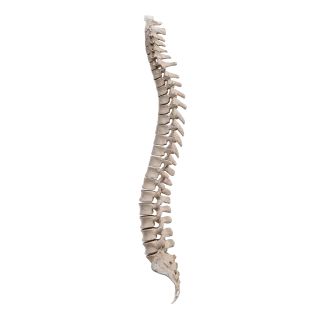
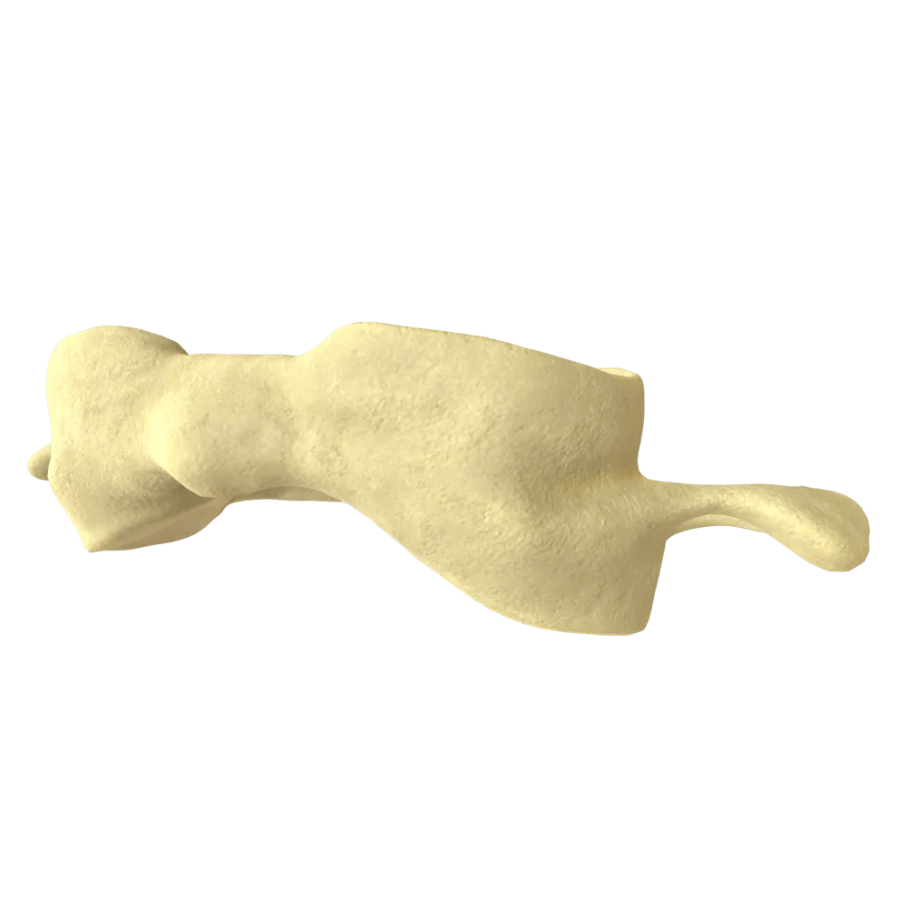
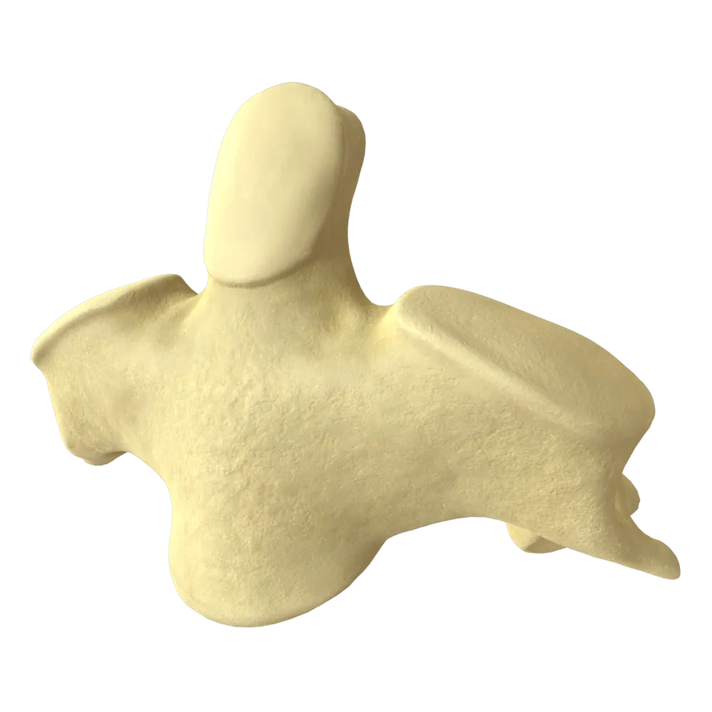
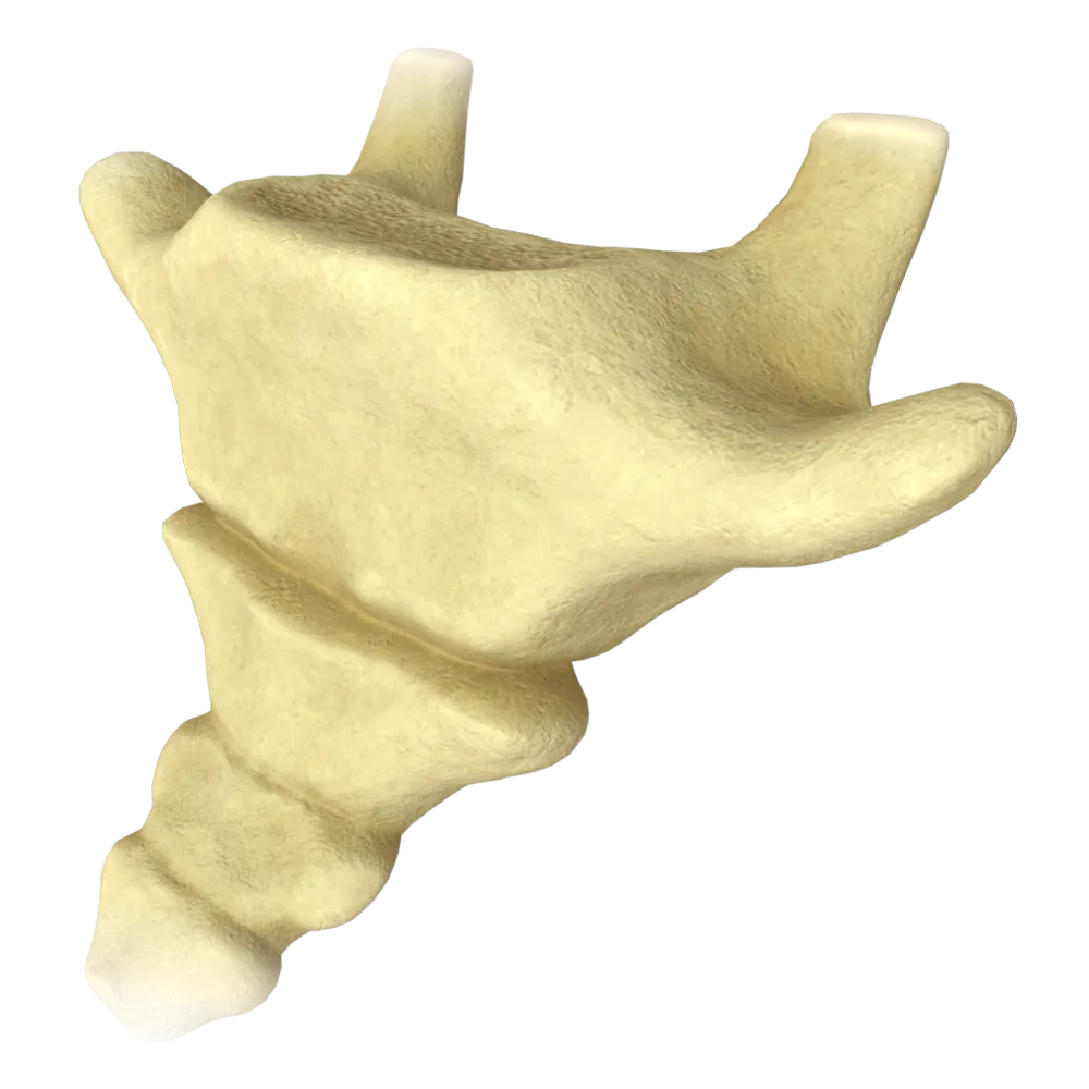
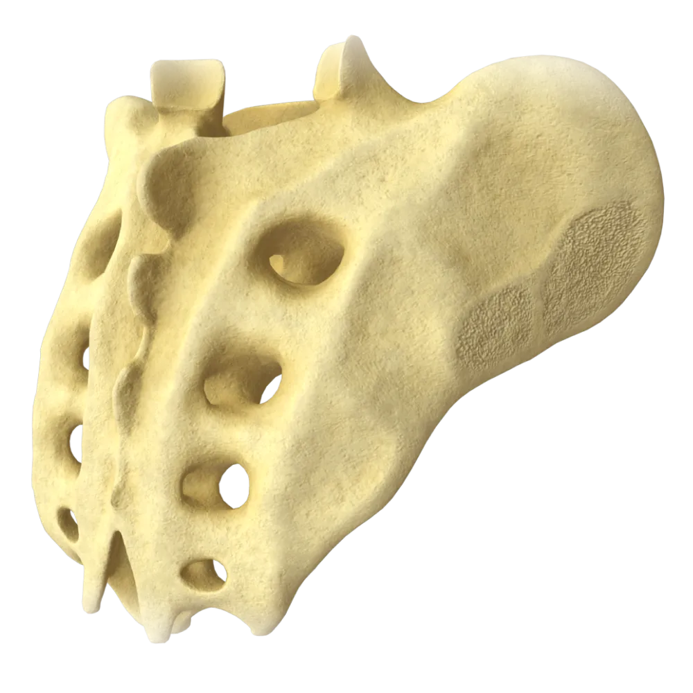
The quiz covers the structure of the sacrum, its parts, canals, foramina, articular surfaces, ligamentous apparatus, and anatomical relationships.
1/20
bold
text
1. How many vertebrae form the sacrum in an adult?
-
3 vertebrae
The sacrum (os sacrum) is formed by the fusion of 5 sacral vertebrae (S1–S5).
-
4 vertebrae
The sacrum (os sacrum) is formed by the fusion of 5 sacral vertebrae (S1–S5).
-
5 vertebrae
The sacrum (os sacrum) is formed by the fusion of 5 sacral vertebrae (S1–S5).
-
6 vertebrae
The sacrum (os sacrum) is formed by the fusion of 5 sacral vertebrae (S1–S5).
-
I find it difficult to answer
The sacrum (os sacrum) is formed by the fusion of 5 sacral vertebrae (S1–S5).
2. What is the superior border of the sacrum that projects into the lesser pelvis called?
-
Median sacral crest
Promontorium (promontory) is the anteriorly projecting superior border of the body of S1, forming a characteristic prominence at the junction of the lumbar spine and sacrum and serving as an important landmark in the topography of the lesser pelvis.
-
Promontory
Promontorium (promontory) is the anteriorly projecting superior border of the body of S1, forming a characteristic prominence at the junction of the lumbar spine and sacrum and serving as an important landmark in the topography of the lesser pelvis.
-
Base of sacrum
Promontorium (promontory) is the anteriorly projecting superior border of the body of S1, forming a characteristic prominence at the junction of the lumbar spine and sacrum and serving as an important landmark in the topography of the lesser pelvis.
-
Apex of sacrum
Promontorium (promontory) is the anteriorly projecting superior border of the body of S1, forming a characteristic prominence at the junction of the lumbar spine and sacrum and serving as an important landmark in the topography of the lesser pelvis.
-
I find it difficult to answer
Promontorium (promontory) is the anteriorly projecting superior border of the body of S1, forming a characteristic prominence at the junction of the lumbar spine and sacrum and serving as an important landmark in the topography of the lesser pelvis.
3. What is the smooth, concave surface of the sacrum that faces anteriorly into the pelvic cavity called?
-
Dorsal surface
Facies pelvica (pelvic surface) is the concave anterior surface of the sacrum facing the cavity of the lesser pelvis. The anterior sacral foramina open on this surface.
-
Lateral surface
Facies pelvica (pelvic surface) is the concave anterior surface of the sacrum facing the cavity of the lesser pelvis. The anterior sacral foramina open on this surface.
-
Pelvic surface
Facies pelvica (pelvic surface) is the concave anterior surface of the sacrum facing the cavity of the lesser pelvis. The anterior sacral foramina open on this surface.
-
Auricular surface
Facies pelvica (pelvic surface) is the concave anterior surface of the sacrum facing the cavity of the lesser pelvis. The anterior sacral foramina open on this surface.
-
I find it difficult to answer
Facies pelvica (pelvic surface) is the concave anterior surface of the sacrum facing the cavity of the lesser pelvis. The anterior sacral foramina open on this surface.
4. How many pairs of anterior sacral foramina (foramina sacralia anteriora) are present on the pelvic surface of the sacrum?
-
3 pairs
On the pelvic surface of the sacrum, there are 4 pairs of anterior sacral foramina (foramina sacralia anteriora), through which the anterior rami of the sacral spinal nerves and vessels exit.
-
4 pairs
On the pelvic surface of the sacrum, there are 4 pairs of anterior sacral foramina (foramina sacralia anteriora), through which the anterior rami of the sacral spinal nerves and vessels exit.
-
5 pairs
On the pelvic surface of the sacrum, there are 4 pairs of anterior sacral foramina (foramina sacralia anteriora), through which the anterior rami of the sacral spinal nerves and vessels exit.
-
2 pairs
On the pelvic surface of the sacrum, there are 4 pairs of anterior sacral foramina (foramina sacralia anteriora), through which the anterior rami of the sacral spinal nerves and vessels exit.
-
I find it difficult to answer
On the pelvic surface of the sacrum, there are 4 pairs of anterior sacral foramina (foramina sacralia anteriora), through which the anterior rami of the sacral spinal nerves and vessels exit.
5. The following pass through the posterior sacral foramina (foramina sacralia posteriora):
-
Anterior rami of the sacral nerves
The posterior rami of the sacral spinal nerves (rami dorsales nn. sacrales), as well as their accompanying vessels, pass through the foramina sacralia posteriora.
-
Posterior rami of lumbar nerves
The posterior rami of the sacral spinal nerves (rami dorsales nn. sacrales), as well as their accompanying vessels, pass through the foramina sacralia posteriora.
-
Internal iliac vessels
The posterior rami of the sacral spinal nerves (rami dorsales nn. sacrales), as well as their accompanying vessels, pass through the foramina sacralia posteriora.
-
Lymphatic trunks
The posterior rami of the sacral spinal nerves (rami dorsales nn. sacrales), as well as their accompanying vessels, pass through the foramina sacralia posteriora.
-
I find it difficult to answer
The posterior rami of the sacral spinal nerves (rami dorsales nn. sacrales), as well as their accompanying vessels, pass through the foramina sacralia posteriora.
6. What is the canal that passes within the sacrum and is a continuation of the vertebral canal called?
-
Canalis obturatorius
Canalis sacralis (sacral canal) is a continuation of the canalis vertebralis and contains the cauda equina, filum terminale, and spinal cord meninges with their contents.
-
Pudendal canal
Canalis sacralis (sacral canal) is a continuation of the canalis vertebralis and contains the cauda equina, filum terminale, and spinal cord meninges with their contents.
-
Sacral canal
Canalis sacralis (sacral canal) is a continuation of the canalis vertebralis and contains the cauda equina, filum terminale, and spinal cord meninges with their contents.
-
Lumbar vertebral canal
Canalis sacralis (sacral canal) is a continuation of the canalis vertebralis and contains the cauda equina, filum terminale, and spinal cord meninges with their contents.
-
I find it difficult to answer
Canalis sacralis (sacral canal) is a continuation of the canalis vertebralis and contains the cauda equina, filum terminale, and spinal cord meninges with their contents.
7. What terminates the sacral canal inferiorly?
-
Intervertebral foramen
The sacral canal terminates inferiorly at the hiatus sacralis—the sacral hiatus.
-
Sacrococcygeal symphysis
The sacral canal terminates inferiorly at the hiatus sacralis—the sacral hiatus.
-
Sacral hiatus
The sacral canal terminates inferiorly at the hiatus sacralis—the sacral hiatus.
-
Fourth anterior sacral foramen
The sacral canal terminates inferiorly at the hiatus sacralis—the sacral hiatus.
-
I find it difficult to answer
The sacral canal terminates inferiorly at the hiatus sacralis—the sacral hiatus.
8. What are the lateral parts of the sacrum called?
-
Transverse processes
Partes laterales—the lateral parts of the sacrum.
-
Lateral parts
Partes laterales—the lateral parts of the sacrum.
-
Laminae of the vertebral arches
Partes laterales—the lateral parts of the sacrum.
-
Lateral sacral crests
Partes laterales—the lateral parts of the sacrum.
-
I find it difficult to answer
Partes laterales—the lateral parts of the sacrum.
9. What is the articular surface of the sacrum that articulates with the ilium called?
-
Pelvic surface
Facies auricularis (auricular surface) is the cartilage-covered articular surface of the lateral part of the sacrum that forms the sacroiliac joint (articulatio sacroiliaca) with the corresponding surface of the ilium.
-
Dorsal surface
Facies auricularis (auricular surface) is the cartilage-covered articular surface of the lateral part of the sacrum that forms the sacroiliac joint (articulatio sacroiliaca) with the corresponding surface of the ilium.
-
Auricular surface
Facies auricularis (auricular surface) is the cartilage-covered articular surface of the lateral part of the sacrum that forms the sacroiliac joint (articulatio sacroiliaca) with the corresponding surface of the ilium.
-
Symphyseal surface
Facies auricularis (auricular surface) is the cartilage-covered articular surface of the lateral part of the sacrum that forms the sacroiliac joint (articulatio sacroiliaca) with the corresponding surface of the ilium.
-
I find it difficult to answer
Facies auricularis (auricular surface) is the cartilage-covered articular surface of the lateral part of the sacrum that forms the sacroiliac joint (articulatio sacroiliaca) with the corresponding surface of the ilium.
10. What is the tuberositas sacralis?
-
Apex of the sacrum
Tuberositas sacralis—the sacral tuberosity, a rough surface located poster to the facies auricularis on the posterior surface of the lateral part of the sacrum. It serves as the attachment site for the strong sacroiliac ligaments.
-
A rough surface poster to the auricular surface for ligament attachment
Tuberositas sacralis—the sacral tuberosity, a rough surface located poster to the facies auricularis on the posterior surface of the lateral part of the sacrum. It serves as the attachment site for the strong sacroiliac ligaments.
-
A crest on the pelvic surface
Tuberositas sacralis—the sacral tuberosity, a rough surface located poster to the facies auricularis on the posterior surface of the lateral part of the sacrum. It serves as the attachment site for the strong sacroiliac ligaments.
-
Lateral border of the sacrum
Tuberositas sacralis—the sacral tuberosity, a rough surface located poster to the facies auricularis on the posterior surface of the lateral part of the sacrum. It serves as the attachment site for the strong sacroiliac ligaments.
-
I find it difficult to answer
Tuberositas sacralis—the sacral tuberosity, a rough surface located poster to the facies auricularis on the posterior surface of the lateral part of the sacrum. It serves as the attachment site for the strong sacroiliac ligaments.
11. Which crest is located along the midline of the dorsal surface of the sacrum and is formed by the fusion of the spinous processes?
-
Lateral sacral crest
Crista sacralis mediana (median sacral crest) is located along the midline of the dorsal surface of the sacrum and is formed by the fusion of the rudimentary spinous processes of the sacral vertebrae.
-
Intermediate sacral crest
Crista sacralis mediana (median sacral crest) is located along the midline of the dorsal surface of the sacrum and is formed by the fusion of the rudimentary spinous processes of the sacral vertebrae.
-
Median sacral crest
Crista sacralis mediana (median sacral crest) is located along the midline of the dorsal surface of the sacrum and is formed by the fusion of the rudimentary spinous processes of the sacral vertebrae.
-
Crista iliaca
Crista sacralis mediana (median sacral crest) is located along the midline of the dorsal surface of the sacrum and is formed by the fusion of the rudimentary spinous processes of the sacral vertebrae.
-
I find it difficult to answer
Crista sacralis mediana (median sacral crest) is located along the midline of the dorsal surface of the sacrum and is formed by the fusion of the rudimentary spinous processes of the sacral vertebrae.
12. Crista sacralis medialis is formed by the fusion of:
-
Spinous processes
Crista sacralis medialis (intermediate sacral crest) is formed by the fusion of the articular processes (processus articulares) of the sacral vertebrae and is located between the median and lateral crests.
-
Articular processes
Crista sacralis medialis (intermediate sacral crest) is formed by the fusion of the articular processes (processus articulares) of the sacral vertebrae and is located between the median and lateral crests.
-
Transverse processes
Crista sacralis medialis (intermediate sacral crest) is formed by the fusion of the articular processes (processus articulares) of the sacral vertebrae and is located between the median and lateral crests.
-
Vertebral arches
Crista sacralis medialis (intermediate sacral crest) is formed by the fusion of the articular processes (processus articulares) of the sacral vertebrae and is located between the median and lateral crests.
-
I find it difficult to answer
Crista sacralis medialis (intermediate sacral crest) is formed by the fusion of the articular processes (processus articulares) of the sacral vertebrae and is located between the median and lateral crests.
13. What is the inferior tapered end of the sacrum that articulates with the coccyx called?
-
Base of sacrum
Apex ossis sacri (apex of the sacrum) is the inferior narrowed end of the sacrum bearing an articular surface for articulation with the first coccygeal vertebra at the sacrococcygeal joint (junctio sacrococcygea).
-
Promontory
Apex ossis sacri (apex of the sacrum) is the inferior narrowed end of the sacrum bearing an articular surface for articulation with the first coccygeal vertebra at the sacrococcygeal joint (junctio sacrococcygea).
-
Apex of sacrum
Apex ossis sacri (apex of the sacrum) is the inferior narrowed end of the sacrum bearing an articular surface for articulation with the first coccygeal vertebra at the sacrococcygeal joint (junctio sacrococcygea).
-
Sacral hiatus
Apex ossis sacri (apex of the sacrum) is the inferior narrowed end of the sacrum bearing an articular surface for articulation with the first coccygeal vertebra at the sacrococcygeal joint (junctio sacrococcygea).
-
I find it difficult to answer
Apex ossis sacri (apex of the sacrum) is the inferior narrowed end of the sacrum bearing an articular surface for articulation with the first coccygeal vertebra at the sacrococcygeal joint (junctio sacrococcygea).
14. Crista sacralis lateralis is formed by the fusion of:
-
Articular processes
Crista sacralis lateralis (lateral sacral crest) is formed by the fusion of the transverse processes (processus transversi) of the sacral vertebrae. It is located lateral to the posterior sacral foramina.
-
Spinous processes
Crista sacralis lateralis (lateral sacral crest) is formed by the fusion of the transverse processes (processus transversi) of the sacral vertebrae. It is located lateral to the posterior sacral foramina.
-
Transverse processes
Crista sacralis lateralis (lateral sacral crest) is formed by the fusion of the transverse processes (processus transversi) of the sacral vertebrae. It is located lateral to the posterior sacral foramina.
-
Vertebral bodies
Crista sacralis lateralis (lateral sacral crest) is formed by the fusion of the transverse processes (processus transversi) of the sacral vertebrae. It is located lateral to the posterior sacral foramina.
-
I find it difficult to answer
Crista sacralis lateralis (lateral sacral crest) is formed by the fusion of the transverse processes (processus transversi) of the sacral vertebrae. It is located lateral to the posterior sacral foramina.
15. The base of the sacrum (basis ossis sacri) faces:
-
Downward and anteriorly
Basis ossis sacri is the broad superior part of the sacrum, which faces upward and anteriorly. It articulates with the body of the fifth lumbar vertebra through the intervertebral disc.
-
Upward and anteriorly
Basis ossis sacri is the broad superior part of the sacrum, which faces upward and anteriorly. It articulates with the body of the fifth lumbar vertebra through the intervertebral disc.
-
Posteriorly and downward
Basis ossis sacri is the broad superior part of the sacrum, which faces upward and anteriorly. It articulates with the body of the fifth lumbar vertebra through the intervertebral disc.
-
Laterally
Basis ossis sacri is the broad superior part of the sacrum, which faces upward and anteriorly. It articulates with the body of the fifth lumbar vertebra through the intervertebral disc.
-
I find it difficult to answer
Basis ossis sacri is the broad superior part of the sacrum, which faces upward and anteriorly. It articulates with the body of the fifth lumbar vertebra through the intervertebral disc.
16. By what age do the sacral vertebrae completely fuse with one another to form a single bone—the sacrum (os sacrum)?
-
By 5–7 years of age
By 18–25 years of age, the complete replacement of cartilage by bone is completed, and the sacrum becomes a single solid bone.
-
By 18–25 years of age
By 18–25 years of age, the complete replacement of cartilage by bone is completed, and the sacrum becomes a single solid bone.
-
By 12–14 years of age
By 18–25 years of age, the complete replacement of cartilage by bone is completed, and the sacrum becomes a single solid bone.
-
By 35–40 years of age
By 18–25 years of age, the complete replacement of cartilage by bone is completed, and the sacrum becomes a single solid bone.
-
I find it difficult to answer
By 18–25 years of age, the complete replacement of cartilage by bone is completed, and the sacrum becomes a single solid bone.
17. What do the transverse lines (lineae transversae) located on the pelvic surface of the sacrum represent anatomically?
-
Attachment sites for the sacrotuberous and sacrospinous ligaments
They form at the sites of the former cartilaginous intervertebral discs as a result of the osseous fusion of the bodies of the 5 sacral vertebrae.
-
Grooves through which the anterior rami of the sacral nerves pass
They form at the sites of the former cartilaginous intervertebral discs as a result of the osseous fusion of the bodies of the 5 sacral vertebrae.
-
Traces of osseous fusion of the bodies of the five sacral vertebrae
They form at the sites of the former cartilaginous intervertebral discs as a result of the osseous fusion of the bodies of the 5 sacral vertebrae.
-
Rudiments of the costal processes of the sacral region
They form at the sites of the former cartilaginous intervertebral discs as a result of the osseous fusion of the bodies of the 5 sacral vertebrae.
-
I find it difficult to answer
They form at the sites of the former cartilaginous intervertebral discs as a result of the osseous fusion of the bodies of the 5 sacral vertebrae.
18. Does the sacrum (os sacrum) have superior articular processes, and what is their function?
-
Yes, two superior articular processes are present on the base of the sacrum and articulate with the inferior articular processes of L5
On the base of the sacrum (basis ossis sacri), on either side of the entrance to the sacral canal, there are two superior articular processes (processus articulares superiores). They are required to form paired zygapophysial joints with the inferior articular processes of the 5th lumbar vertebra.
-
No, the sacrum has no articular processes; articulation occurs only through the intervertebral disc
On the base of the sacrum (basis ossis sacri), on either side of the entrance to the sacral canal, there are two superior articular processes (processus articulares superiores). They are required to form paired zygapophysial joints with the inferior articular processes of the 5th lumbar vertebra.
-
No, only inferior articular processes directed toward the coccyx are present
On the base of the sacrum (basis ossis sacri), on either side of the entrance to the sacral canal, there are two superior articular processes (processus articulares superiores). They are required to form paired zygapophysial joints with the inferior articular processes of the 5th lumbar vertebra.
-
Articular processes are present only in embryos; in adults, they regress into the lateral masses
On the base of the sacrum (basis ossis sacri), on either side of the entrance to the sacral canal, there are two superior articular processes (processus articulares superiores). They are required to form paired zygapophysial joints with the inferior articular processes of the 5th lumbar vertebra.
-
I find it difficult to answer
On the base of the sacrum (basis ossis sacri), on either side of the entrance to the sacral canal, there are two superior articular processes (processus articulares superiores). They are required to form paired zygapophysial joints with the inferior articular processes of the 5th lumbar vertebra.
19. Cornua sacralia are:
-
Rudiments of the transverse processes of S5
Cornua sacralia (sacral horns) are the inferiorly directed rudimentary inferior articular processes of S5. They bound the hiatus sacralis laterally and serve as landmarks for determining its location.
-
Rudiments of the inferior articular processes of S5 that bound the hiatus sacralis laterally
Cornua sacralia (sacral horns) are the inferiorly directed rudimentary inferior articular processes of S5. They bound the hiatus sacralis laterally and serve as landmarks for determining its location.
-
Lateral borders of the sacrum
Cornua sacralia (sacral horns) are the inferiorly directed rudimentary inferior articular processes of S5. They bound the hiatus sacralis laterally and serve as landmarks for determining its location.
-
Projections of the body of S1
Cornua sacralia (sacral horns) are the inferiorly directed rudimentary inferior articular processes of S5. They bound the hiatus sacralis laterally and serve as landmarks for determining its location.
-
I find it difficult to answer
Cornua sacralia (sacral horns) are the inferiorly directed rudimentary inferior articular processes of S5. They bound the hiatus sacralis laterally and serve as landmarks for determining its location.
20. At which sacral vertebral levels are complete arches absent, resulting in the formation of the hiatus sacralis?
-
S1–S2
The hiatus sacralis forms due to underdevelopment (non-fusion) of the arches of S4 and S5. This non-fusion is a variant of normal sacral development and forms the inferior opening of the sacral canal.
-
S2–S3
The hiatus sacralis forms due to underdevelopment (non-fusion) of the arches of S4 and S5. This non-fusion is a variant of normal sacral development and forms the inferior opening of the sacral canal.
-
S3–S4
The hiatus sacralis forms due to underdevelopment (non-fusion) of the arches of S4 and S5. This non-fusion is a variant of normal sacral development and forms the inferior opening of the sacral canal.
-
S4–S5
The hiatus sacralis forms due to underdevelopment (non-fusion) of the arches of S4 and S5. This non-fusion is a variant of normal sacral development and forms the inferior opening of the sacral canal.
-
I find it difficult to answer
The hiatus sacralis forms due to underdevelopment (non-fusion) of the arches of S4 and S5. This non-fusion is a variant of normal sacral development and forms the inferior opening of the sacral canal.
Retake this quiz?
Your current progress will be reset.
