
Atlas anatomy quiz
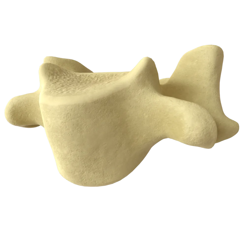
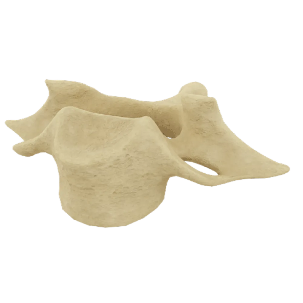
The quiz covers the structure of the atlas: its parts, processes, joints, ligaments, topography, and functional relationships within the cervical spine.
1/20
bold
text
1. What is the first cervical vertebra (C1) called?
-
Axis
The first cervical vertebra is called the atlas (atlas), after the mythical Titan who supported the celestial sphere on his shoulders. The atlas articulates with the occipital bone and supports the skull.
-
Vertebra prominens
The first cervical vertebra is called the atlas (atlas), after the mythical Titan who supported the celestial sphere on his shoulders. The atlas articulates with the occipital bone and supports the skull.
-
Atlas
The first cervical vertebra is called the atlas (atlas), after the mythical Titan who supported the celestial sphere on his shoulders. The atlas articulates with the occipital bone and supports the skull.
-
Odontoid vertebra
The first cervical vertebra is called the atlas (atlas), after the mythical Titan who supported the celestial sphere on his shoulders. The atlas articulates with the occipital bone and supports the skull.
-
I find it difficult to answer
The first cervical vertebra is called the atlas (atlas), after the mythical Titan who supported the celestial sphere on his shoulders. The atlas articulates with the occipital bone and supports the skull.
2. Which structure is absent from the atlas, unlike typical cervical vertebrae?
-
Vertebral arch
The atlas has no vertebral body. Instead of a body, it has an anterior tubercle and an anterior arch. During development, the body of the atlas fused with the body of the second cervical vertebra, forming its dens (dens axis).
-
Transverse processes
The atlas has no vertebral body. Instead of a body, it has an anterior tubercle and an anterior arch. During development, the body of the atlas fused with the body of the second cervical vertebra, forming its dens (dens axis).
-
Vertebral body
The atlas has no vertebral body. Instead of a body, it has an anterior tubercle and an anterior arch. During development, the body of the atlas fused with the body of the second cervical vertebra, forming its dens (dens axis).
-
Articular surfaces
The atlas has no vertebral body. Instead of a body, it has an anterior tubercle and an anterior arch. During development, the body of the atlas fused with the body of the second cervical vertebra, forming its dens (dens axis).
-
I find it difficult to answer
The atlas has no vertebral body. Instead of a body, it has an anterior tubercle and an anterior arch. During development, the body of the atlas fused with the body of the second cervical vertebra, forming its dens (dens axis).
3. What is the anterior part of the ring of the atlas called?
-
Anterior arch of the atlas
The anterior part of the ring of the atlas is called the anterior arch—arcus anterior atlantis.
-
Posterior arch of the atlas
The anterior part of the ring of the atlas is called the anterior arch—arcus anterior atlantis.
-
Lateral mass of the atlas
The anterior part of the ring of the atlas is called the anterior arch—arcus anterior atlantis.
-
Posterior tubercle of the atlas
The anterior part of the ring of the atlas is called the anterior arch—arcus anterior atlantis.
-
I find it difficult to answer
The anterior part of the ring of the atlas is called the anterior arch—arcus anterior atlantis.
4. Where is the fovea dentis located on the atlas?
-
On the anterior surface of the anterior arch
The fovea dentis (facet for dens) is located on the posterior surface of the anterior arch of the atlas and serves as an articular surface for articulation with the dens of the axis at the median atlantoaxial joint.
-
On the posterior surface of the anterior arch
The fovea dentis (facet for dens) is located on the posterior surface of the anterior arch of the atlas and serves as an articular surface for articulation with the dens of the axis at the median atlantoaxial joint.
-
On the superior surface of the lateral mass
The fovea dentis (facet for dens) is located on the posterior surface of the anterior arch of the atlas and serves as an articular surface for articulation with the dens of the axis at the median atlantoaxial joint.
-
On the posterior surface of the posterior arch
The fovea dentis (facet for dens) is located on the posterior surface of the anterior arch of the atlas and serves as an articular surface for articulation with the dens of the axis at the median atlantoaxial joint.
-
I find it difficult to answer
The fovea dentis (facet for dens) is located on the posterior surface of the anterior arch of the atlas and serves as an articular surface for articulation with the dens of the axis at the median atlantoaxial joint.
5. What are the thickened lateral parts of the atlas that bear the articular surfaces called?
-
Pedicles of the vertebral arch
The thickened lateral parts of the atlas are called the lateral masses (massae laterales atlantis). They bear the superior and inferior articular surfaces and constitute the primary supporting structure of the atlas.
-
Transverse processes
The thickened lateral parts of the atlas are called the lateral masses (massae laterales atlantis). They bear the superior and inferior articular surfaces and constitute the primary supporting structure of the atlas.
-
Lateral masses of the atlas
The thickened lateral parts of the atlas are called the lateral masses (massae laterales atlantis). They bear the superior and inferior articular surfaces and constitute the primary supporting structure of the atlas.
-
Posterior tubercle
The thickened lateral parts of the atlas are called the lateral masses (massae laterales atlantis). They bear the superior and inferior articular surfaces and constitute the primary supporting structure of the atlas.
-
I find it difficult to answer
The thickened lateral parts of the atlas are called the lateral masses (massae laterales atlantis). They bear the superior and inferior articular surfaces and constitute the primary supporting structure of the atlas.
6. What is the shape of the superior articular surfaces of the atlas (foveae articulares superiores)?
-
Plane
The superior articular surfaces of the atlas are elliptical and concave. They articulate with the occipital condyles, forming the Atlantooccipital joint (articulatio atlantooccipitalis).
-
Elliptical and concave
The superior articular surfaces of the atlas are elliptical and concave. They articulate with the occipital condyles, forming the Atlantooccipital joint (articulatio atlantooccipitalis).
-
Saddle-shaped
The superior articular surfaces of the atlas are elliptical and concave. They articulate with the occipital condyles, forming the Atlantooccipital joint (articulatio atlantooccipitalis).
-
Cylindrical
The superior articular surfaces of the atlas are elliptical and concave. They articulate with the occipital condyles, forming the Atlantooccipital joint (articulatio atlantooccipitalis).
-
I find it difficult to answer
The superior articular surfaces of the atlas are elliptical and concave. They articulate with the occipital condyles, forming the Atlantooccipital joint (articulatio atlantooccipitalis).
7. With which structure do the inferior articular surfaces of the atlas (foveae articulares inferiores) articulate?
-
With the occipital condyles
The inferior articular surfaces of the atlas (foveae articulares inferiores) articulate with the superior articular surfaces of the axis (C2), forming the lateral atlantoaxial joints (articulationes atlantoaxiales laterales).
-
With the superior articular surfaces of the axis (C2)
The inferior articular surfaces of the atlas (foveae articulares inferiores) articulate with the superior articular surfaces of the axis (C2), forming the lateral atlantoaxial joints (articulationes atlantoaxiales laterales).
-
With the dens of the axis (C2)
The inferior articular surfaces of the atlas (foveae articulares inferiores) articulate with the superior articular surfaces of the axis (C2), forming the lateral atlantoaxial joints (articulationes atlantoaxiales laterales).
-
With the body of the C3 vertebra
The inferior articular surfaces of the atlas (foveae articulares inferiores) articulate with the superior articular surfaces of the axis (C2), forming the lateral atlantoaxial joints (articulationes atlantoaxiales laterales).
-
I find it difficult to answer
The inferior articular surfaces of the atlas (foveae articulares inferiores) articulate with the superior articular surfaces of the axis (C2), forming the lateral atlantoaxial joints (articulationes atlantoaxiales laterales).
8. What is the foramen in the transverse process of the atlas through which the vertebral artery passes called?
-
Intervertebral foramen
The vertebral artery (a. vertebralis) passes through the foramen transversarium (transverse foramen). This foramen is present in all cervical vertebrae.
-
Vertebral foramen
The vertebral artery (a. vertebralis) passes through the foramen transversarium (transverse foramen). This foramen is present in all cervical vertebrae.
-
Transverse foramen
The vertebral artery (a. vertebralis) passes through the foramen transversarium (transverse foramen). This foramen is present in all cervical vertebrae.
-
Hypoglossal canal
The vertebral artery (a. vertebralis) passes through the foramen transversarium (transverse foramen). This foramen is present in all cervical vertebrae.
-
I find it difficult to answer
The vertebral artery (a. vertebralis) passes through the foramen transversarium (transverse foramen). This foramen is present in all cervical vertebrae.
9. Which structure is located on the posterior arch of the atlas at the midline?
-
Anterior tubercle
The posterior tubercle (tuberculum posterius), a rudiment of the spinous process, is located on the posterior arch of the atlas at the midline. The atlas lacks a true spinous process.
-
Spinous process
The posterior tubercle (tuberculum posterius), a rudiment of the spinous process, is located on the posterior arch of the atlas at the midline. The atlas lacks a true spinous process.
-
Posterior tubercle
The posterior tubercle (tuberculum posterius), a rudiment of the spinous process, is located on the posterior arch of the atlas at the midline. The atlas lacks a true spinous process.
-
Mastoid process
The posterior tubercle (tuberculum posterius), a rudiment of the spinous process, is located on the posterior arch of the atlas at the midline. The atlas lacks a true spinous process.
-
I find it difficult to answer
The posterior tubercle (tuberculum posterius), a rudiment of the spinous process, is located on the posterior arch of the atlas at the midline. The atlas lacks a true spinous process.
10. Which groove is located on the superior surface of the posterior arch of the atlas?
-
Groove for the spinal nerve
The groove for the vertebral artery (sulcus arteriae vertebralis) is located on the superior surface of the posterior arch of the atlas; the vertebral artery passes along this groove before entering through the foramen magnum.
-
Groove for the vertebral artery
The groove for the vertebral artery (sulcus arteriae vertebralis) is located on the superior surface of the posterior arch of the atlas; the vertebral artery passes along this groove before entering through the foramen magnum.
-
Carotid groove
The groove for the vertebral artery (sulcus arteriae vertebralis) is located on the superior surface of the posterior arch of the atlas; the vertebral artery passes along this groove before entering through the foramen magnum.
-
Groove for the hypoglossal nerve
The groove for the vertebral artery (sulcus arteriae vertebralis) is located on the superior surface of the posterior arch of the atlas; the vertebral artery passes along this groove before entering through the foramen magnum.
-
I find it difficult to answer
The groove for the vertebral artery (sulcus arteriae vertebralis) is located on the superior surface of the posterior arch of the atlas; the vertebral artery passes along this groove before entering through the foramen magnum.
11. Which joint is formed by the facet for dens of the atlas and the dens of the axis?
-
Atlanto-occipital joint
The median atlantoaxial joint (articulatio atlantoaxialis mediana) is formed by the facet for dens on the anterior arch of the atlas and the dens of the axis. It is a cylindrical joint that enables rotation of the head.
-
Lateral atlanto-axial joint
The median atlantoaxial joint (articulatio atlantoaxialis mediana) is formed by the facet for dens on the anterior arch of the atlas and the dens of the axis. It is a cylindrical joint that enables rotation of the head.
-
Median atlanto-axial joint
The median atlantoaxial joint (articulatio atlantoaxialis mediana) is formed by the facet for dens on the anterior arch of the atlas and the dens of the axis. It is a cylindrical joint that enables rotation of the head.
-
Zygapophyseal joint
The median atlantoaxial joint (articulatio atlantoaxialis mediana) is formed by the facet for dens on the anterior arch of the atlas and the dens of the axis. It is a cylindrical joint that enables rotation of the head.
-
I find it difficult to answer
The median atlantoaxial joint (articulatio atlantoaxialis mediana) is formed by the facet for dens on the anterior arch of the atlas and the dens of the axis. It is a cylindrical joint that enables rotation of the head.
12. Which ligament holds the dens of the axis against the posterior surface of the anterior arch of the atlas?
-
Ligamentum nuchae
The transverse ligament of the atlas (ligamentum transversum atlantis) extends between the lateral masses of the atlas and holds the dens of the axis against the fovea dentis, preventing its posterior displacement into the vertebral canal.
-
Transverse ligament of the atlas
The transverse ligament of the atlas (ligamentum transversum atlantis) extends between the lateral masses of the atlas and holds the dens of the axis against the fovea dentis, preventing its posterior displacement into the vertebral canal.
-
Apical ligament of the dens
The transverse ligament of the atlas (ligamentum transversum atlantis) extends between the lateral masses of the atlas and holds the dens of the axis against the fovea dentis, preventing its posterior displacement into the vertebral canal.
-
Tectorial membrane
The transverse ligament of the atlas (ligamentum transversum atlantis) extends between the lateral masses of the atlas and holds the dens of the axis against the fovea dentis, preventing its posterior displacement into the vertebral canal.
-
I find it difficult to answer
The transverse ligament of the atlas (ligamentum transversum atlantis) extends between the lateral masses of the atlas and holds the dens of the axis against the fovea dentis, preventing its posterior displacement into the vertebral canal.
13. What is the primary type of movement at the median atlantoaxial joint?
-
Flexion and extension
At the median atlantoaxial joint (articulatio atlantoaxialis mediana), the atlas rotates together with the skull around the vertical axis of the dens of the axis. This joint is cylindrical in shape.
-
Rotation around the vertical axis
At the median atlantoaxial joint (articulatio atlantoaxialis mediana), the atlas rotates together with the skull around the vertical axis of the dens of the axis. This joint is cylindrical in shape.
-
Lateral flexion
At the median atlantoaxial joint (articulatio atlantoaxialis mediana), the atlas rotates together with the skull around the vertical axis of the dens of the axis. This joint is cylindrical in shape.
-
Circumduction
At the median atlantoaxial joint (articulatio atlantoaxialis mediana), the atlas rotates together with the skull around the vertical axis of the dens of the axis. This joint is cylindrical in shape.
-
I find it difficult to answer
At the median atlantoaxial joint (articulatio atlantoaxialis mediana), the atlas rotates together with the skull around the vertical axis of the dens of the axis. This joint is cylindrical in shape.
14. What type of joint is the articulatio atlantooccipitalis?
-
Cylinder-shaped
The Atlantooccipital joint (articulatio atlantooccipitalis) is a condylar (ellipsoid) joint. It is formed by two sets of elliptical articular surfaces: the occipital condyles and the superior articular surfaces of the atlas.
-
Flat
The Atlantooccipital joint (articulatio atlantooccipitalis) is a condylar (ellipsoid) joint. It is formed by two sets of elliptical articular surfaces: the occipital condyles and the superior articular surfaces of the atlas.
-
Condylar (ellipsoid).
The Atlantooccipital joint (articulatio atlantooccipitalis) is a condylar (ellipsoid) joint. It is formed by two sets of elliptical articular surfaces: the occipital condyles and the superior articular surfaces of the atlas.
-
Ball-and-socket
The Atlantooccipital joint (articulatio atlantooccipitalis) is a condylar (ellipsoid) joint. It is formed by two sets of elliptical articular surfaces: the occipital condyles and the superior articular surfaces of the atlas.
-
I find it difficult to answer
The Atlantooccipital joint (articulatio atlantooccipitalis) is a condylar (ellipsoid) joint. It is formed by two sets of elliptical articular surfaces: the occipital condyles and the superior articular surfaces of the atlas.
15. Which membrane spans the space between the posterior arch of the atlas and the posterior margin of the foramen magnum?
-
Tectorial membrane
The Posterior atlantooccipital membrane (membrana atlantooccipitalis posterior) extends between the posterior arch of the atlas and the posterior margin of the foramen magnum. It is homologous to the Ligamenta flava.
-
Ligamentum flavum
The Posterior atlantooccipital membrane (membrana atlantooccipitalis posterior) extends between the posterior arch of the atlas and the posterior margin of the foramen magnum. It is homologous to the Ligamenta flava.
-
Posterior atlanto-occipital membrane
The Posterior atlantooccipital membrane (membrana atlantooccipitalis posterior) extends between the posterior arch of the atlas and the posterior margin of the foramen magnum. It is homologous to the Ligamenta flava.
-
Anterior atlanto-occipital membrane
The Posterior atlantooccipital membrane (membrana atlantooccipitalis posterior) extends between the posterior arch of the atlas and the posterior margin of the foramen magnum. It is homologous to the Ligamenta flava.
-
I find it difficult to answer
The Posterior atlantooccipital membrane (membrana atlantooccipitalis posterior) extends between the posterior arch of the atlas and the posterior margin of the foramen magnum. It is homologous to the Ligamenta flava.
16. Which artery pierces (passes through) the lateral parts of the posterior atlantooccipital membrane?
-
Internal carotid artery
The lateral parts of the posterior atlantooccipital membrane (membrana atlantooccipitalis posterior) contain openings through which the vertebral artery (a. vertebralis) passes as it enters the cranial cavity through the foramen magnum.
-
Vertebral artery
The lateral parts of the posterior atlantooccipital membrane (membrana atlantooccipitalis posterior) contain openings through which the vertebral artery (a. vertebralis) passes as it enters the cranial cavity through the foramen magnum.
-
Subclavian artery
The lateral parts of the posterior atlantooccipital membrane (membrana atlantooccipitalis posterior) contain openings through which the vertebral artery (a. vertebralis) passes as it enters the cranial cavity through the foramen magnum.
-
Ascending pharyngeal artery
The lateral parts of the posterior atlantooccipital membrane (membrana atlantooccipitalis posterior) contain openings through which the vertebral artery (a. vertebralis) passes as it enters the cranial cavity through the foramen magnum.
-
I find it difficult to answer
The lateral parts of the posterior atlantooccipital membrane (membrana atlantooccipitalis posterior) contain openings through which the vertebral artery (a. vertebralis) passes as it enters the cranial cavity through the foramen magnum.
17. Which ligament is located between the anterior arch of the atlas and the anterior margin of the foramen magnum?
-
Posterior atlanto-occipital membrane
The anterior atlantooccipital membrane (membrana atlantooccipitalis anterior) extends between the anterior arch of the atlas and the anterior margin of the foramen magnum. It is derived from the Anterior longitudinal ligament.
-
Transverse ligament of the atlas
The anterior atlantooccipital membrane (membrana atlantooccipitalis anterior) extends between the anterior arch of the atlas and the anterior margin of the foramen magnum. It is derived from the Anterior longitudinal ligament.
-
Anterior atlanto-occipital membrane
The anterior atlantooccipital membrane (membrana atlantooccipitalis anterior) extends between the anterior arch of the atlas and the anterior margin of the foramen magnum. It is derived from the Anterior longitudinal ligament.
-
Apical ligament of the dens
The anterior atlantooccipital membrane (membrana atlantooccipitalis anterior) extends between the anterior arch of the atlas and the anterior margin of the foramen magnum. It is derived from the Anterior longitudinal ligament.
-
I find it difficult to answer
The anterior atlantooccipital membrane (membrana atlantooccipitalis anterior) extends between the anterior arch of the atlas and the anterior margin of the foramen magnum. It is derived from the Anterior longitudinal ligament.
18. What is the diameter of the foramen vertebrale of the atlas compared with that of other cervical vertebrae?
-
Smaller because there is no vertebral body
The vertebral foramen of the atlas is considerably larger than that of the other cervical vertebrae. According to Steele's rule, it is divided into three equal parts: one is occupied by the dens of the axis, another by the spinal cord, and the third remains unoccupied (buffer space).
-
Comparable to that of the middle cervical vertebrae
The vertebral foramen of the atlas is considerably larger than that of the other cervical vertebrae. According to Steele's rule, it is divided into three equal parts: one is occupied by the dens of the axis, another by the spinal cord, and the third remains unoccupied (buffer space).
-
Significantly more
The vertebral foramen of the atlas is considerably larger than that of the other cervical vertebrae. According to Steele's rule, it is divided into three equal parts: one is occupied by the dens of the axis, another by the spinal cord, and the third remains unoccupied (buffer space).
-
Triangular in shape and the smallest among the cervical vertebrae
The vertebral foramen of the atlas is considerably larger than that of the other cervical vertebrae. According to Steele's rule, it is divided into three equal parts: one is occupied by the dens of the axis, another by the spinal cord, and the third remains unoccupied (buffer space).
-
I find it difficult to answer
The vertebral foramen of the atlas is considerably larger than that of the other cervical vertebrae. According to Steele's rule, it is divided into three equal parts: one is occupied by the dens of the axis, another by the spinal cord, and the third remains unoccupied (buffer space).
19. Which nerve passes through the posterior atlantooccipital membrane together with the vertebral artery?
-
Accessory nerve (XI)
The first cervical spinal nerve (C1) passes through the posterior atlantooccipital membrane together with the vertebral artery.
-
Hypoglossal nerve (XII)
The first cervical spinal nerve (C1) passes through the posterior atlantooccipital membrane together with the vertebral artery.
-
First cervical spinal nerve (C1)
The first cervical spinal nerve (C1) passes through the posterior atlantooccipital membrane together with the vertebral artery.
-
Greater occipital nerve
The first cervical spinal nerve (C1) passes through the posterior atlantooccipital membrane together with the vertebral artery.
-
I find it difficult to answer
The first cervical spinal nerve (C1) passes through the posterior atlantooccipital membrane together with the vertebral artery.
20. Which of the following statements about the transverse process of the atlas is anatomically correct?
-
The transverse process of the atlas has anterior and posterior tubercles separated by a groove
The transverse process of the atlas comprises anterior and posterior tubercula separated by a groove (sulcus nervi spinalis). It contains the foramen transversarium, is the longest among the cervical vertebrae, and serves as an attachment site for several muscles (m. obliquus capitis inferior, m. rectus capitis lateralis, etc.).
-
The transverse process of the atlas lacks a foramen transversarium
The transverse process of the atlas comprises anterior and posterior tubercula separated by a groove (sulcus nervi spinalis). It contains the foramen transversarium, is the longest among the cervical vertebrae, and serves as an attachment site for several muscles (m. obliquus capitis inferior, m. rectus capitis lateralis, etc.).
-
The transverse process of the atlas is the shortest among the cervical vertebrae
The transverse process of the atlas comprises anterior and posterior tubercula separated by a groove (sulcus nervi spinalis). It contains the foramen transversarium, is the longest among the cervical vertebrae, and serves as an attachment site for several muscles (m. obliquus capitis inferior, m. rectus capitis lateralis, etc.).
-
No muscles attach to the transverse process of the atlas
The transverse process of the atlas comprises anterior and posterior tubercula separated by a groove (sulcus nervi spinalis). It contains the foramen transversarium, is the longest among the cervical vertebrae, and serves as an attachment site for several muscles (m. obliquus capitis inferior, m. rectus capitis lateralis, etc.).
-
I find it difficult to answer
The transverse process of the atlas comprises anterior and posterior tubercula separated by a groove (sulcus nervi spinalis). It contains the foramen transversarium, is the longest among the cervical vertebrae, and serves as an attachment site for several muscles (m. obliquus capitis inferior, m. rectus capitis lateralis, etc.).
Retake this quiz?
Your current progress will be reset.
