
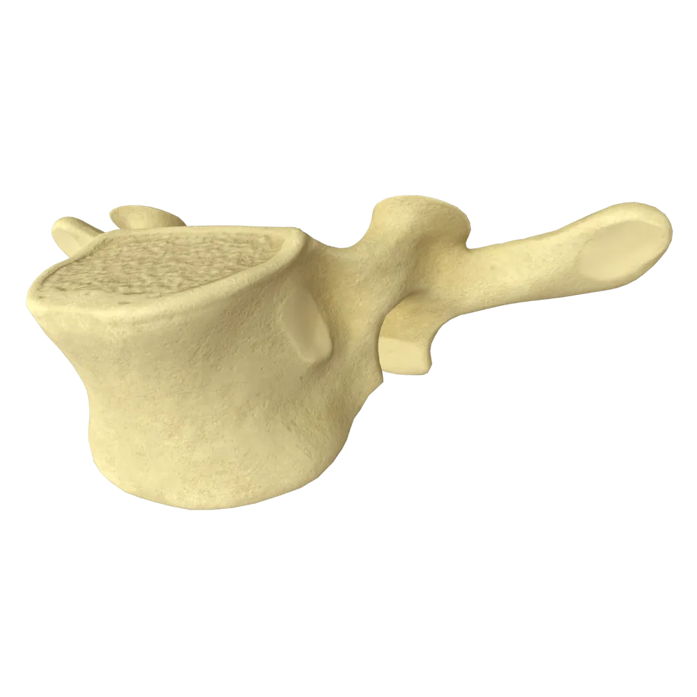
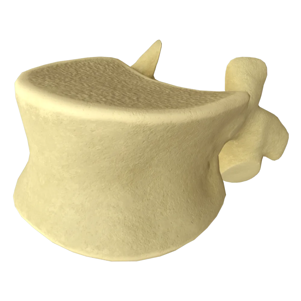
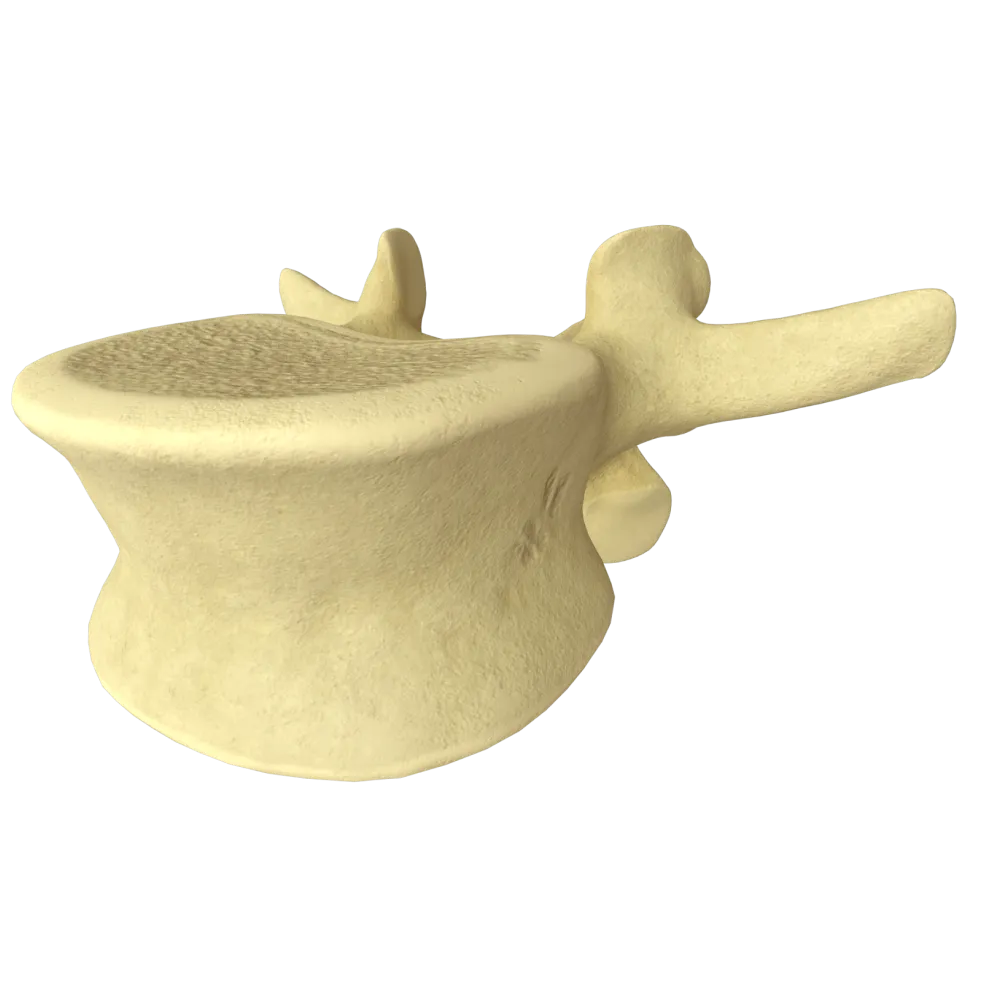
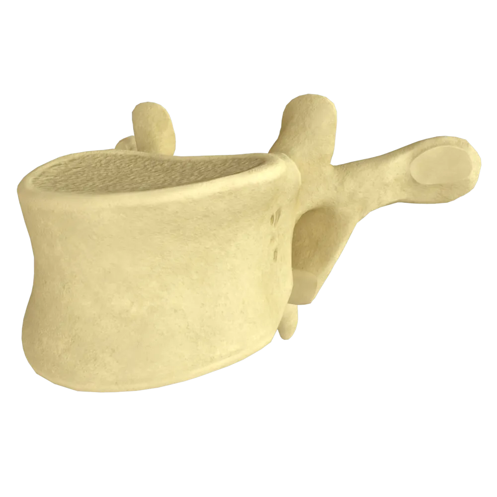
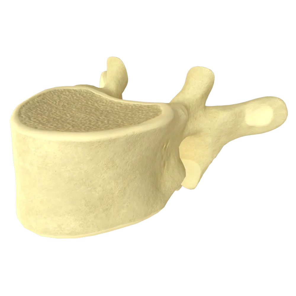
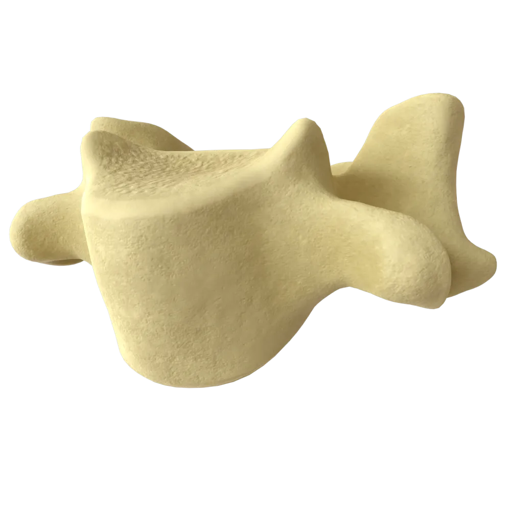
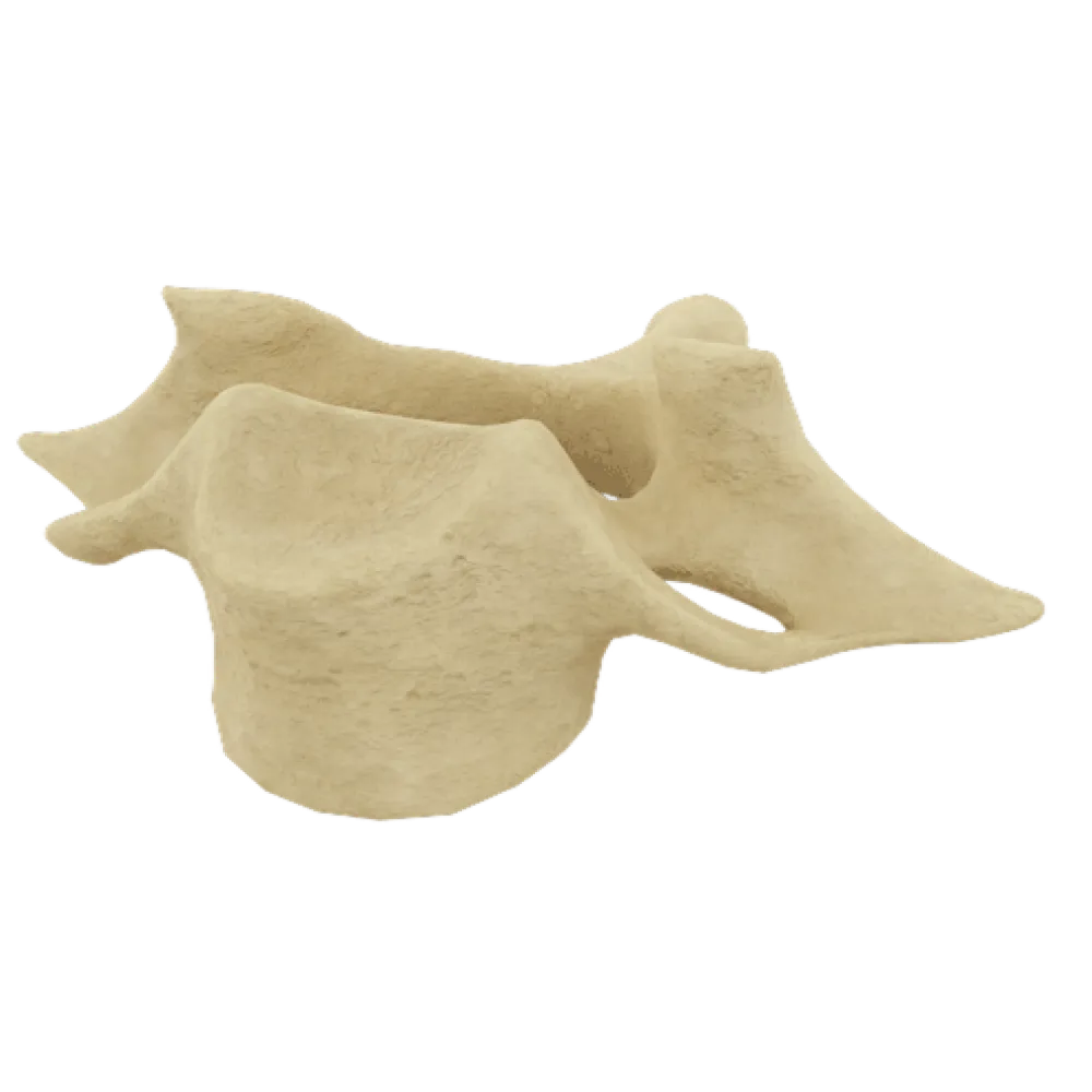
Anatomy quiz of a typical thoracic vertebra
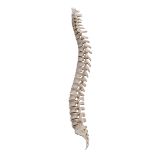
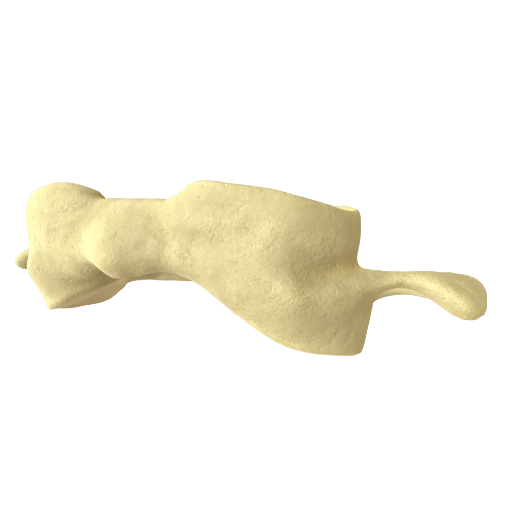
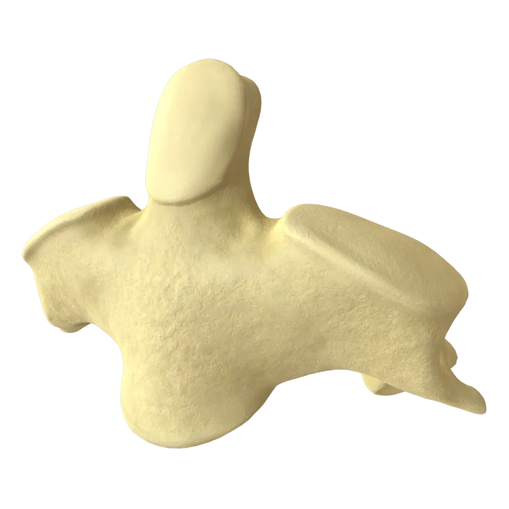
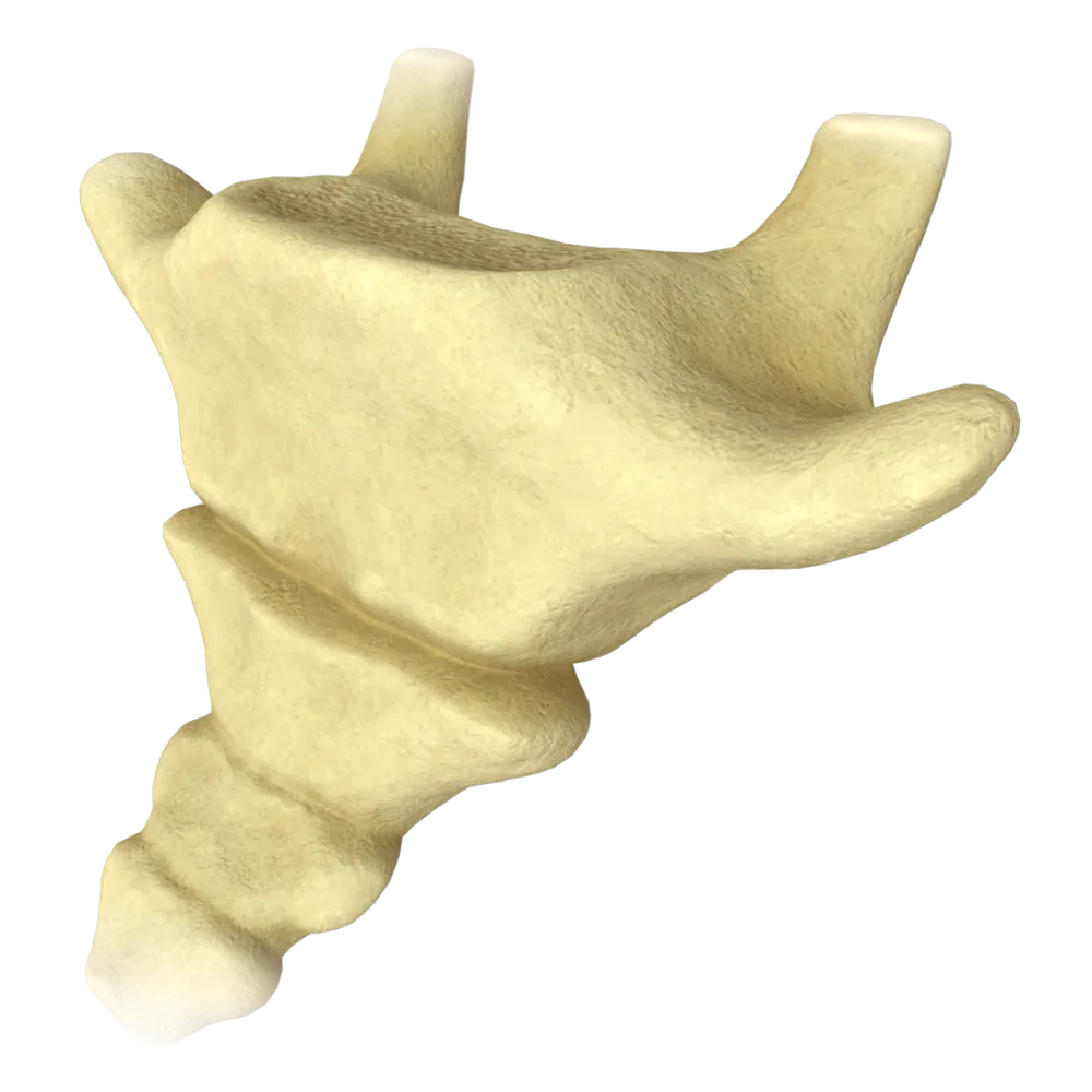
Normal anatomy quiz on the typical thoracic vertebra: structure, processes, articular surfaces, and topographic relationships.
1/20
bold
text
1. Which structure is a distinguishing feature of a typical thoracic vertebra that is absent in cervical and lumbar vertebrae?
-
Bifid spinous process
A typical thoracic vertebra comprises costal facets (foveae costales) on the lateral surfaces of the body for articulation with the heads of the ribs—a structure unique to the thoracic region of the vertebral column.
-
Costal facet on the vertebral body
A typical thoracic vertebra comprises costal facets (foveae costales) on the lateral surfaces of the body for articulation with the heads of the ribs—a structure unique to the thoracic region of the vertebral column.
-
Transverse foramen in the transverse process
A typical thoracic vertebra comprises costal facets (foveae costales) on the lateral surfaces of the body for articulation with the heads of the ribs—a structure unique to the thoracic region of the vertebral column.
-
Accessory process
A typical thoracic vertebra comprises costal facets (foveae costales) on the lateral surfaces of the body for articulation with the heads of the ribs—a structure unique to the thoracic region of the vertebral column.
-
I find it difficult to answer
A typical thoracic vertebra comprises costal facets (foveae costales) on the lateral surfaces of the body for articulation with the heads of the ribs—a structure unique to the thoracic region of the vertebral column.
2. What is the shape of the body of a typical thoracic vertebra in cross-section?
-
Triangular
The body of a typical thoracic vertebra is rounded or slightly heart-shaped in cross-section.
-
Reniform
The body of a typical thoracic vertebra is rounded or slightly heart-shaped in cross-section.
-
Rounded
The body of a typical thoracic vertebra is rounded or slightly heart-shaped in cross-section.
-
Rectangular
The body of a typical thoracic vertebra is rounded or slightly heart-shaped in cross-section.
-
I find it difficult to answer
The body of a typical thoracic vertebra is rounded or slightly heart-shaped in cross-section.
3. How many costal facets (complete and incomplete) are located on the body of one typical thoracic vertebra?
-
One complete facet on each side
Each lateral surface of the body of a typical thoracic vertebra comprises a superior costal demifacet (fovea costalis superior) and an inferior costal demifacet (fovea costalis inferior)—two demifacets on each side.
-
Two demifacets (superior and inferior) on each side
Each lateral surface of the body of a typical thoracic vertebra comprises a superior costal demifacet (fovea costalis superior) and an inferior costal demifacet (fovea costalis inferior)—two demifacets on each side.
-
Three facets on each side
Each lateral surface of the body of a typical thoracic vertebra comprises a superior costal demifacet (fovea costalis superior) and an inferior costal demifacet (fovea costalis inferior)—two demifacets on each side.
-
One demifacet only in the superior part of the body
Each lateral surface of the body of a typical thoracic vertebra comprises a superior costal demifacet (fovea costalis superior) and an inferior costal demifacet (fovea costalis inferior)—two demifacets on each side.
-
I find it difficult to answer
Each lateral surface of the body of a typical thoracic vertebra comprises a superior costal demifacet (fovea costalis superior) and an inferior costal demifacet (fovea costalis inferior)—two demifacets on each side.
4. Where is the costal facet of the transverse process (fovea costalis processus transversi) of a typical thoracic vertebra located?
-
On the anterior surface of the transverse process
The costal facet of the transverse process is located on its anterior surface and articulates with the tubercle of the corresponding rib, forming the costotransverse joint.
-
On the posterior surface of the transverse process
The costal facet of the transverse process is located on its anterior surface and articulates with the tubercle of the corresponding rib, forming the costotransverse joint.
-
At the tip of the transverse process
The costal facet of the transverse process is located on its anterior surface and articulates with the tubercle of the corresponding rib, forming the costotransverse joint.
-
At the base of the transverse process on the side of the vertebral arch
The costal facet of the transverse process is located on its anterior surface and articulates with the tubercle of the corresponding rib, forming the costotransverse joint.
-
I find it difficult to answer
The costal facet of the transverse process is located on its anterior surface and articulates with the tubercle of the corresponding rib, forming the costotransverse joint.
5. How is the spinous process of a typical thoracic vertebra directed?
-
Horizontally, almost perpendicular to the axis of the vertebral column
The spinous processes of typical thoracic vertebrae (particularly Th5–Th8) are directed steeply downward and overlap the arch of the subjacent vertebra like roof tiles, thereby limiting extension in the thoracic region.
-
Steeply downward, overlapping the subjacent vertebra like roof tiles
The spinous processes of typical thoracic vertebrae (particularly Th5–Th8) are directed steeply downward and overlap the arch of the subjacent vertebra like roof tiles, thereby limiting extension in the thoracic region.
-
Bifid at its tip and directed horizontally
The spinous processes of typical thoracic vertebrae (particularly Th5–Th8) are directed steeply downward and overlap the arch of the subjacent vertebra like roof tiles, thereby limiting extension in the thoracic region.
-
Upward and backward at an acute angle
The spinous processes of typical thoracic vertebrae (particularly Th5–Th8) are directed steeply downward and overlap the arch of the subjacent vertebra like roof tiles, thereby limiting extension in the thoracic region.
-
I find it difficult to answer
The spinous processes of typical thoracic vertebrae (particularly Th5–Th8) are directed steeply downward and overlap the arch of the subjacent vertebra like roof tiles, thereby limiting extension in the thoracic region.
6. What is the foramen bounded by the vertebral arch and body called?
-
Intervertebral foramen (foramen intervertebrale)
The vertebral foramen (foramen vertebrale) is formed by the body anteriorly and the vertebral arch posteriorly. In the thoracic region, it is rounded and relatively small, corresponding to the diameter of the thoracic region of the spinal cord.
-
Vertebral foramen (foramen vertebrale)
The vertebral foramen (foramen vertebrale) is formed by the body anteriorly and the vertebral arch posteriorly. In the thoracic region, it is rounded and relatively small, corresponding to the diameter of the thoracic region of the spinal cord.
-
Transverse foramen (foramen transversarium)
The vertebral foramen (foramen vertebrale) is formed by the body anteriorly and the vertebral arch posteriorly. In the thoracic region, it is rounded and relatively small, corresponding to the diameter of the thoracic region of the spinal cord.
-
Nutrient foramen (foramen nutricium)
The vertebral foramen (foramen vertebrale) is formed by the body anteriorly and the vertebral arch posteriorly. In the thoracic region, it is rounded and relatively small, corresponding to the diameter of the thoracic region of the spinal cord.
-
I find it difficult to answer
The vertebral foramen (foramen vertebrale) is formed by the body anteriorly and the vertebral arch posteriorly. In the thoracic region, it is rounded and relatively small, corresponding to the diameter of the thoracic region of the spinal cord.
7. What is the size of the vertebral foramen of a typical thoracic vertebra compared with those of cervical and lumbar vertebrae?
-
The largest among all vertebrae
The vertebral foramen of thoracic vertebrae is the smallest in the vertebral column, corresponding to the smaller cross-sectional area of the thoracic region of the spinal cord compared with the cervical and lumbar enlargements.
-
Intermediate, larger than in lumbar vertebrae
The vertebral foramen of thoracic vertebrae is the smallest in the vertebral column, corresponding to the smaller cross-sectional area of the thoracic region of the spinal cord compared with the cervical and lumbar enlargements.
-
The smallest, smaller than in cervical and lumbar vertebrae
The vertebral foramen of thoracic vertebrae is the smallest in the vertebral column, corresponding to the smaller cross-sectional area of the thoracic region of the spinal cord compared with the cervical and lumbar enlargements.
-
Equal in size to that of lumbar vertebrae
The vertebral foramen of thoracic vertebrae is the smallest in the vertebral column, corresponding to the smaller cross-sectional area of the thoracic region of the spinal cord compared with the cervical and lumbar enlargements.
-
I find it difficult to answer
The vertebral foramen of thoracic vertebrae is the smallest in the vertebral column, corresponding to the smaller cross-sectional area of the thoracic region of the spinal cord compared with the cervical and lumbar enlargements.
8. What shape is the vertebral foramen of a typical thoracic vertebra?
-
Triangular
The vertebral foramen of a typical thoracic vertebra is rounded.
-
Large and oval
The vertebral foramen of a typical thoracic vertebra is rounded.
-
Rounded
The vertebral foramen of a typical thoracic vertebra is rounded.
-
Reniform with a ventral notch
The vertebral foramen of a typical thoracic vertebra is rounded.
-
I find it difficult to answer
The vertebral foramen of a typical thoracic vertebra is rounded.
9. How are the superior articular processes (processus articulares superiores) of a typical thoracic vertebra oriented?
-
In the sagittal plane, with the articular surfaces facing medially
The articular surfaces of the superior articular processes of thoracic vertebrae are oriented almost in the frontal plane and face dorsally and slightly laterally, permitting rotation (rotational movement) and limiting flexion-extension.
-
In the frontal plane, with the articular surfaces facing posteriorly and slightly laterally
The articular surfaces of the superior articular processes of thoracic vertebrae are oriented almost in the frontal plane and face dorsally and slightly laterally, permitting rotation (rotational movement) and limiting flexion-extension.
-
In the horizontal plane, with the articular surfaces facing superiorly
The articular surfaces of the superior articular processes of thoracic vertebrae are oriented almost in the frontal plane and face dorsally and slightly laterally, permitting rotation (rotational movement) and limiting flexion-extension.
-
At an angle of 45° to the frontal plane, with the articular surfaces facing inferiorly
The articular surfaces of the superior articular processes of thoracic vertebrae are oriented almost in the frontal plane and face dorsally and slightly laterally, permitting rotation (rotational movement) and limiting flexion-extension.
-
I find it difficult to answer
The articular surfaces of the superior articular processes of thoracic vertebrae are oriented almost in the frontal plane and face dorsally and slightly laterally, permitting rotation (rotational movement) and limiting flexion-extension.
10. What is distinctive about the transverse processes of a typical thoracic vertebra compared with the vertebrae of other regions of the vertebral column?
-
They are the shortest among all vertebrae
The transverse processes of thoracic vertebrae are longer and more massive than those of lumbar vertebrae and bear a costal facet on their anterior surface for articulation with the tubercle of the corresponding rib.
-
They are longer than those of lumbar vertebrae and comprise a costal facet
The transverse processes of thoracic vertebrae are longer and more massive than those of lumbar vertebrae and bear a costal facet on their anterior surface for articulation with the tubercle of the corresponding rib.
-
They are equal in length to those of lumbar vertebrae but comprise a costal facet
The transverse processes of thoracic vertebrae are longer and more massive than those of lumbar vertebrae and bear a costal facet on their anterior surface for articulation with the tubercle of the corresponding rib.
-
They are equal in length to those of cervical vertebrae
The transverse processes of thoracic vertebrae are longer and more massive than those of lumbar vertebrae and bear a costal facet on their anterior surface for articulation with the tubercle of the corresponding rib.
-
I find it difficult to answer
The transverse processes of thoracic vertebrae are longer and more massive than those of lumbar vertebrae and bear a costal facet on their anterior surface for articulation with the tubercle of the corresponding rib.
11. Which structures form the intervertebral foramen (foramen intervertebrale) in the thoracic region?
-
The bodies of two adjacent vertebrae and the intervertebral disc
The intervertebral foramen is formed by the inferior vertebral notch (incisura vertebralis inferior) of the superior vertebra, the superior vertebral notch (incisura vertebralis superior) of the inferior vertebra, as well as the vertebral bodies and the intervertebral disc.
-
The vertebral notches (incisurae vertebrales) of two adjacent vertebrae
The intervertebral foramen is formed by the inferior vertebral notch (incisura vertebralis inferior) of the superior vertebra, the superior vertebral notch (incisura vertebralis superior) of the inferior vertebra, as well as the vertebral bodies and the intervertebral disc.
-
Only the inferior vertebral notch of the superior vertebra and the body of the inferior vertebra
The intervertebral foramen is formed by the inferior vertebral notch (incisura vertebralis inferior) of the superior vertebra, the superior vertebral notch (incisura vertebralis superior) of the inferior vertebra, as well as the vertebral bodies and the intervertebral disc.
-
The articular processes of two adjacent vertebrae and the vertebral arches
The intervertebral foramen is formed by the inferior vertebral notch (incisura vertebralis inferior) of the superior vertebra, the superior vertebral notch (incisura vertebralis superior) of the inferior vertebra, as well as the vertebral bodies and the intervertebral disc.
-
I find it difficult to answer
The intervertebral foramen is formed by the inferior vertebral notch (incisura vertebralis inferior) of the superior vertebra, the superior vertebral notch (incisura vertebralis superior) of the inferior vertebra, as well as the vertebral bodies and the intervertebral disc.
12. Where are the nutrient foramina (foramina nutricia) located on the vertebral body?
-
Only on the posterior surface of the body
The nutrient foramina (foramina nutricia) are located predominantly on the anterior and lateral surfaces of the vertebral body and transmit the nutrient arteries and veins of the vertebral body.
-
On the anterior and lateral surfaces of the body
The nutrient foramina (foramina nutricia) are located predominantly on the anterior and lateral surfaces of the vertebral body and transmit the nutrient arteries and veins of the vertebral body.
-
Exclusively on the superior endplate
The nutrient foramina (foramina nutricia) are located predominantly on the anterior and lateral surfaces of the vertebral body and transmit the nutrient arteries and veins of the vertebral body.
-
On the articular processes
The nutrient foramina (foramina nutricia) are located predominantly on the anterior and lateral surfaces of the vertebral body and transmit the nutrient arteries and veins of the vertebral body.
-
I find it difficult to answer
The nutrient foramina (foramina nutricia) are located predominantly on the anterior and lateral surfaces of the vertebral body and transmit the nutrient arteries and veins of the vertebral body.
13. What is the horizontal plate connecting the pedicle of the vertebral arch to the spinous and articular processes called?
-
Pedicle of the vertebral arch (pediculus arcus vertebrae)
The vertebral arch consists of two parts: the pedicles (pediculi arcus vertebrae), which arise from the body, and the laminae (laminae arcus vertebrae), which extend posteriorly and medially from the pedicles, enclosing the vertebral foramen and giving rise to the processes.
-
Lamina of the vertebral arch (lamina arcus vertebrae)
The vertebral arch consists of two parts: the pedicles (pediculi arcus vertebrae), which arise from the body, and the laminae (laminae arcus vertebrae), which extend posteriorly and medially from the pedicles, enclosing the vertebral foramen and giving rise to the processes.
-
Base of the vertebral arch (basis arcus vertebrae)
The vertebral arch consists of two parts: the pedicles (pediculi arcus vertebrae), which arise from the body, and the laminae (laminae arcus vertebrae), which extend posteriorly and medially from the pedicles, enclosing the vertebral foramen and giving rise to the processes.
-
Transverse process (processus transversus)
The vertebral arch consists of two parts: the pedicles (pediculi arcus vertebrae), which arise from the body, and the laminae (laminae arcus vertebrae), which extend posteriorly and medially from the pedicles, enclosing the vertebral foramen and giving rise to the processes.
-
I find it difficult to answer
The vertebral arch consists of two parts: the pedicles (pediculi arcus vertebrae), which arise from the body, and the laminae (laminae arcus vertebrae), which extend posteriorly and medially from the pedicles, enclosing the vertebral foramen and giving rise to the processes.
14. Which process of a typical thoracic vertebra is unpaired?
-
Superior articular process
The spinous process (processus spinosus) is the only unpaired process of a vertebra and arises in the midline from the junction of the laminae of the vertebral arch.
-
Inferior articular process
The spinous process (processus spinosus) is the only unpaired process of a vertebra and arises in the midline from the junction of the laminae of the vertebral arch.
-
Spinous process
The spinous process (processus spinosus) is the only unpaired process of a vertebra and arises in the midline from the junction of the laminae of the vertebral arch.
-
Transverse process
The spinous process (processus spinosus) is the only unpaired process of a vertebra and arises in the midline from the junction of the laminae of the vertebral arch.
-
I find it difficult to answer
The spinous process (processus spinosus) is the only unpaired process of a vertebra and arises in the midline from the junction of the laminae of the vertebral arch.
15. The superior costal demifacet (fovea costalis superior) of a typical thoracic vertebra is located:
-
On the posterosuperior margin of the lateral surface of the vertebral body
The superior costal demifacet is located on the posterosuperior margin of the lateral surface of the vertebral body. Together with the inferior costal demifacet of the superior vertebra and the intervertebral disc, it forms a complete articular facet for the head of the rib.
-
On the anterior surface of the transverse process
The superior costal demifacet is located on the posterosuperior margin of the lateral surface of the vertebral body. Together with the inferior costal demifacet of the superior vertebra and the intervertebral disc, it forms a complete articular facet for the head of the rib.
-
On the superior endplate of the body
The superior costal demifacet is located on the posterosuperior margin of the lateral surface of the vertebral body. Together with the inferior costal demifacet of the superior vertebra and the intervertebral disc, it forms a complete articular facet for the head of the rib.
-
At the base of the superior articular process
The superior costal demifacet is located on the posterosuperior margin of the lateral surface of the vertebral body. Together with the inferior costal demifacet of the superior vertebra and the intervertebral disc, it forms a complete articular facet for the head of the rib.
-
I find it difficult to answer
The superior costal demifacet is located on the posterosuperior margin of the lateral surface of the vertebral body. Together with the inferior costal demifacet of the superior vertebra and the intervertebral disc, it forms a complete articular facet for the head of the rib.
16. With the head of which rib does the inferior costal demifacet of the typical thoracic vertebra Th5 articulate?
-
With the head of rib V
The inferior costal demifacet of Th5, together with the superior costal demifacet of Th6 and the Th5–Th6 intervertebral disc, forms an articular facet for the head of rib VI.
-
With the head of rib VI
The inferior costal demifacet of Th5, together with the superior costal demifacet of Th6 and the Th5–Th6 intervertebral disc, forms an articular facet for the head of rib VI.
-
With the head of rib IV
The inferior costal demifacet of Th5, together with the superior costal demifacet of Th6 and the Th5–Th6 intervertebral disc, forms an articular facet for the head of rib VI.
-
With the heads of ribs V and VI jointly
The inferior costal demifacet of Th5, together with the superior costal demifacet of Th6 and the Th5–Th6 intervertebral disc, forms an articular facet for the head of rib VI.
-
I find it difficult to answer
The inferior costal demifacet of Th5, together with the superior costal demifacet of Th6 and the Th5–Th6 intervertebral disc, forms an articular facet for the head of rib VI.
17. The pedicle of the vertebral arch (pediculus arcus vertebrae) arises from the vertebral body:
-
From the anteroinferior angle of the body
The pedicles of the vertebral arch arise from the posterosuperior part of the lateral surface of the vertebral body. This is why the inferior vertebral notch is deeper than the superior notch.
-
From the posterosuperior part of the lateral surface of the body
The pedicles of the vertebral arch arise from the posterosuperior part of the lateral surface of the vertebral body. This is why the inferior vertebral notch is deeper than the superior notch.
-
From the center of the posterior surface of the body
The pedicles of the vertebral arch arise from the posterosuperior part of the lateral surface of the vertebral body. This is why the inferior vertebral notch is deeper than the superior notch.
-
From the superior endplate of the body
The pedicles of the vertebral arch arise from the posterosuperior part of the lateral surface of the vertebral body. This is why the inferior vertebral notch is deeper than the superior notch.
-
I find it difficult to answer
The pedicles of the vertebral arch arise from the posterosuperior part of the lateral surface of the vertebral body. This is why the inferior vertebral notch is deeper than the superior notch.
18. What relationship between the heights of the anterior and posterior surfaces of the body of a typical thoracic vertebra determines physiological thoracic kyphosis?
-
The anterior height of the body is greater than the posterior height
The bodies of typical thoracic vertebrae are slightly lower anteriorly than posteriorly (wedge-shaped), which, together with the intervertebral discs, forms physiological thoracic kyphosis.
-
The anterior and posterior heights of the body are equal
The bodies of typical thoracic vertebrae are slightly lower anteriorly than posteriorly (wedge-shaped), which, together with the intervertebral discs, forms physiological thoracic kyphosis.
-
The anterior height of the body is less than the posterior height
The bodies of typical thoracic vertebrae are slightly lower anteriorly than posteriorly (wedge-shaped), which, together with the intervertebral discs, forms physiological thoracic kyphosis.
-
The lateral height of the body is less than the anterior and posterior heights
The bodies of typical thoracic vertebrae are slightly lower anteriorly than posteriorly (wedge-shaped), which, together with the intervertebral discs, forms physiological thoracic kyphosis.
-
I find it difficult to answer
The bodies of typical thoracic vertebrae are slightly lower anteriorly than posteriorly (wedge-shaped), which, together with the intervertebral discs, forms physiological thoracic kyphosis.
19. Which statement about a typical thoracic vertebra is INCORRECT?
-
The body comprises superior and inferior costal demifacets
In thoracic vertebrae, the spinous process is long and directed obliquely downward (in an overlapping, roof tile-like arrangement), rather than horizontally.
-
The spinous process is directed strictly horizontally backward
In thoracic vertebrae, the spinous process is long and directed obliquely downward (in an overlapping, roof tile-like arrangement), rather than horizontally.
-
The transverse processes comprise costal facets
In thoracic vertebrae, the spinous process is long and directed obliquely downward (in an overlapping, roof tile-like arrangement), rather than horizontally.
-
The articular processes are located in the frontal plane
In thoracic vertebrae, the spinous process is long and directed obliquely downward (in an overlapping, roof tile-like arrangement), rather than horizontally.
-
I find it difficult to answer
In thoracic vertebrae, the spinous process is long and directed obliquely downward (in an overlapping, roof tile-like arrangement), rather than horizontally.
20. How many processes in total arise from the arch of a typical thoracic vertebra?
-
Three (one spinous and two transverse)
Seven (7) processes arise from the arch of a typical thoracic vertebra: one spinous process (processus spinosus), two transverse processes (processus transversi), two superior articular processes (processus articulares superiores), and two inferior articular processes (processus articulares inferiores).
-
Five (one spinous, two transverse, and two superior articular)
Seven (7) processes arise from the arch of a typical thoracic vertebra: one spinous process (processus spinosus), two transverse processes (processus transversi), two superior articular processes (processus articulares superiores), and two inferior articular processes (processus articulares inferiores).
-
Seven (one spinous, two transverse, two superior articular, and two inferior articular)
Seven (7) processes arise from the arch of a typical thoracic vertebra: one spinous process (processus spinosus), two transverse processes (processus transversi), two superior articular processes (processus articulares superiores), and two inferior articular processes (processus articulares inferiores).
-
Nine (including the costal processes)
Seven (7) processes arise from the arch of a typical thoracic vertebra: one spinous process (processus spinosus), two transverse processes (processus transversi), two superior articular processes (processus articulares superiores), and two inferior articular processes (processus articulares inferiores).
-
I find it difficult to answer
Seven (7) processes arise from the arch of a typical thoracic vertebra: one spinous process (processus spinosus), two transverse processes (processus transversi), two superior articular processes (processus articulares superiores), and two inferior articular processes (processus articulares inferiores).
Retake this quiz?
Your current progress will be reset.
