
Vertebral anatomy quiz (typical structure)
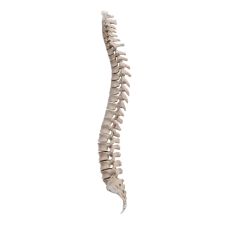
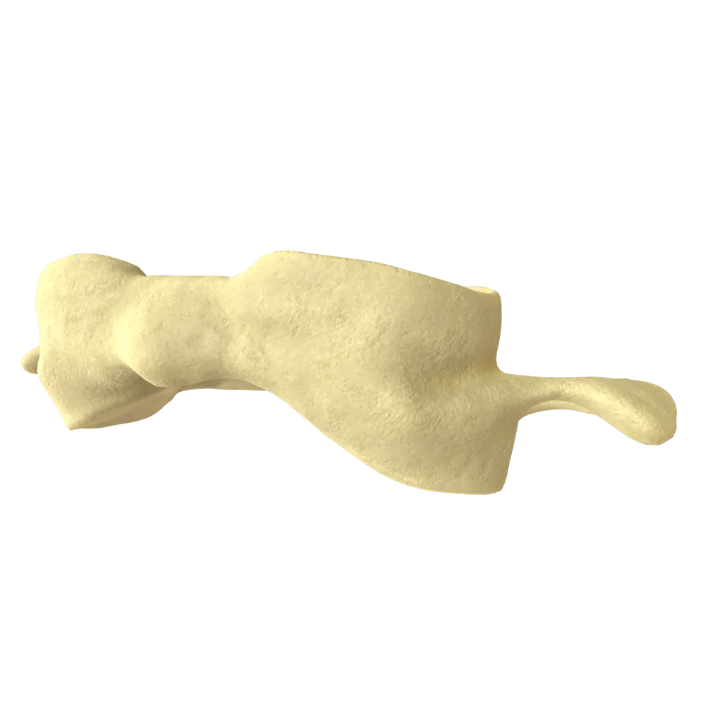
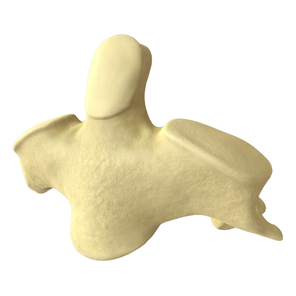
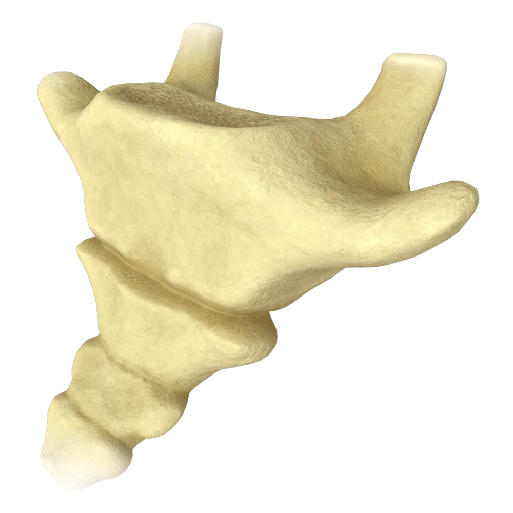
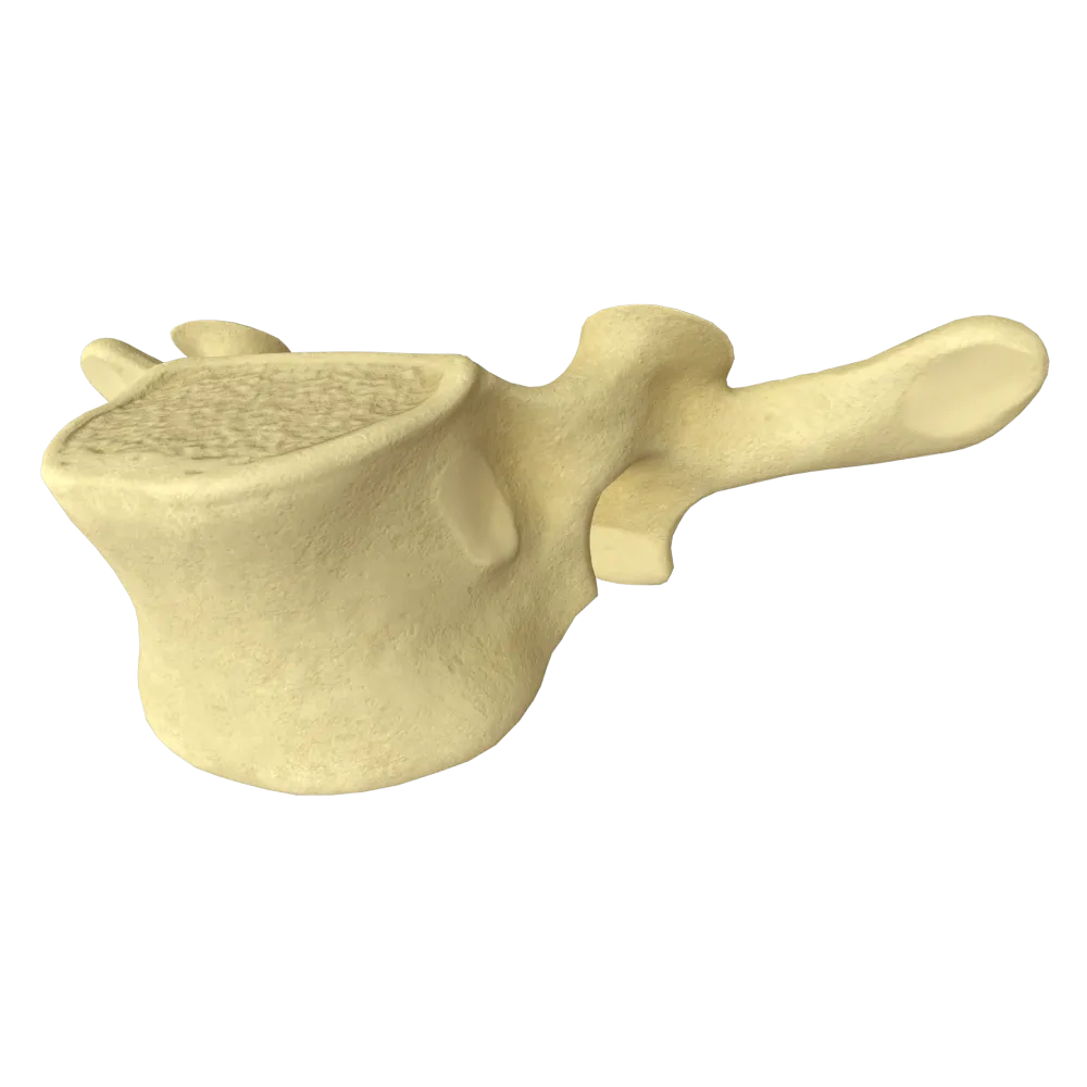
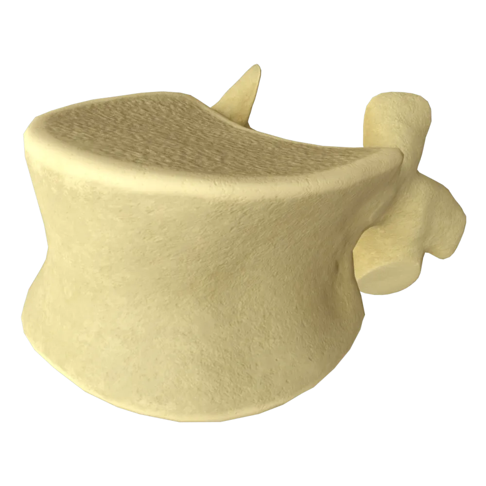
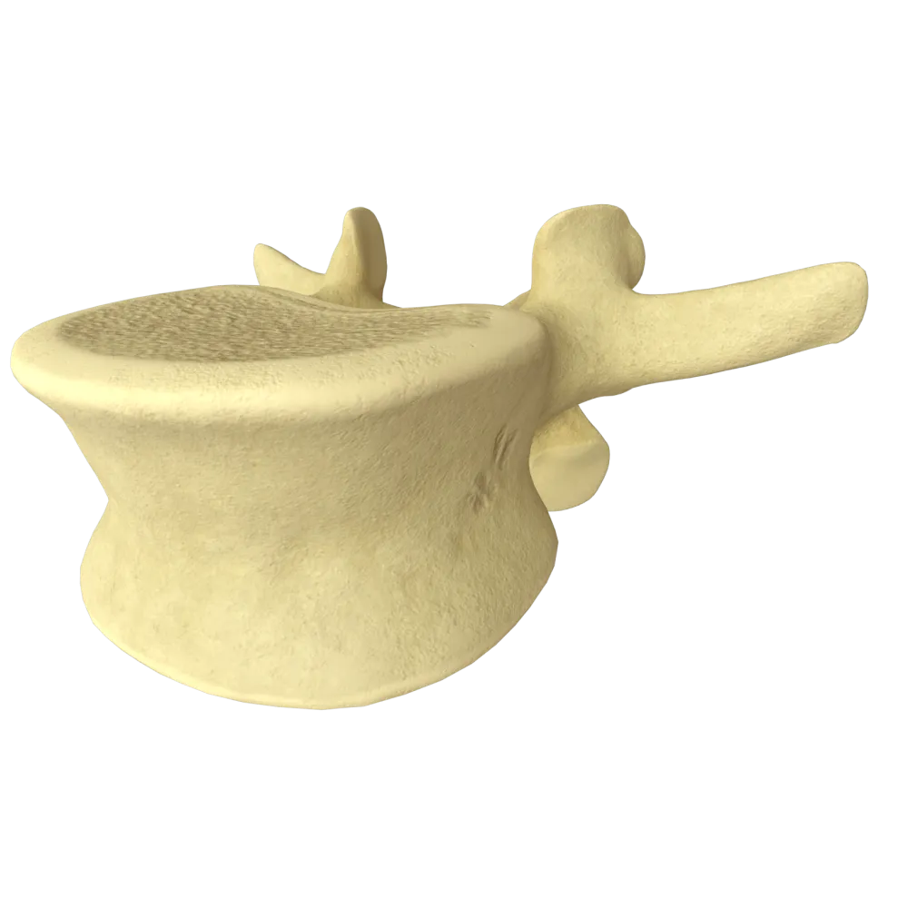
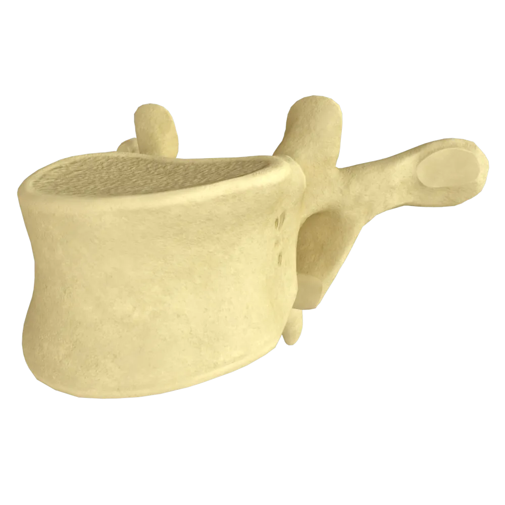
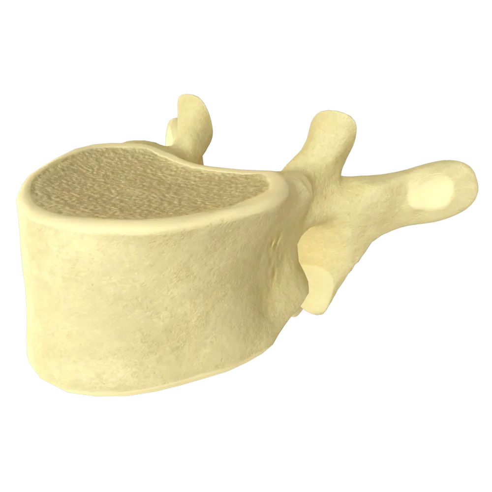
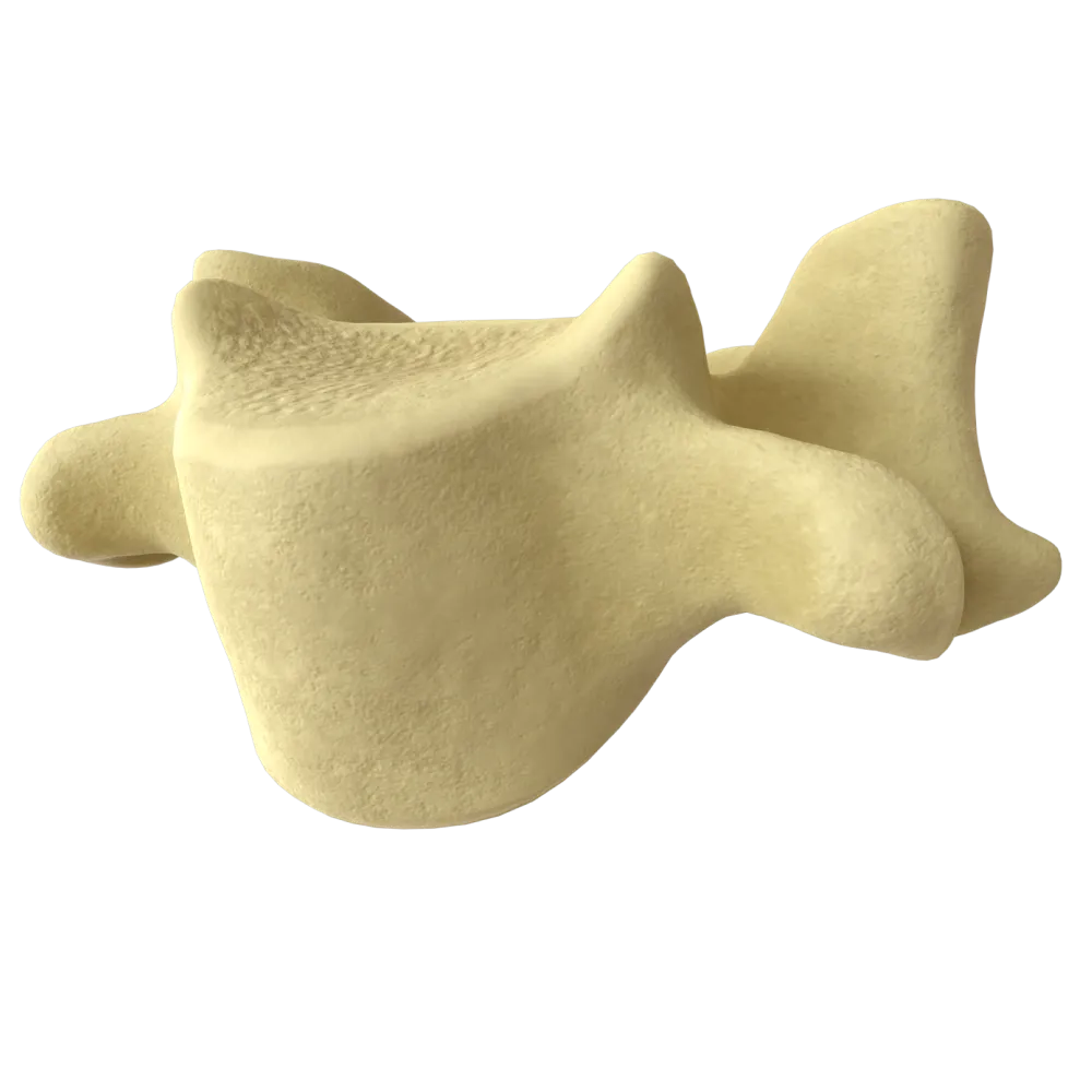
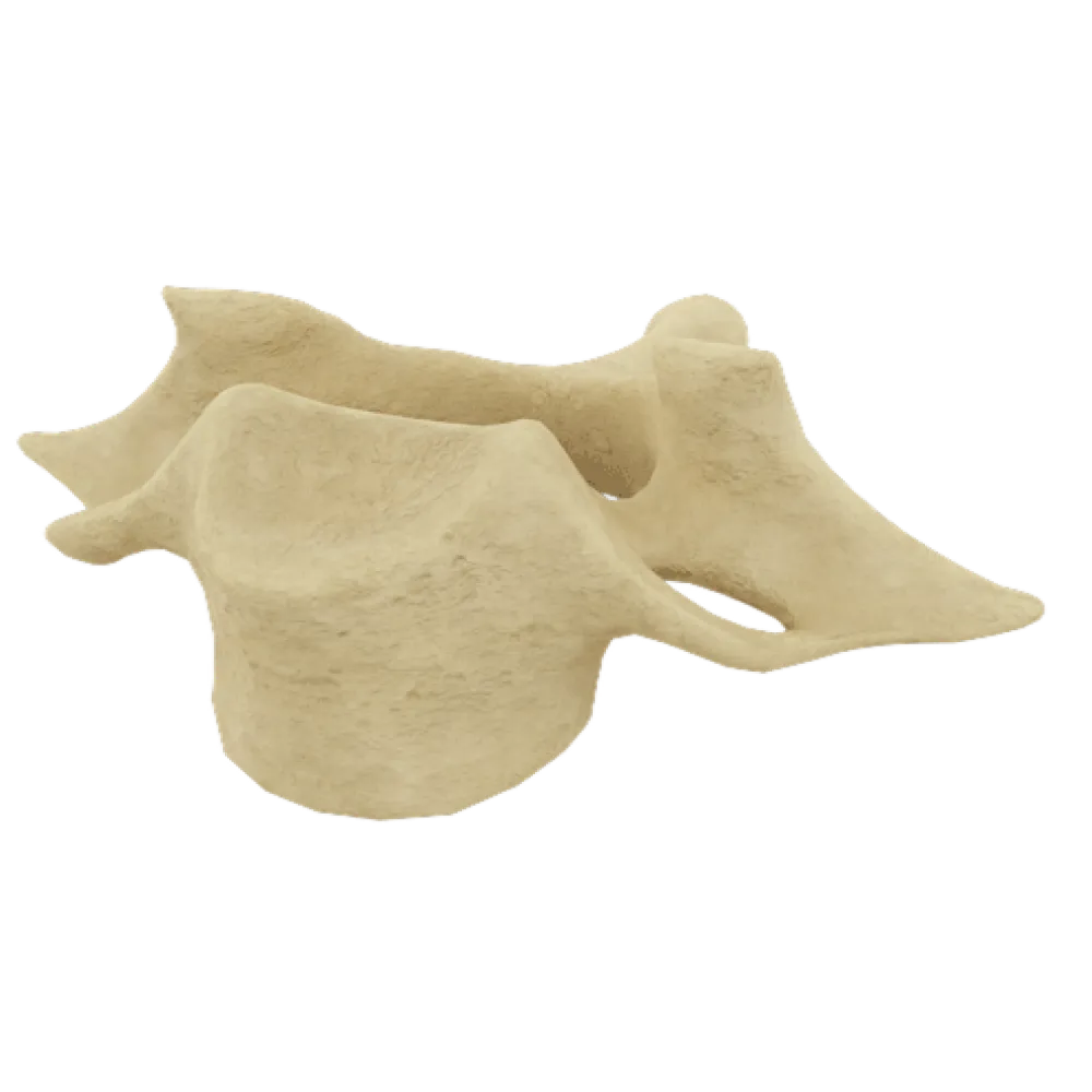
The quiz covers the typical structure of a vertebra: the body, arch, processes, notches, and foramina—within the framework of systemic osteology.
1/20
bold
text
1. Which part of a typical vertebra is its largest, load-bearing structure?
-
Vertebral arch
The vertebral body (corpus vertebrae) is the largest part, located in an anteverted position; it bears the principal axial load. The arch and processes perform other functions.
-
Spinous process
The vertebral body (corpus vertebrae) is the largest part, located in an anteverted position; it bears the principal axial load. The arch and processes perform other functions.
-
Vertebral body
The vertebral body (corpus vertebrae) is the largest part, located in an anteverted position; it bears the principal axial load. The arch and processes perform other functions.
-
Transverse process
The vertebral body (corpus vertebrae) is the largest part, located in an anteverted position; it bears the principal axial load. The arch and processes perform other functions.
-
I find it difficult to answer
The vertebral body (corpus vertebrae) is the largest part, located in an anteverted position; it bears the principal axial load. The arch and processes perform other functions.
2. What is the foramen formed by the body and arch of a typical vertebra called?
-
Intervertebral foramen
The vertebral foramen (foramen vertebrale) is bounded anteriorly by the body and laterally and posteriorly by the vertebral arch. Collectively, these foramina form the vertebral canal.
-
Vertebral foramen
The vertebral foramen (foramen vertebrale) is bounded anteriorly by the body and laterally and posteriorly by the vertebral arch. Collectively, these foramina form the vertebral canal.
-
Foramen transversarium
The vertebral foramen (foramen vertebrale) is bounded anteriorly by the body and laterally and posteriorly by the vertebral arch. Collectively, these foramina form the vertebral canal.
-
Accessory foramen
The vertebral foramen (foramen vertebrale) is bounded anteriorly by the body and laterally and posteriorly by the vertebral arch. Collectively, these foramina form the vertebral canal.
-
I find it difficult to answer
The vertebral foramen (foramen vertebrale) is bounded anteriorly by the body and laterally and posteriorly by the vertebral arch. Collectively, these foramina form the vertebral canal.
3. How many processes does a typical vertebra comprise?
-
5
A typical vertebra comprises 7 processes: 1 spinous, 2 transverse, and 4 articular (2 superior and 2 inferior).
-
7
A typical vertebra comprises 7 processes: 1 spinous, 2 transverse, and 4 articular (2 superior and 2 inferior).
-
6
A typical vertebra comprises 7 processes: 1 spinous, 2 transverse, and 4 articular (2 superior and 2 inferior).
-
4
A typical vertebra comprises 7 processes: 1 spinous, 2 transverse, and 4 articular (2 superior and 2 inferior).
-
I find it difficult to answer
A typical vertebra comprises 7 processes: 1 spinous, 2 transverse, and 4 articular (2 superior and 2 inferior).
4. What are the parts of the vertebral arch that connect it to the body called?
-
Laminae of the arch
The pedicles of the vertebral arch (pediculi arcus vertebrae) are paired, short, cylindrical portions of the arch that attach to the body on both sides. The laminae (laminae) are the posterior portions of the arch.
-
Pedicles of the vertebral arch
The pedicles of the vertebral arch (pediculi arcus vertebrae) are paired, short, cylindrical portions of the arch that attach to the body on both sides. The laminae (laminae) are the posterior portions of the arch.
-
Roots of the articular processes
The pedicles of the vertebral arch (pediculi arcus vertebrae) are paired, short, cylindrical portions of the arch that attach to the body on both sides. The laminae (laminae) are the posterior portions of the arch.
-
Transverse parts of the arch
The pedicles of the vertebral arch (pediculi arcus vertebrae) are paired, short, cylindrical portions of the arch that attach to the body on both sides. The laminae (laminae) are the posterior portions of the arch.
-
I find it difficult to answer
The pedicles of the vertebral arch (pediculi arcus vertebrae) are paired, short, cylindrical portions of the arch that attach to the body on both sides. The laminae (laminae) are the posterior portions of the arch.
5. The superior and inferior vertebral notches (incisura vertebralis superior et inferior) are located on the:
-
Lateral surfaces of the vertebral body
The vertebral notches are located on the pedicles of the arch (pediculi arcus vertebrae): the superior notch is at the superior border, and the inferior notch is at the inferior border. When adjacent vertebrae articulate, they form the intervertebral foramen.
-
Laminae of the vertebral arch
The vertebral notches are located on the pedicles of the arch (pediculi arcus vertebrae): the superior notch is at the superior border, and the inferior notch is at the inferior border. When adjacent vertebrae articulate, they form the intervertebral foramen.
-
Pedicles of the vertebral arch
The vertebral notches are located on the pedicles of the arch (pediculi arcus vertebrae): the superior notch is at the superior border, and the inferior notch is at the inferior border. When adjacent vertebrae articulate, they form the intervertebral foramen.
-
Articular processes of the vertebra
The vertebral notches are located on the pedicles of the arch (pediculi arcus vertebrae): the superior notch is at the superior border, and the inferior notch is at the inferior border. When adjacent vertebrae articulate, they form the intervertebral foramen.
-
I find it difficult to answer
The vertebral notches are located on the pedicles of the arch (pediculi arcus vertebrae): the superior notch is at the superior border, and the inferior notch is at the inferior border. When adjacent vertebrae articulate, they form the intervertebral foramen.
6. Where does the earliest (primary) ossification center appear during the development of a typical vertebra?
-
In the spinous process
During intrauterine development (at weeks 7–8 of embryogenesis), the first ossification centers arise at the bases of the vertebral arches (one on each side). In the vertebral body itself, an ossification center appears slightly later—closer to weeks 9–10.
-
In the vertebral arch
During intrauterine development (at weeks 7–8 of embryogenesis), the first ossification centers arise at the bases of the vertebral arches (one on each side). In the vertebral body itself, an ossification center appears slightly later—closer to weeks 9–10.
-
In the vertebral body
During intrauterine development (at weeks 7–8 of embryogenesis), the first ossification centers arise at the bases of the vertebral arches (one on each side). In the vertebral body itself, an ossification center appears slightly later—closer to weeks 9–10.
-
In the intervertebral disc
During intrauterine development (at weeks 7–8 of embryogenesis), the first ossification centers arise at the bases of the vertebral arches (one on each side). In the vertebral body itself, an ossification center appears slightly later—closer to weeks 9–10.
-
I find it difficult to answer
During intrauterine development (at weeks 7–8 of embryogenesis), the first ossification centers arise at the bases of the vertebral arches (one on each side). In the vertebral body itself, an ossification center appears slightly later—closer to weeks 9–10.
7. The spinous process (processus spinosus) of a typical vertebra arises from the:
-
Junction of the pedicle and body
The spinous process is unpaired and arises in the midline from the site of fusion (union) of the two laminae of the arch (laminae arcus vertebrae), extending posteriorly and inferiorly.
-
Apex of the articular process
The spinous process is unpaired and arises in the midline from the site of fusion (union) of the two laminae of the arch (laminae arcus vertebrae), extending posteriorly and inferiorly.
-
Site where both laminae of the arch fuse in the midline
The spinous process is unpaired and arises in the midline from the site of fusion (union) of the two laminae of the arch (laminae arcus vertebrae), extending posteriorly and inferiorly.
-
Transverse process
The spinous process is unpaired and arises in the midline from the site of fusion (union) of the two laminae of the arch (laminae arcus vertebrae), extending posteriorly and inferiorly.
-
I find it difficult to answer
The spinous process is unpaired and arises in the midline from the site of fusion (union) of the two laminae of the arch (laminae arcus vertebrae), extending posteriorly and inferiorly.
8. The articular processes of a typical vertebra bear articular surfaces covered with:
-
Fibrocartilage
The articular surfaces (facies articulares) on the articular processes of the vertebrae are covered with hyaline cartilage, which is characteristic of the zygapophyseal (facet) joints.
-
Hyaline cartilage
The articular surfaces (facies articulares) on the articular processes of the vertebrae are covered with hyaline cartilage, which is characteristic of the zygapophyseal (facet) joints.
-
Elastic cartilage
The articular surfaces (facies articulares) on the articular processes of the vertebrae are covered with hyaline cartilage, which is characteristic of the zygapophyseal (facet) joints.
-
Perichondrium without cartilage
The articular surfaces (facies articulares) on the articular processes of the vertebrae are covered with hyaline cartilage, which is characteristic of the zygapophyseal (facet) joints.
-
I find it difficult to answer
The articular surfaces (facies articulares) on the articular processes of the vertebrae are covered with hyaline cartilage, which is characteristic of the zygapophyseal (facet) joints.
9. The intervertebral foramen (foramen intervertebrale) is formed by the:
-
Superior vertebral notch of the overlying vertebra only
The intervertebral foramen is formed by the inferior notch of the pedicle of the arch of the overlying vertebra and the superior notch of the pedicle of the arch of the underlying vertebra. A spinal nerve and blood vessels pass through it.
-
Inferior notch of the overlying vertebra and superior notch of the underlying vertebra
The intervertebral foramen is formed by the inferior notch of the pedicle of the arch of the overlying vertebra and the superior notch of the pedicle of the arch of the underlying vertebra. A spinal nerve and blood vessels pass through it.
-
Bodies of two adjacent vertebrae and the disc between them
The intervertebral foramen is formed by the inferior notch of the pedicle of the arch of the overlying vertebra and the superior notch of the pedicle of the arch of the underlying vertebra. A spinal nerve and blood vessels pass through it.
-
Articular processes of two adjacent vertebrae
The intervertebral foramen is formed by the inferior notch of the pedicle of the arch of the overlying vertebra and the superior notch of the pedicle of the arch of the underlying vertebra. A spinal nerve and blood vessels pass through it.
-
I find it difficult to answer
The intervertebral foramen is formed by the inferior notch of the pedicle of the arch of the overlying vertebra and the superior notch of the pedicle of the arch of the underlying vertebra. A spinal nerve and blood vessels pass through it.
10. Which structure forms the posterior wall of the vertebral canal?
-
Vertebral bodies
The posterior wall of the vertebral canal is formed by the laminae of the arches (laminae arcus vertebrae) and the ligaments between them (ligg. flava). The bodies form the anterior wall, and the pedicles form the lateral walls.
-
Pedicles of the vertebral arches
The posterior wall of the vertebral canal is formed by the laminae of the arches (laminae arcus vertebrae) and the ligaments between them (ligg. flava). The bodies form the anterior wall, and the pedicles form the lateral walls.
-
Laminae of the vertebral arches
The posterior wall of the vertebral canal is formed by the laminae of the arches (laminae arcus vertebrae) and the ligaments between them (ligg. flava). The bodies form the anterior wall, and the pedicles form the lateral walls.
-
Transverse processes
The posterior wall of the vertebral canal is formed by the laminae of the arches (laminae arcus vertebrae) and the ligaments between them (ligg. flava). The bodies form the anterior wall, and the pedicles form the lateral walls.
-
I find it difficult to answer
The posterior wall of the vertebral canal is formed by the laminae of the arches (laminae arcus vertebrae) and the ligaments between them (ligg. flava). The bodies form the anterior wall, and the pedicles form the lateral walls.
11. The transverse processes (processus transversi) of a typical vertebra arise from the:
-
Vertebral body laterally
The transverse processes extend laterally from the junction of the pedicle and lamina of the vertebral arch (junctio pediculolaminar). They serve as sites of attachment for muscles and ligaments.
-
Junction of the pedicle and lamina of the arch
The transverse processes extend laterally from the junction of the pedicle and lamina of the vertebral arch (junctio pediculolaminar). They serve as sites of attachment for muscles and ligaments.
-
Inferior articular processes
The transverse processes extend laterally from the junction of the pedicle and lamina of the vertebral arch (junctio pediculolaminar). They serve as sites of attachment for muscles and ligaments.
-
Spinous process on both sides
The transverse processes extend laterally from the junction of the pedicle and lamina of the vertebral arch (junctio pediculolaminar). They serve as sites of attachment for muscles and ligaments.
-
I find it difficult to answer
The transverse processes extend laterally from the junction of the pedicle and lamina of the vertebral arch (junctio pediculolaminar). They serve as sites of attachment for muscles and ligaments.
12. The articular surfaces of the superior articular processes (processus articulares superiores) of a typical vertebra face:
-
Downward and anteverted
In a typical thoracic vertebra, the superior articular processes bear articular surfaces facing posteriorly and superiorly (posteriorly and medially), which provides the specific kinematics of the zygapophyseal joints.
-
Upward and posteriorly
In a typical thoracic vertebra, the superior articular processes bear articular surfaces facing posteriorly and superiorly (posteriorly and medially), which provides the specific kinematics of the zygapophyseal joints.
-
Laterally and anteverted
In a typical thoracic vertebra, the superior articular processes bear articular surfaces facing posteriorly and superiorly (posteriorly and medially), which provides the specific kinematics of the zygapophyseal joints.
-
Medially and inferiorly
In a typical thoracic vertebra, the superior articular processes bear articular surfaces facing posteriorly and superiorly (posteriorly and medially), which provides the specific kinematics of the zygapophyseal joints.
-
I find it difficult to answer
In a typical thoracic vertebra, the superior articular processes bear articular surfaces facing posteriorly and superiorly (posteriorly and medially), which provides the specific kinematics of the zygapophyseal joints.
13. What is the principal function of the vertebral canal (canalis vertebralis)?
-
Conduction of spinal nerves to the periphery
The vertebral canal (canalis vertebralis), formed by the collective vertebral foramina, serves as a protective sheath for the spinal cord, its roots, the vascular network, and the surrounding meninges (dura mater, arachnoid mater, and pia mater).
-
Protection of the spinal cord and nerve roots
The vertebral canal (canalis vertebralis), formed by the collective vertebral foramina, serves as a protective sheath for the spinal cord, its roots, the vascular network, and the surrounding meninges (dura mater, arachnoid mater, and pia mater).
-
Site of attachment for the ligaments of the vertebral column
The vertebral canal (canalis vertebralis), formed by the collective vertebral foramina, serves as a protective sheath for the spinal cord, its roots, the vascular network, and the surrounding meninges (dura mater, arachnoid mater, and pia mater).
-
Canal for the vertebral arteries
The vertebral canal (canalis vertebralis), formed by the collective vertebral foramina, serves as a protective sheath for the spinal cord, its roots, the vascular network, and the surrounding meninges (dura mater, arachnoid mater, and pia mater).
-
I find it difficult to answer
The vertebral canal (canalis vertebralis), formed by the collective vertebral foramina, serves as a protective sheath for the spinal cord, its roots, the vascular network, and the surrounding meninges (dura mater, arachnoid mater, and pia mater).
14. With what structures do the inferior articular processes articulate in relation to the underlying vertebra?
-
They articulate with the body of the underlying vertebra
The inferior articular processes (processus articulares inferiores) of the overlying vertebra articulate with the superior articular processes (processus articulares superiores) of the underlying vertebra, forming paired zygapophyseal joints.
-
They articulate with the superior articular processes of the underlying vertebra
The inferior articular processes (processus articulares inferiores) of the overlying vertebra articulate with the superior articular processes (processus articulares superiores) of the underlying vertebra, forming paired zygapophyseal joints.
-
They articulate with the transverse processes of the underlying vertebra
The inferior articular processes (processus articulares inferiores) of the overlying vertebra articulate with the superior articular processes (processus articulares superiores) of the underlying vertebra, forming paired zygapophyseal joints.
-
They articulate with the pedicles of the arch of the underlying vertebra
The inferior articular processes (processus articulares inferiores) of the overlying vertebra articulate with the superior articular processes (processus articulares superiores) of the underlying vertebra, forming paired zygapophyseal joints.
-
I find it difficult to answer
The inferior articular processes (processus articulares inferiores) of the overlying vertebra articulate with the superior articular processes (processus articulares superiores) of the underlying vertebra, forming paired zygapophyseal joints.
15. To which structural type of bone does a vertebra belong?
-
Long tubular bone
A vertebra is classified as a short (spongy) bone (os breve) because its body consists predominantly of spongy bone covered by a thin layer of compact bone.
-
Short (spongy) bone
A vertebra is classified as a short (spongy) bone (os breve) because its body consists predominantly of spongy bone covered by a thin layer of compact bone.
-
Flat bone
A vertebra is classified as a short (spongy) bone (os breve) because its body consists predominantly of spongy bone covered by a thin layer of compact bone.
-
Mixed bone
A vertebra is classified as a short (spongy) bone (os breve) because its body consists predominantly of spongy bone covered by a thin layer of compact bone.
-
I find it difficult to answer
A vertebra is classified as a short (spongy) bone (os breve) because its body consists predominantly of spongy bone covered by a thin layer of compact bone.
16. The laminae of the vertebral arch (laminae arcus vertebrae) join in the midline, forming the:
-
Foramen transversarium
At the site where the two laminae of the arch fuse in the posterior midline, a base is formed from which the unpaired spinous process (processus spinosus) arises.
-
Base of the spinous process
At the site where the two laminae of the arch fuse in the posterior midline, a base is formed from which the unpaired spinous process (processus spinosus) arises.
-
Root of the articular process
At the site where the two laminae of the arch fuse in the posterior midline, a base is formed from which the unpaired spinous process (processus spinosus) arises.
-
Pedicle of the arch
At the site where the two laminae of the arch fuse in the posterior midline, a base is formed from which the unpaired spinous process (processus spinosus) arises.
-
I find it difficult to answer
At the site where the two laminae of the arch fuse in the posterior midline, a base is formed from which the unpaired spinous process (processus spinosus) arises.
17. What covers the external surfaces of the vertebral body (except the superior and inferior surfaces)?
-
Hyaline cartilage
The external (anterior and lateral) surfaces of the vertebral body are covered by periosteum (periosteum). The central portions of the superior and inferior surfaces are covered with hyaline cartilage, while their peripheries are covered by the ring apophysis.
-
Fibrocartilage
The external (anterior and lateral) surfaces of the vertebral body are covered by periosteum (periosteum). The central portions of the superior and inferior surfaces are covered with hyaline cartilage, while their peripheries are covered by the ring apophysis.
-
Periosteum (periosteum)
The external (anterior and lateral) surfaces of the vertebral body are covered by periosteum (periosteum). The central portions of the superior and inferior surfaces are covered with hyaline cartilage, while their peripheries are covered by the ring apophysis.
-
Synovial membrane
The external (anterior and lateral) surfaces of the vertebral body are covered by periosteum (periosteum). The central portions of the superior and inferior surfaces are covered with hyaline cartilage, while their peripheries are covered by the ring apophysis.
-
I find it difficult to answer
The external (anterior and lateral) surfaces of the vertebral body are covered by periosteum (periosteum). The central portions of the superior and inferior surfaces are covered with hyaline cartilage, while their peripheries are covered by the ring apophysis.
18. To which anatomical structure does the yellow ligament (ligamentum flavum) attach?
-
To the bodies of adjacent vertebrae
The yellow ligament (ligamentum flavum) extends between the laminae of the arches (laminae) of adjacent vertebrae and forms part of the posterior wall of the vertebral canal. It is rich in elastic fibers, which accounts for its yellow color.
-
To the spinous processes of adjacent vertebrae
The yellow ligament (ligamentum flavum) extends between the laminae of the arches (laminae) of adjacent vertebrae and forms part of the posterior wall of the vertebral canal. It is rich in elastic fibers, which accounts for its yellow color.
-
To the laminae of the arches of adjacent vertebrae
The yellow ligament (ligamentum flavum) extends between the laminae of the arches (laminae) of adjacent vertebrae and forms part of the posterior wall of the vertebral canal. It is rich in elastic fibers, which accounts for its yellow color.
-
To the transverse processes of adjacent vertebrae
The yellow ligament (ligamentum flavum) extends between the laminae of the arches (laminae) of adjacent vertebrae and forms part of the posterior wall of the vertebral canal. It is rich in elastic fibers, which accounts for its yellow color.
-
I find it difficult to answer
The yellow ligament (ligamentum flavum) extends between the laminae of the arches (laminae) of adjacent vertebrae and forms part of the posterior wall of the vertebral canal. It is rich in elastic fibers, which accounts for its yellow color.
19. What is the collective set of vertebral foramina of all vertebrae within the vertebral column called?
-
Canalis centralis
The collective vertebral foramina (foramina vertebralia) of all vertebrae joined to form the vertebral column constitute the vertebral canal (canalis vertebralis), which contains the spinal cord and its meninges.
-
Canalis vertebralis
The collective vertebral foramina (foramina vertebralia) of all vertebrae joined to form the vertebral column constitute the vertebral canal (canalis vertebralis), which contains the spinal cord and its meninges.
-
Canalis nervi spinalis
The collective vertebral foramina (foramina vertebralia) of all vertebrae joined to form the vertebral column constitute the vertebral canal (canalis vertebralis), which contains the spinal cord and its meninges.
-
Canalis transversarius
The collective vertebral foramina (foramina vertebralia) of all vertebrae joined to form the vertebral column constitute the vertebral canal (canalis vertebralis), which contains the spinal cord and its meninges.
-
I find it difficult to answer
The collective vertebral foramina (foramina vertebralia) of all vertebrae joined to form the vertebral column constitute the vertebral canal (canalis vertebralis), which contains the spinal cord and its meninges.
20. Which of the following structures is NOT a component of a typical human vertebra?
-
Vertebral body
A typical vertebra consists of a body (the massive anterior part), an arch (enclosing the vertebral foramen), and 7 processes (one spinous, two transverse, and four articular).
-
Coronoid process
A typical vertebra consists of a body (the massive anterior part), an arch (enclosing the vertebral foramen), and 7 processes (one spinous, two transverse, and four articular).
-
Vertebral arch
A typical vertebra consists of a body (the massive anterior part), an arch (enclosing the vertebral foramen), and 7 processes (one spinous, two transverse, and four articular).
-
Spinous process
A typical vertebra consists of a body (the massive anterior part), an arch (enclosing the vertebral foramen), and 7 processes (one spinous, two transverse, and four articular).
-
I find it difficult to answer
A typical vertebra consists of a body (the massive anterior part), an arch (enclosing the vertebral foramen), and 7 processes (one spinous, two transverse, and four articular).
Retake this quiz?
Your current progress will be reset.
