
1/20
Anesthesia
Pain management and sedation techniques
Angiology
Arterial and venous pathologies
Cardiology
Acquired and congenital heart diseases
Dentistry
Diseases of teeth, gums, and the oral cavity
Dermatology
Disorders of the skin and subcutaneous tissue
Embryology
Stages of embryogenesis and congenital anomalies
Endocrinology
Disorders of the glands and hormonal imbalance
Gastroenterology
Stomach, intestinal, and digestive diseases
Gynecology
Diseases of female reproductive organs
Hematology
Hematopoiesis and blood-related disorders
Hepatology
Liver, gallbladder, and biliary tract diseases
Histology
Microscopic tissue and cell structures
Infectious diseases
Bacterial, viral, and parasitic infections
Neurology
Brain, spinal cord, and peripheral nerve disorders
Obstetrics
Pregnancy complications and abnormal fetal positions
Oncology
Cancer types, benign and malignant tumors
Ophthalmology
Conditions affecting the eyes and vision
Orthopedics
Bone, joint, and soft tissue disorders
Otorhinolaryngology
Ear, nose, and throat diseases
Pediatrics
Child health, development, and clinical conditions
Physiology
Biological processes within organs and systems
Pulmonology
Lung and respiratory tract diseases
Traumatology
Acute injuries and musculoskeletal trauma
Urology
Urinary tract and male reproductive disorders
Anesthesia
Pain management and sedation techniques
Angiology
Arterial and venous pathologies
Cardiology
Acquired and congenital heart diseases
Dentistry
Diseases of teeth, gums, and the oral cavity
Dermatology
Disorders of the skin and subcutaneous tissue
Embryology
Stages of embryogenesis and congenital anomalies
Endocrinology
Disorders of the glands and hormonal imbalance
Gastroenterology
Stomach, intestinal, and digestive diseases
Gynecology
Diseases of female reproductive organs
Hematology
Hematopoiesis and blood-related disorders
Hepatology
Liver, gallbladder, and biliary tract diseases
Histology
Microscopic tissue and cell structures
Infectious diseases
Bacterial, viral, and parasitic infections
Neurology
Brain, spinal cord, and peripheral nerve disorders
Obstetrics
Pregnancy complications and abnormal fetal positions
Oncology
Cancer types, benign and malignant tumors
Ophthalmology
Conditions affecting the eyes and vision
Orthopedics
Bone, joint, and soft tissue disorders
Otorhinolaryngology
Ear, nose, and throat diseases
Pediatrics
Child health, development, and clinical conditions
Physiology
Biological processes within organs and systems
Pulmonology
Lung and respiratory tract diseases
Traumatology
Acute injuries and musculoskeletal trauma
Urology
Urinary tract and male reproductive disorders
Specialties
AnesthesiaAngiologyCardiologyDentistryDermatologyEmbryologyEndocrinologyGastroenterologyGynecologyHematologyHepatologyHistologyInfectious diseasesNeurologyObstetricsOncologyOphthalmologyOrthopedicsOtorhinolaryngologyPediatricsPhysiologyPulmonologyTraumatologyUrologyA quiz on the anatomy of bone connections: classification and the normal structure of joints, ligaments, and cartilage.
1/20
bold
text
1. In the classification of bone connections, which type is characterized by a joint cavity lined with synovial membrane?
Syndesmosis
A synovial joint, or diarthrosis, is the only type of bone connection that has a joint cavity filled with synovial fluid and lined by synovial membrane. Syndesmoses and synchondroses are continuous bone connections, or synarthroses, whereas synostosis is bony fusion.
Synchondrosis
A synovial joint, or diarthrosis, is the only type of bone connection that has a joint cavity filled with synovial fluid and lined by synovial membrane. Syndesmoses and synchondroses are continuous bone connections, or synarthroses, whereas synostosis is bony fusion.
Synovial joint (diarthrosis)
A synovial joint, or diarthrosis, is the only type of bone connection that has a joint cavity filled with synovial fluid and lined by synovial membrane. Syndesmoses and synchondroses are continuous bone connections, or synarthroses, whereas synostosis is bony fusion.
Synostosis
A synovial joint, or diarthrosis, is the only type of bone connection that has a joint cavity filled with synovial fluid and lined by synovial membrane. Syndesmoses and synchondroses are continuous bone connections, or synarthroses, whereas synostosis is bony fusion.
I find it difficult to answer
A synovial joint, or diarthrosis, is the only type of bone connection that has a joint cavity filled with synovial fluid and lined by synovial membrane. Syndesmoses and synchondroses are continuous bone connections, or synarthroses, whereas synostosis is bony fusion.
2. What tissue forms the glenoid labrum of the shoulder joint?
Hyaline cartilage
The glenoid labrum of the shoulder joint is formed by fibrocartilage. It attaches to the rim of the glenoid cavity of the scapula, increasing its depth. Hyaline cartilage covers the articular surfaces of bones, whereas elastic cartilage is found in the auricle and epiglottis.
Fibrocartilage
The glenoid labrum of the shoulder joint is formed by fibrocartilage. It attaches to the rim of the glenoid cavity of the scapula, increasing its depth. Hyaline cartilage covers the articular surfaces of bones, whereas elastic cartilage is found in the auricle and epiglottis.
Elastic cartilage
The glenoid labrum of the shoulder joint is formed by fibrocartilage. It attaches to the rim of the glenoid cavity of the scapula, increasing its depth. Hyaline cartilage covers the articular surfaces of bones, whereas elastic cartilage is found in the auricle and epiglottis.
Dense regular connective tissue
The glenoid labrum of the shoulder joint is formed by fibrocartilage. It attaches to the rim of the glenoid cavity of the scapula, increasing its depth. Hyaline cartilage covers the articular surfaces of bones, whereas elastic cartilage is found in the auricle and epiglottis.
I find it difficult to answer
The glenoid labrum of the shoulder joint is formed by fibrocartilage. It attaches to the rim of the glenoid cavity of the scapula, increasing its depth. Hyaline cartilage covers the articular surfaces of bones, whereas elastic cartilage is found in the auricle and epiglottis.
3. Based on the shape of its articular surfaces, what type of joint is the atlantooccipital joint?
Ball-and-socket
The atlantooccipital joint is a condylar joint. The two occipital condyles articulate with the superior articular facets of the atlas. Movements are possible around two axes: the frontal axis for flexion and extension, and the sagittal axis for lateral flexion.
Hinge
The atlantooccipital joint is a condylar joint. The two occipital condyles articulate with the superior articular facets of the atlas. Movements are possible around two axes: the frontal axis for flexion and extension, and the sagittal axis for lateral flexion.
Condylar joint
The atlantooccipital joint is a condylar joint. The two occipital condyles articulate with the superior articular facets of the atlas. Movements are possible around two axes: the frontal axis for flexion and extension, and the sagittal axis for lateral flexion.
Saddle-shaped
The atlantooccipital joint is a condylar joint. The two occipital condyles articulate with the superior articular facets of the atlas. Movements are possible around two axes: the frontal axis for flexion and extension, and the sagittal axis for lateral flexion.
I find it difficult to answer
The atlantooccipital joint is a condylar joint. The two occipital condyles articulate with the superior articular facets of the atlas. Movements are possible around two axes: the frontal axis for flexion and extension, and the sagittal axis for lateral flexion.
4. Which ligament limits excessive extension of the knee joint?
Fibular collateral ligament
The posterior cruciate ligament becomes taut during knee extension. It limits excessive extension of the knee and helps prevent posterior displacement of the tibia. The anterior cruciate ligament limits anterior displacement of the tibia.
Posterior cruciate ligament
The posterior cruciate ligament becomes taut during knee extension. It limits excessive extension of the knee and helps prevent posterior displacement of the tibia. The anterior cruciate ligament limits anterior displacement of the tibia.
Anterior cruciate ligament
The posterior cruciate ligament becomes taut during knee extension. It limits excessive extension of the knee and helps prevent posterior displacement of the tibia. The anterior cruciate ligament limits anterior displacement of the tibia.
Patellar ligament
The posterior cruciate ligament becomes taut during knee extension. It limits excessive extension of the knee and helps prevent posterior displacement of the tibia. The anterior cruciate ligament limits anterior displacement of the tibia.
I find it difficult to answer
The posterior cruciate ligament becomes taut during knee extension. It limits excessive extension of the knee and helps prevent posterior displacement of the tibia. The anterior cruciate ligament limits anterior displacement of the tibia.
5. What is a symphysis in the anatomical classification of bone connections?
Synovial joint with an articular disc
A symphysis, or hemiarthrosis, is a transitional form between continuous and discontinuous bone connections. Its fibrocartilage contains a narrow slit-like cavity that is not lined by synovial membrane. Example: the pubic symphysis.
A hemiarthrosis (a transitional form between continuous and discontinuous bone connections) with a narrow slit-like cavity in the fibrocartilage
A symphysis, or hemiarthrosis, is a transitional form between continuous and discontinuous bone connections. Its fibrocartilage contains a narrow slit-like cavity that is not lined by synovial membrane. Example: the pubic symphysis.
A type of syndesmosis with an interosseous membrane
A symphysis, or hemiarthrosis, is a transitional form between continuous and discontinuous bone connections. Its fibrocartilage contains a narrow slit-like cavity that is not lined by synovial membrane. Example: the pubic symphysis.
Synostosis with remnants of cartilage tissue
A symphysis, or hemiarthrosis, is a transitional form between continuous and discontinuous bone connections. Its fibrocartilage contains a narrow slit-like cavity that is not lined by synovial membrane. Example: the pubic symphysis.
I find it difficult to answer
A symphysis, or hemiarthrosis, is a transitional form between continuous and discontinuous bone connections. Its fibrocartilage contains a narrow slit-like cavity that is not lined by synovial membrane. Example: the pubic symphysis.
6. What is the primary movement at the median atlantoaxial joint?
Flexion and extension
The median atlantoaxial joint is a pivot, or rotary, joint. In this joint, the atlas rotates with the skull around the dens of the axis, along a vertical axis. This movement allows the head to turn to the right and left.
Lateral flexion
The median atlantoaxial joint is a pivot, or rotary, joint. In this joint, the atlas rotates with the skull around the dens of the axis, along a vertical axis. This movement allows the head to turn to the right and left.
Rotation around a vertical axis
The median atlantoaxial joint is a pivot, or rotary, joint. In this joint, the atlas rotates with the skull around the dens of the axis, along a vertical axis. This movement allows the head to turn to the right and left.
Circumduction
The median atlantoaxial joint is a pivot, or rotary, joint. In this joint, the atlas rotates with the skull around the dens of the axis, along a vertical axis. This movement allows the head to turn to the right and left.
I find it difficult to answer
The median atlantoaxial joint is a pivot, or rotary, joint. In this joint, the atlas rotates with the skull around the dens of the axis, along a vertical axis. This movement allows the head to turn to the right and left.
7. What lines the inside of the capsule of a synovial joint?
Periosteum
The joint capsule has two layers: an outer fibrous membrane and an inner synovial membrane. The synovial membrane lines the capsule internally and produces synovial fluid. The articular surfaces of bones are covered with hyaline cartilage, but the capsule is not.
Hyaline cartilage
The joint capsule has two layers: an outer fibrous membrane and an inner synovial membrane. The synovial membrane lines the capsule internally and produces synovial fluid. The articular surfaces of bones are covered with hyaline cartilage, but the capsule is not.
Synovial membrane
The joint capsule has two layers: an outer fibrous membrane and an inner synovial membrane. The synovial membrane lines the capsule internally and produces synovial fluid. The articular surfaces of bones are covered with hyaline cartilage, but the capsule is not.
Fibrous membrane
The joint capsule has two layers: an outer fibrous membrane and an inner synovial membrane. The synovial membrane lines the capsule internally and produces synovial fluid. The articular surfaces of bones are covered with hyaline cartilage, but the capsule is not.
I find it difficult to answer
The joint capsule has two layers: an outer fibrous membrane and an inner synovial membrane. The synovial membrane lines the capsule internally and produces synovial fluid. The articular surfaces of bones are covered with hyaline cartilage, but the capsule is not.
8. What type of bone connection is a suture between skull bones?
Diarthrosis
A suture is a type of syndesmosis in which bones are connected by a thin layer of fibrous connective tissue. Sutures occur only between skull bones. Based on their shape, sutures are classified as serrate, squamous, or plane sutures.
Amphiarthrosis
A suture is a type of syndesmosis in which bones are connected by a thin layer of fibrous connective tissue. Sutures occur only between skull bones. Based on their shape, sutures are classified as serrate, squamous, or plane sutures.
Syndesmosis
A suture is a type of syndesmosis in which bones are connected by a thin layer of fibrous connective tissue. Sutures occur only between skull bones. Based on their shape, sutures are classified as serrate, squamous, or plane sutures.
Synchondrosis
A suture is a type of syndesmosis in which bones are connected by a thin layer of fibrous connective tissue. Sutures occur only between skull bones. Based on their shape, sutures are classified as serrate, squamous, or plane sutures.
I find it difficult to answer
A suture is a type of syndesmosis in which bones are connected by a thin layer of fibrous connective tissue. Sutures occur only between skull bones. Based on their shape, sutures are classified as serrate, squamous, or plane sutures.
9. Which structure lubricates articular surfaces and nourishes articular cartilage?
Periosteum
Synovial fluid is secreted by the synovial membrane. It lubricates articular surfaces, reduces friction, and nourishes articular cartilage, which has no blood vessels of its own. Synovial fluid also helps absorb mechanical loads.
Synovial fluid
Synovial fluid is secreted by the synovial membrane. It lubricates articular surfaces, reduces friction, and nourishes articular cartilage, which has no blood vessels of its own. Synovial fluid also helps absorb mechanical loads.
Joint capsule
Synovial fluid is secreted by the synovial membrane. It lubricates articular surfaces, reduces friction, and nourishes articular cartilage, which has no blood vessels of its own. Synovial fluid also helps absorb mechanical loads.
Intraarticular ligaments
Synovial fluid is secreted by the synovial membrane. It lubricates articular surfaces, reduces friction, and nourishes articular cartilage, which has no blood vessels of its own. Synovial fluid also helps absorb mechanical loads.
I find it difficult to answer
Synovial fluid is secreted by the synovial membrane. It lubricates articular surfaces, reduces friction, and nourishes articular cartilage, which has no blood vessels of its own. Synovial fluid also helps absorb mechanical loads.
10. What structure is located between the vertebral bodies in the thoracic spine?
Plane synovial joint
The bodies of adjacent vertebrae are connected by intervertebral discs, which are a type of synchondrosis. Each disc consists of a fibrous ring, the annulus fibrosus, and a gelatinous core, the nucleus pulposus.
Synostosis
The bodies of adjacent vertebrae are connected by intervertebral discs, which are a type of synchondrosis. Each disc consists of a fibrous ring, the annulus fibrosus, and a gelatinous core, the nucleus pulposus.
Intervertebral disc
The bodies of adjacent vertebrae are connected by intervertebral discs, which are a type of synchondrosis. Each disc consists of a fibrous ring, the annulus fibrosus, and a gelatinous core, the nucleus pulposus.
Syndesmosis
The bodies of adjacent vertebrae are connected by intervertebral discs, which are a type of synchondrosis. Each disc consists of a fibrous ring, the annulus fibrosus, and a gelatinous core, the nucleus pulposus.
I find it difficult to answer
The bodies of adjacent vertebrae are connected by intervertebral discs, which are a type of synchondrosis. Each disc consists of a fibrous ring, the annulus fibrosus, and a gelatinous core, the nucleus pulposus.
11. What type of connection is formed between the first costal cartilage and the sternum?
Synovial joint
The first costal cartilage is directly joined to the sternum by hyaline cartilage, forming a synchondrosis, a continuous cartilaginous connection. By contrast, ribs 2–7 form true synovial joints with the sternum.
Synchondrosis
The first costal cartilage is directly joined to the sternum by hyaline cartilage, forming a synchondrosis, a continuous cartilaginous connection. By contrast, ribs 2–7 form true synovial joints with the sternum.
Syndesmosis
The first costal cartilage is directly joined to the sternum by hyaline cartilage, forming a synchondrosis, a continuous cartilaginous connection. By contrast, ribs 2–7 form true synovial joints with the sternum.
Symphysis
The first costal cartilage is directly joined to the sternum by hyaline cartilage, forming a synchondrosis, a continuous cartilaginous connection. By contrast, ribs 2–7 form true synovial joints with the sternum.
I find it difficult to answer
The first costal cartilage is directly joined to the sternum by hyaline cartilage, forming a synchondrosis, a continuous cartilaginous connection. By contrast, ribs 2–7 form true synovial joints with the sternum.
12. Around which axis does movement occur in a hinge joint?
Vertical axis only
A hinge joint is a uniaxial joint in which movements occur only around the frontal axis: flexion and extension. The trochlear articular surface guides movement strictly along one axis. Examples: interphalangeal joints and the humeroulnar joint.
Frontal axis only
A hinge joint is a uniaxial joint in which movements occur only around the frontal axis: flexion and extension. The trochlear articular surface guides movement strictly along one axis. Examples: interphalangeal joints and the humeroulnar joint.
Sagittal axis only
A hinge joint is a uniaxial joint in which movements occur only around the frontal axis: flexion and extension. The trochlear articular surface guides movement strictly along one axis. Examples: interphalangeal joints and the humeroulnar joint.
Around both the frontal and sagittal axes
A hinge joint is a uniaxial joint in which movements occur only around the frontal axis: flexion and extension. The trochlear articular surface guides movement strictly along one axis. Examples: interphalangeal joints and the humeroulnar joint.
I find it difficult to answer
A hinge joint is a uniaxial joint in which movements occur only around the frontal axis: flexion and extension. The trochlear articular surface guides movement strictly along one axis. Examples: interphalangeal joints and the humeroulnar joint.
13. Which ligament connects the acromion of the scapula to the coracoid process and forms the arch over the shoulder joint?
Coracohumeral ligament
The coracoacromial ligament extends between the coracoid process and the acromion of the scapula, forming an arch over the shoulder joint. It limits shoulder abduction above the horizontal and protects the joint from superior dislocation.
Acromioclavicular ligament
The coracoacromial ligament extends between the coracoid process and the acromion of the scapula, forming an arch over the shoulder joint. It limits shoulder abduction above the horizontal and protects the joint from superior dislocation.
Coracoacromial ligament
The coracoacromial ligament extends between the coracoid process and the acromion of the scapula, forming an arch over the shoulder joint. It limits shoulder abduction above the horizontal and protects the joint from superior dislocation.
Superior glenohumeral ligament
The coracoacromial ligament extends between the coracoid process and the acromion of the scapula, forming an arch over the shoulder joint. It limits shoulder abduction above the horizontal and protects the joint from superior dislocation.
I find it difficult to answer
The coracoacromial ligament extends between the coracoid process and the acromion of the scapula, forming an arch over the shoulder joint. It limits shoulder abduction above the horizontal and protects the joint from superior dislocation.
14. What is the knee meniscus composed of histologically?
Hyaline cartilage
The medial and lateral menisci of the knee joint are composed of fibrocartilage. They help improve congruence of the articular surfaces, cushion loads, and stabilize the joint. Hyaline cartilage covers the articular surfaces of the condyles.
Elastic cartilage
The medial and lateral menisci of the knee joint are composed of fibrocartilage. They help improve congruence of the articular surfaces, cushion loads, and stabilize the joint. Hyaline cartilage covers the articular surfaces of the condyles.
Fibrocartilage
The medial and lateral menisci of the knee joint are composed of fibrocartilage. They help improve congruence of the articular surfaces, cushion loads, and stabilize the joint. Hyaline cartilage covers the articular surfaces of the condyles.
Dense regular connective tissue without chondrocytes
The medial and lateral menisci of the knee joint are composed of fibrocartilage. They help improve congruence of the articular surfaces, cushion loads, and stabilize the joint. Hyaline cartilage covers the articular surfaces of the condyles.
I find it difficult to answer
The medial and lateral menisci of the knee joint are composed of fibrocartilage. They help improve congruence of the articular surfaces, cushion loads, and stabilize the joint. Hyaline cartilage covers the articular surfaces of the condyles.
15. Which of the listed bone connections is a synostosis?
Connection of vertebral bodies via intervertebral discs
Synostosis is a connection of bones by bone tissue. The adult hip bone is a classic example of synostosis. The ilium, pubis, and ischium, which in children are joined by cartilage as a cartilaginous synchondrosis, fuse completely into a single bone by 16–20 years of age.
Connection between pelvic bones in an adult
Synostosis is a connection of bones by bone tissue. The adult hip bone is a classic example of synostosis. The ilium, pubis, and ischium, which in children are joined by cartilage as a cartilaginous synchondrosis, fuse completely into a single bone by 16–20 years of age.
Connection of ribs to the sternum via costal cartilages
Synostosis is a connection of bones by bone tissue. The adult hip bone is a classic example of synostosis. The ilium, pubis, and ischium, which in children are joined by cartilage as a cartilaginous synchondrosis, fuse completely into a single bone by 16–20 years of age.
Connection of tarsal bones by interosseous ligaments
Synostosis is a connection of bones by bone tissue. The adult hip bone is a classic example of synostosis. The ilium, pubis, and ischium, which in children are joined by cartilage as a cartilaginous synchondrosis, fuse completely into a single bone by 16–20 years of age.
I find it difficult to answer
Synostosis is a connection of bones by bone tissue. The adult hip bone is a classic example of synostosis. The ilium, pubis, and ischium, which in children are joined by cartilage as a cartilaginous synchondrosis, fuse completely into a single bone by 16–20 years of age.
16. Which joint is multiaxial in structure and provides the greatest range of motion among the joints of the upper limb?
Glenohumeral joint (right)
The shoulder joint is a ball-and-socket, multiaxial joint with the greatest range of motion: flexion, extension, abduction, adduction, rotation, and circumduction.
Elbow joint
The shoulder joint is a ball-and-socket, multiaxial joint with the greatest range of motion: flexion, extension, abduction, adduction, rotation, and circumduction.
Radiocarpal joint
The shoulder joint is a ball-and-socket, multiaxial joint with the greatest range of motion: flexion, extension, abduction, adduction, rotation, and circumduction.
Acromioclavicular joint
The shoulder joint is a ball-and-socket, multiaxial joint with the greatest range of motion: flexion, extension, abduction, adduction, rotation, and circumduction.
I find it difficult to answer
The shoulder joint is a ball-and-socket, multiaxial joint with the greatest range of motion: flexion, extension, abduction, adduction, rotation, and circumduction.
17. Which function is characteristic of the nucleus pulposus of the intervertebral disc?
Providing rigid fixation of vertebral bodies
The nucleus pulposus is a remnant of the notochord located at the center of the intervertebral disc. Its high water content and turgor allow it to cushion axial loads and distribute pressure evenly across the adjacent surfaces of the vertebral bodies.
Conducting nerve impulses between vertebrae
The nucleus pulposus is a remnant of the notochord located at the center of the intervertebral disc. Its high water content and turgor allow it to cushion axial loads and distribute pressure evenly across the adjacent surfaces of the vertebral bodies.
Absorbing vertical loads and redistributing pressure
The nucleus pulposus is a remnant of the notochord located at the center of the intervertebral disc. Its high water content and turgor allow it to cushion axial loads and distribute pressure evenly across the adjacent surfaces of the vertebral bodies.
Production of synovial fluid
The nucleus pulposus is a remnant of the notochord located at the center of the intervertebral disc. Its high water content and turgor allow it to cushion axial loads and distribute pressure evenly across the adjacent surfaces of the vertebral bodies.
I find it difficult to answer
The nucleus pulposus is a remnant of the notochord located at the center of the intervertebral disc. Its high water content and turgor allow it to cushion axial loads and distribute pressure evenly across the adjacent surfaces of the vertebral bodies.
18. Which ligament of the hip joint originates from the acetabular fossa and attaches to the fovea of the femoral head?
Iliofemoral ligament.
The ligament of the head of the femur is an intraarticular ligament extending from the acetabular fossa to the fovea of the femoral head. It is covered by synovial membrane and contains blood vessels that supply the femoral head. The iliofemoral, pubofemoral, and ischiofemoral ligaments are extracapsular ligaments.
Pubofemoral ligament.
The ligament of the head of the femur is an intraarticular ligament extending from the acetabular fossa to the fovea of the femoral head. It is covered by synovial membrane and contains blood vessels that supply the femoral head. The iliofemoral, pubofemoral, and ischiofemoral ligaments are extracapsular ligaments.
Ligament of head of femur.
The ligament of the head of the femur is an intraarticular ligament extending from the acetabular fossa to the fovea of the femoral head. It is covered by synovial membrane and contains blood vessels that supply the femoral head. The iliofemoral, pubofemoral, and ischiofemoral ligaments are extracapsular ligaments.
Ischiofemoral ligament.
The ligament of the head of the femur is an intraarticular ligament extending from the acetabular fossa to the fovea of the femoral head. It is covered by synovial membrane and contains blood vessels that supply the femoral head. The iliofemoral, pubofemoral, and ischiofemoral ligaments are extracapsular ligaments.
I find it difficult to answer
The ligament of the head of the femur is an intraarticular ligament extending from the acetabular fossa to the fovea of the femoral head. It is covered by synovial membrane and contains blood vessels that supply the femoral head. The iliofemoral, pubofemoral, and ischiofemoral ligaments are extracapsular ligaments.
19. Which of the listed joints is an example of a plane joint?
Glenohumeral joint (right)
Intercarpal joints are plane joints. Their articular surfaces are nearly flat, allowing only slight gliding movements. Plane joints are multiaxial, but their range of movement is minimal.
Intercarpal joints
Intercarpal joints are plane joints. Their articular surfaces are nearly flat, allowing only slight gliding movements. Plane joints are multiaxial, but their range of movement is minimal.
Hip joint
Intercarpal joints are plane joints. Their articular surfaces are nearly flat, allowing only slight gliding movements. Plane joints are multiaxial, but their range of movement is minimal.
Proximal radioulnar joint
Intercarpal joints are plane joints. Their articular surfaces are nearly flat, allowing only slight gliding movements. Plane joints are multiaxial, but their range of movement is minimal.
I find it difficult to answer
Intercarpal joints are plane joints. Their articular surfaces are nearly flat, allowing only slight gliding movements. Plane joints are multiaxial, but their range of movement is minimal.
20. Which type of bone connection is characterized by an interosseous membrane?
Synchondrosis
A syndesmosis is a type of continuous bone connection in which bones are connected by fibrous connective tissue. Types of syndesmosis include cranial sutures, gomphoses of the teeth, and interosseous membranes, such as those between the bones of the forearm or leg.
Synostosis
A syndesmosis is a type of continuous bone connection in which bones are connected by fibrous connective tissue. Types of syndesmosis include cranial sutures, gomphoses of the teeth, and interosseous membranes, such as those between the bones of the forearm or leg.
Syndesmosis
A syndesmosis is a type of continuous bone connection in which bones are connected by fibrous connective tissue. Types of syndesmosis include cranial sutures, gomphoses of the teeth, and interosseous membranes, such as those between the bones of the forearm or leg.
Symphysis
A syndesmosis is a type of continuous bone connection in which bones are connected by fibrous connective tissue. Types of syndesmosis include cranial sutures, gomphoses of the teeth, and interosseous membranes, such as those between the bones of the forearm or leg.
I find it difficult to answer
A syndesmosis is a type of continuous bone connection in which bones are connected by fibrous connective tissue. Types of syndesmosis include cranial sutures, gomphoses of the teeth, and interosseous membranes, such as those between the bones of the forearm or leg.
Retake this quiz?
Your current progress will be reset.
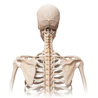
General quiz
Bone Connections
A quiz on the anatomy of bone connections: classification and the normal structure of joints, ligaments, and cartilage.
Fibrous joints of the vertebral column, ligamenta flava, cartilaginous joints of the vertebral column, and others.
0/9
Joints of vertebral column
0/20
By topic
Atlantoaxial joints (median and lateral)
0/20
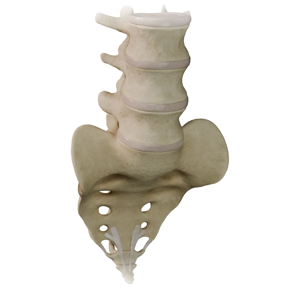
Cartilaginous joints (synchondroses) of the vertebral column
0/20
Fibrous joints of the vertebral column
0/20
Joints of thoracolumbar vertebral column
0/20
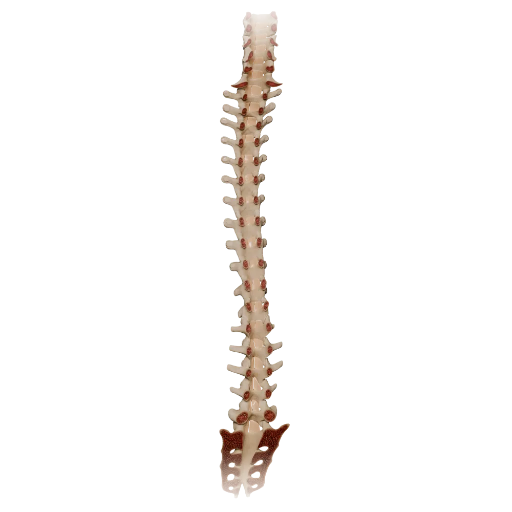
Ligamenta flava
0/20
Synovial joints of vertebral column
0/20
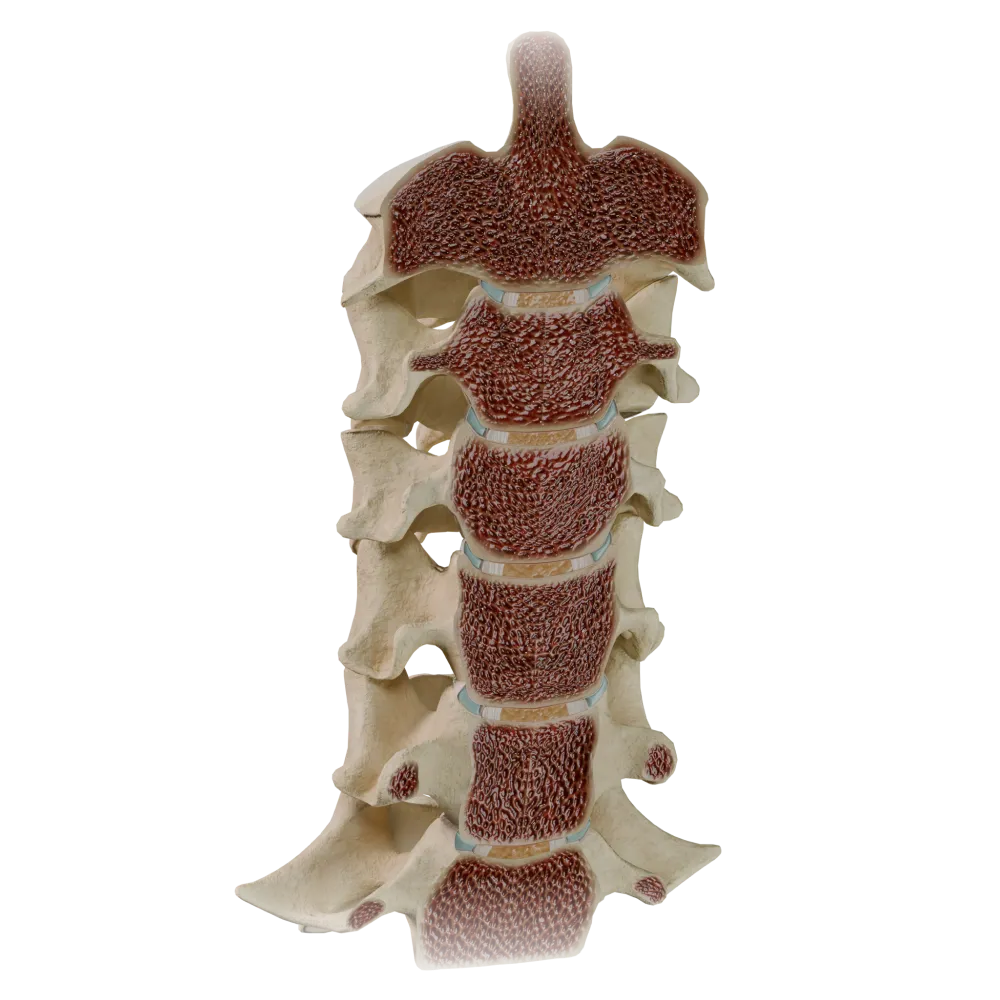
Uncovertebral joints
0/20
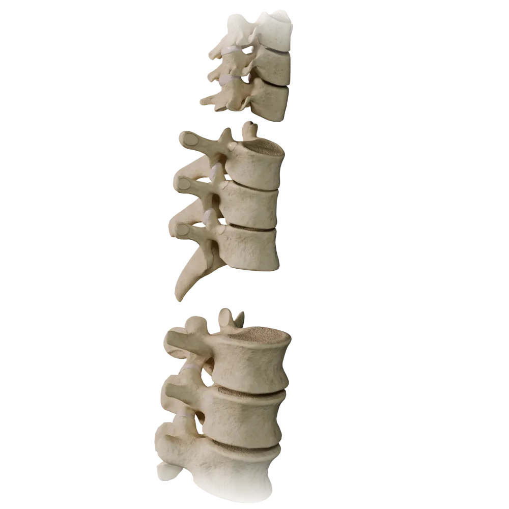
Zygapophyseal joints
0/20

We use cookies to enhance your browsing experience, analyze site traffic, and deliver content. Please choose whether you accept all cookies or wish to reject non-essential tracking.
Manage your cookie preferences below:
Essential cookies enable basic functions and are necessary for the proper function of the website.
Google reCAPTCHA helps protect websites from spam and abuse by verifying user interactions through challenges.
Statistics cookies collect information anonymously. This information helps us understand how visitors use our website.
Google Analytics is a powerful tool that tracks and analyzes website traffic for informed marketing decisions.
Service URL: policies.google.com (opens in a new window)
Clarity is a web analytics service that tracks and reports website traffic.
Service URL: clarity.microsoft.com (opens in a new window)