
Joints of vertebral column
0/20
By topic

Atlantoaxial joints (median and lateral)
0/20
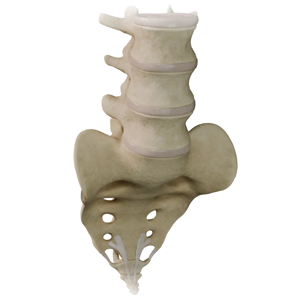
Cartilaginous joints (synchondroses) of the vertebral column
0/20

Fibrous joints of the vertebral column
0/20

Joints of thoracolumbar vertebral column
0/20
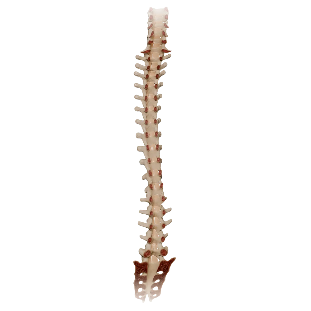
Ligamenta flava
0/20

Synovial joints of vertebral column
0/20
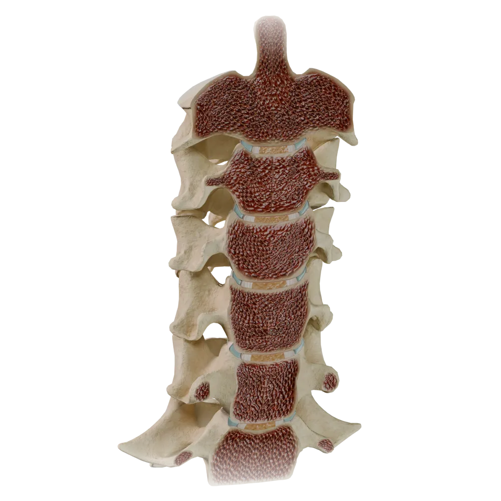
Uncovertebral joints
0/20
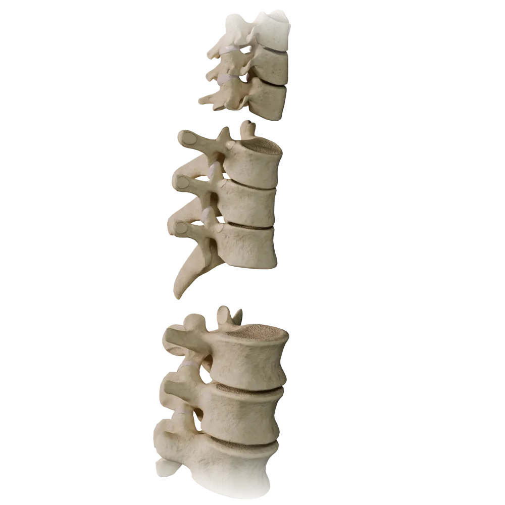
Zygapophyseal joints
0/20
Anatomy quiz on Fibrous joints of vertebral column
The quiz covers the anatomy of the Fibrous joints of vertebral column: ligaments, membranes, intervertebral discs, and their structural components.
1/20
bold
text
1. Which ligament runs along the anterior surfaces of the vertebral bodies and intervertebral discs throughout the Vertebral column?
-
Posterior longitudinal ligament
The Anterior longitudinal ligament (ligamentum longitudinale anterius) is located on the anterior surfaces of the vertebral bodies and intervertebral discs, extending from the cranial base to the sacrum. It restricts extension of the spine.
-
Anterior longitudinal ligament
The Anterior longitudinal ligament (ligamentum longitudinale anterius) is located on the anterior surfaces of the vertebral bodies and intervertebral discs, extending from the cranial base to the sacrum. It restricts extension of the spine.
-
Nuchal ligament
The Anterior longitudinal ligament (ligamentum longitudinale anterius) is located on the anterior surfaces of the vertebral bodies and intervertebral discs, extending from the cranial base to the sacrum. It restricts extension of the spine.
-
Supraspinous ligament
The Anterior longitudinal ligament (ligamentum longitudinale anterius) is located on the anterior surfaces of the vertebral bodies and intervertebral discs, extending from the cranial base to the sacrum. It restricts extension of the spine.
-
I find it difficult to answer
The Anterior longitudinal ligament (ligamentum longitudinale anterius) is located on the anterior surfaces of the vertebral bodies and intervertebral discs, extending from the cranial base to the sacrum. It restricts extension of the spine.
2. What are the two main components of the Intervertebral disc (discus intervertebralis)?
-
The nucleus pulposus and anulus fibrosus
The Intervertebral disc consists of the nucleus pulposus (nucleus pulposus) centrally and the anulus fibrosus (anulus fibrosus) peripherally. The nucleus pulposus is a remnant of the notochord, whereas the anulus fibrosus is formed by concentric layers of fibrocartilage.
-
Hyaline cartilage and perichondrium
The Intervertebral disc consists of the nucleus pulposus (nucleus pulposus) centrally and the anulus fibrosus (anulus fibrosus) peripherally. The nucleus pulposus is a remnant of the notochord, whereas the anulus fibrosus is formed by concentric layers of fibrocartilage.
-
Cancellous bone and compact bone
The Intervertebral disc consists of the nucleus pulposus (nucleus pulposus) centrally and the anulus fibrosus (anulus fibrosus) peripherally. The nucleus pulposus is a remnant of the notochord, whereas the anulus fibrosus is formed by concentric layers of fibrocartilage.
-
The pulpal ring and fibrous nucleus
The Intervertebral disc consists of the nucleus pulposus (nucleus pulposus) centrally and the anulus fibrosus (anulus fibrosus) peripherally. The nucleus pulposus is a remnant of the notochord, whereas the anulus fibrosus is formed by concentric layers of fibrocartilage.
-
I find it difficult to answer
The Intervertebral disc consists of the nucleus pulposus (nucleus pulposus) centrally and the anulus fibrosus (anulus fibrosus) peripherally. The nucleus pulposus is a remnant of the notochord, whereas the anulus fibrosus is formed by concentric layers of fibrocartilage.
3. Where does the Posterior longitudinal ligament (ligamentum longitudinale posterius) attach in relation to the vertebral bodies?
-
To the anterior surfaces of the vertebral bodies and the anuli fibrosi of the discs
The Posterior longitudinal ligament (ligamentum longitudinale posterius) is located on the posterior surfaces of the vertebral bodies and the anuli fibrosi of the discs within the vertebral canal. It restricts flexion of the spine and prevents posterior displacement of the discs.
-
To the transverse processes of the vertebrae
The Posterior longitudinal ligament (ligamentum longitudinale posterius) is located on the posterior surfaces of the vertebral bodies and the anuli fibrosi of the discs within the vertebral canal. It restricts flexion of the spine and prevents posterior displacement of the discs.
-
To the posterior surfaces of the vertebral bodies and intervertebral discs, within the vertebral canal
The Posterior longitudinal ligament (ligamentum longitudinale posterius) is located on the posterior surfaces of the vertebral bodies and the anuli fibrosi of the discs within the vertebral canal. It restricts flexion of the spine and prevents posterior displacement of the discs.
-
To the vertebral arches on the side facing the vertebral canal
The Posterior longitudinal ligament (ligamentum longitudinale posterius) is located on the posterior surfaces of the vertebral bodies and the anuli fibrosi of the discs within the vertebral canal. It restricts flexion of the spine and prevents posterior displacement of the discs.
-
I find it difficult to answer
The Posterior longitudinal ligament (ligamentum longitudinale posterius) is located on the posterior surfaces of the vertebral bodies and the anuli fibrosi of the discs within the vertebral canal. It restricts flexion of the spine and prevents posterior displacement of the discs.
4. Which of the following Joints of vertebral column is a syndesmosis (fibrous joint)?
-
Median atlantoaxial joint
The Ligamenta flava (ligg. flava) are typical syndesmoses—fibrous joints formed by elastic connective tissue. They connect the arches of adjacent vertebrae and restrict excessive flexion of the spine.
-
Costovertebral joint
The Ligamenta flava (ligg. flava) are typical syndesmoses—fibrous joints formed by elastic connective tissue. They connect the arches of adjacent vertebrae and restrict excessive flexion of the spine.
-
Intervertebral symphysis
The Ligamenta flava (ligg. flava) are typical syndesmoses—fibrous joints formed by elastic connective tissue. They connect the arches of adjacent vertebrae and restrict excessive flexion of the spine.
-
Ligamenta flava (ligg. flava)
The Ligamenta flava (ligg. flava) are typical syndesmoses—fibrous joints formed by elastic connective tissue. They connect the arches of adjacent vertebrae and restrict excessive flexion of the spine.
-
I find it difficult to answer
The Ligamenta flava (ligg. flava) are typical syndesmoses—fibrous joints formed by elastic connective tissue. They connect the arches of adjacent vertebrae and restrict excessive flexion of the spine.
5. What tissue predominantly forms the Ligamenta flava (ligg. flava)?
-
Dense irregular connective tissue
The Ligamenta flava are composed predominantly of elastic fibers, which account for their yellow color and high elasticity. This allows them to return the spine to its original position after flexion without forming folds in the vertebral canal.
-
Hyaline cartilage
The Ligamenta flava are composed predominantly of elastic fibers, which account for their yellow color and high elasticity. This allows them to return the spine to its original position after flexion without forming folds in the vertebral canal.
-
Elastic connective tissue
The Ligamenta flava are composed predominantly of elastic fibers, which account for their yellow color and high elasticity. This allows them to return the spine to its original position after flexion without forming folds in the vertebral canal.
-
Fibrocartilage
The Ligamenta flava are composed predominantly of elastic fibers, which account for their yellow color and high elasticity. This allows them to return the spine to its original position after flexion without forming folds in the vertebral canal.
-
I find it difficult to answer
The Ligamenta flava are composed predominantly of elastic fibers, which account for their yellow color and high elasticity. This allows them to return the spine to its original position after flexion without forming folds in the vertebral canal.
6. In humans, the Nuchal ligament (ligamentum nuchae) is derived from which ligament?
-
Anterior longitudinal ligament
The Nuchal ligament (ligamentum nuchae) is the cranial continuation of the supraspinous ligament (ligamentum supraspinale) in the cervical region of the spine. It is a fibroelastic lamina extending from the external occipital protuberance to the spinous process of C7.
-
Supraspinous ligament
The Nuchal ligament (ligamentum nuchae) is the cranial continuation of the supraspinous ligament (ligamentum supraspinale) in the cervical region of the spine. It is a fibroelastic lamina extending from the external occipital protuberance to the spinous process of C7.
-
Posterior longitudinal ligament
The Nuchal ligament (ligamentum nuchae) is the cranial continuation of the supraspinous ligament (ligamentum supraspinale) in the cervical region of the spine. It is a fibroelastic lamina extending from the external occipital protuberance to the spinous process of C7.
-
Intertransverse ligament
The Nuchal ligament (ligamentum nuchae) is the cranial continuation of the supraspinous ligament (ligamentum supraspinale) in the cervical region of the spine. It is a fibroelastic lamina extending from the external occipital protuberance to the spinous process of C7.
-
I find it difficult to answer
The Nuchal ligament (ligamentum nuchae) is the cranial continuation of the supraspinous ligament (ligamentum supraspinale) in the cervical region of the spine. It is a fibroelastic lamina extending from the external occipital protuberance to the spinous process of C7.
7. The Interspinous ligaments (ligg. interspinalia) connect:
-
Transverse processes of adjacent vertebrae
The Interspinous ligaments (ligg. interspinalia) connect the spinous processes of adjacent vertebrae, filling the space between them. They restrict flexion of the spine and are more highly developed in the lumbar region.
-
The bodies of adjacent vertebrae along their lateral surfaces
The Interspinous ligaments (ligg. interspinalia) connect the spinous processes of adjacent vertebrae, filling the space between them. They restrict flexion of the spine and are more highly developed in the lumbar region.
-
Spinous processes of adjacent vertebrae
The Interspinous ligaments (ligg. interspinalia) connect the spinous processes of adjacent vertebrae, filling the space between them. They restrict flexion of the spine and are more highly developed in the lumbar region.
-
Articular processes of adjacent vertebrae
The Interspinous ligaments (ligg. interspinalia) connect the spinous processes of adjacent vertebrae, filling the space between them. They restrict flexion of the spine and are more highly developed in the lumbar region.
-
I find it difficult to answer
The Interspinous ligaments (ligg. interspinalia) connect the spinous processes of adjacent vertebrae, filling the space between them. They restrict flexion of the spine and are more highly developed in the lumbar region.
8. In which region of the spine are the Intertransverse ligaments (ligg. intertransversaria) most highly developed?
-
Cervical
The Intertransverse ligaments (ligg. intertransversaria) connect the transverse processes of adjacent vertebrae. They are most prominent in the lumbar region. In the cervical region, they are often absent or rudimentary.
-
Thoracic
The Intertransverse ligaments (ligg. intertransversaria) connect the transverse processes of adjacent vertebrae. They are most prominent in the lumbar region. In the cervical region, they are often absent or rudimentary.
-
Sacral
The Intertransverse ligaments (ligg. intertransversaria) connect the transverse processes of adjacent vertebrae. They are most prominent in the lumbar region. In the cervical region, they are often absent or rudimentary.
-
Lumbar
The Intertransverse ligaments (ligg. intertransversaria) connect the transverse processes of adjacent vertebrae. They are most prominent in the lumbar region. In the cervical region, they are often absent or rudimentary.
-
I find it difficult to answer
The Intertransverse ligaments (ligg. intertransversaria) connect the transverse processes of adjacent vertebrae. They are most prominent in the lumbar region. In the cervical region, they are often absent or rudimentary.
9. A synchondrosis (synchondrosis) in the spine is represented by:
-
Ligamenta flava
Intervertebral discs (disci intervertebrales) are classified as symphyses (symphyses), which are a type of synchondrosis containing fibrocartilage. The Ligamenta flava and longitudinal ligaments are syndesmoses, whereas the articular processes form true synovial joints.
-
Intervertebral discs
Intervertebral discs (disci intervertebrales) are classified as symphyses (symphyses), which are a type of synchondrosis containing fibrocartilage. The Ligamenta flava and longitudinal ligaments are syndesmoses, whereas the articular processes form true synovial joints.
-
Supraspinous ligament
Intervertebral discs (disci intervertebrales) are classified as symphyses (symphyses), which are a type of synchondrosis containing fibrocartilage. The Ligamenta flava and longitudinal ligaments are syndesmoses, whereas the articular processes form true synovial joints.
-
Anterior longitudinal ligament
Intervertebral discs (disci intervertebrales) are classified as symphyses (symphyses), which are a type of synchondrosis containing fibrocartilage. The Ligamenta flava and longitudinal ligaments are syndesmoses, whereas the articular processes form true synovial joints.
-
I find it difficult to answer
Intervertebral discs (disci intervertebrales) are classified as symphyses (symphyses), which are a type of synchondrosis containing fibrocartilage. The Ligamenta flava and longitudinal ligaments are syndesmoses, whereas the articular processes form true synovial joints.
10. The anulus fibrosus of the Intervertebral disc (anulus fibrosus) attaches to:
-
Only the Posterior longitudinal ligament
The fibers of the anulus fibrosus attach to the hyaline cartilaginous (end) plates and the cortical rim of the apophyses of the vertebral bodies, forming a strong connection between the bodies of adjacent vertebrae.
-
Only the Anterior longitudinal ligament
The fibers of the anulus fibrosus attach to the hyaline cartilaginous (end) plates and the cortical rim of the apophyses of the vertebral bodies, forming a strong connection between the bodies of adjacent vertebrae.
-
The peripheral vertebral end plates (hyaline cartilaginous plates) and the cortical rim of the vertebral bodies
The fibers of the anulus fibrosus attach to the hyaline cartilaginous (end) plates and the cortical rim of the apophyses of the vertebral bodies, forming a strong connection between the bodies of adjacent vertebrae.
-
Only the perichondrium of the articular surfaces of the vertebrae
The fibers of the anulus fibrosus attach to the hyaline cartilaginous (end) plates and the cortical rim of the apophyses of the vertebral bodies, forming a strong connection between the bodies of adjacent vertebrae.
-
I find it difficult to answer
The fibers of the anulus fibrosus attach to the hyaline cartilaginous (end) plates and the cortical rim of the apophyses of the vertebral bodies, forming a strong connection between the bodies of adjacent vertebrae.
11. The Supraspinous ligament (ligamentum supraspinale) attaches to:
-
The vertebral bodies along their lateral surfaces
The Supraspinous ligament (ligamentum supraspinale) is a dense fibrous cord attached to the apices of the spinous processes from C7 to the sacrum. In the cervical region, it continues as the Nuchal ligament (ligamentum nuchae).
-
The apices of the spinous processes throughout the spine
The Supraspinous ligament (ligamentum supraspinale) is a dense fibrous cord attached to the apices of the spinous processes from C7 to the sacrum. In the cervical region, it continues as the Nuchal ligament (ligamentum nuchae).
-
The bases of the transverse processes
The Supraspinous ligament (ligamentum supraspinale) is a dense fibrous cord attached to the apices of the spinous processes from C7 to the sacrum. In the cervical region, it continues as the Nuchal ligament (ligamentum nuchae).
-
The vertebral arches dorsally
The Supraspinous ligament (ligamentum supraspinale) is a dense fibrous cord attached to the apices of the spinous processes from C7 to the sacrum. In the cervical region, it continues as the Nuchal ligament (ligamentum nuchae).
-
I find it difficult to answer
The Supraspinous ligament (ligamentum supraspinale) is a dense fibrous cord attached to the apices of the spinous processes from C7 to the sacrum. In the cervical region, it continues as the Nuchal ligament (ligamentum nuchae).
12. What is the primary function of the nucleus pulposus (nucleus pulposus) of the Intervertebral disc from the perspective of normal anatomy?
-
Conducting nerve impulses between vertebrae
The nucleus pulposus, which contains predominantly water and proteoglycans, functions as a hydraulic shock absorber, evenly distributing the vertical load to the anulus fibrosus and the end plates of the vertebral bodies.
-
Load distribution and absorption of compressive forces acting on the Vertebral column
The nucleus pulposus, which contains predominantly water and proteoglycans, functions as a hydraulic shock absorber, evenly distributing the vertical load to the anulus fibrosus and the end plates of the vertebral bodies.
-
Providing blood supply to the vertebral bodies
The nucleus pulposus, which contains predominantly water and proteoglycans, functions as a hydraulic shock absorber, evenly distributing the vertical load to the anulus fibrosus and the end plates of the vertebral bodies.
-
Restricting rotation in the zygapophyseal joints
The nucleus pulposus, which contains predominantly water and proteoglycans, functions as a hydraulic shock absorber, evenly distributing the vertical load to the anulus fibrosus and the end plates of the vertebral bodies.
-
I find it difficult to answer
The nucleus pulposus, which contains predominantly water and proteoglycans, functions as a hydraulic shock absorber, evenly distributing the vertical load to the anulus fibrosus and the end plates of the vertebral bodies.
13. The Intertransverse ligaments connect:
-
Transverse processes of adjacent vertebrae
The Intertransverse ligaments connect the transverse processes of adjacent vertebrae, filling the space between them.
-
The bodies of adjacent vertebrae along their lateral surfaces
The Intertransverse ligaments connect the transverse processes of adjacent vertebrae, filling the space between them.
-
Spinous processes of adjacent vertebrae
The Intertransverse ligaments connect the transverse processes of adjacent vertebrae, filling the space between them.
-
Articular processes of adjacent vertebrae
The Intertransverse ligaments connect the transverse processes of adjacent vertebrae, filling the space between them.
-
I find it difficult to answer
The Intertransverse ligaments connect the transverse processes of adjacent vertebrae, filling the space between them.
14. The ligament connecting the apex of the dens of the axis (C2) to the basilar part of the Occipital bone is called:
-
Transverse ligament of atlas
Ligamentum apicis dentis (Apical ligament of dens = Apical dental ligament) is an unpaired ligament connecting the apex of the dens of the axis to the anterior margin of the foramen magnum. It is a vestige of the notochord.
-
Alar ligaments
Ligamentum apicis dentis (Apical ligament of dens = Apical dental ligament) is an unpaired ligament connecting the apex of the dens of the axis to the anterior margin of the foramen magnum. It is a vestige of the notochord.
-
Apical ligament of dens = Apical dental ligament (ligamentum apicis dentis)
Ligamentum apicis dentis (Apical ligament of dens = Apical dental ligament) is an unpaired ligament connecting the apex of the dens of the axis to the anterior margin of the foramen magnum. It is a vestige of the notochord.
-
Anterior atlantooccipital membrane
Ligamentum apicis dentis (Apical ligament of dens = Apical dental ligament) is an unpaired ligament connecting the apex of the dens of the axis to the anterior margin of the foramen magnum. It is a vestige of the notochord.
-
I find it difficult to answer
Ligamentum apicis dentis (Apical ligament of dens = Apical dental ligament) is an unpaired ligament connecting the apex of the dens of the axis to the anterior margin of the foramen magnum. It is a vestige of the notochord.
15. Together with the longitudinal bands, the transverse ligament of the atlas (ligamentum transversum atlantis) forms a structure called the:
-
Cruciform ligament of the atlas (ligamentum cruciforme atlantis)
The transverse ligament of the atlas, together with the vertical bundles (fasciculi longitudinales) extending from it, forms the cruciform ligament of the atlas (ligamentum cruciforme atlantis). The transverse ligament holds the dens of the axis against the anterior arch of the atlas.
-
Tectorial membrane (membrana tectoria)
The transverse ligament of the atlas, together with the vertical bundles (fasciculi longitudinales) extending from it, forms the cruciform ligament of the atlas (ligamentum cruciforme atlantis). The transverse ligament holds the dens of the axis against the anterior arch of the atlas.
-
Anterior atlantoaxial membrane
The transverse ligament of the atlas, together with the vertical bundles (fasciculi longitudinales) extending from it, forms the cruciform ligament of the atlas (ligamentum cruciforme atlantis). The transverse ligament holds the dens of the axis against the anterior arch of the atlas.
-
Posterior atlantooccipital membrane
The transverse ligament of the atlas, together with the vertical bundles (fasciculi longitudinales) extending from it, forms the cruciform ligament of the atlas (ligamentum cruciforme atlantis). The transverse ligament holds the dens of the axis against the anterior arch of the atlas.
-
I find it difficult to answer
The transverse ligament of the atlas, together with the vertical bundles (fasciculi longitudinales) extending from it, forms the cruciform ligament of the atlas (ligamentum cruciforme atlantis). The transverse ligament holds the dens of the axis against the anterior arch of the atlas.
16. The tectorial membrane (membrana tectoria) is the cranial continuation of which ligament?
-
Anterior longitudinal ligament
The tectorial membrane (membrana tectoria) is the cranial continuation of the Posterior longitudinal ligament, covers the cruciform ligament of the atlas and the Alar ligaments posteriorly, and attaches to the basilar part of the Occipital bone.
-
Supraspinous ligament
The tectorial membrane (membrana tectoria) is the cranial continuation of the Posterior longitudinal ligament, covers the cruciform ligament of the atlas and the Alar ligaments posteriorly, and attaches to the basilar part of the Occipital bone.
-
Posterior longitudinal ligament
The tectorial membrane (membrana tectoria) is the cranial continuation of the Posterior longitudinal ligament, covers the cruciform ligament of the atlas and the Alar ligaments posteriorly, and attaches to the basilar part of the Occipital bone.
-
Nuchal ligament
The tectorial membrane (membrana tectoria) is the cranial continuation of the Posterior longitudinal ligament, covers the cruciform ligament of the atlas and the Alar ligaments posteriorly, and attaches to the basilar part of the Occipital bone.
-
I find it difficult to answer
The tectorial membrane (membrana tectoria) is the cranial continuation of the Posterior longitudinal ligament, covers the cruciform ligament of the atlas and the Alar ligaments posteriorly, and attaches to the basilar part of the Occipital bone.
17. The Alar ligaments (ligg. alaria) extend from the lateral surfaces of the dens of the axis to the:
-
Anterior arch of the atlas on both sides
The Alar ligaments (ligg. alaria) run from the lateral surfaces of the dens of the axis to the medial surfaces of the lateral masses of the atlas and the condyles of the Occipital bone. They restrict axial rotation and lateral bending.
-
Medial surfaces of the lateral masses of the atlas and the condyles of the Occipital bone
The Alar ligaments (ligg. alaria) run from the lateral surfaces of the dens of the axis to the medial surfaces of the lateral masses of the atlas and the condyles of the Occipital bone. They restrict axial rotation and lateral bending.
-
Transverse processes of the atlas
The Alar ligaments (ligg. alaria) run from the lateral surfaces of the dens of the axis to the medial surfaces of the lateral masses of the atlas and the condyles of the Occipital bone. They restrict axial rotation and lateral bending.
-
Posterior arch of the atlas
The Alar ligaments (ligg. alaria) run from the lateral surfaces of the dens of the axis to the medial surfaces of the lateral masses of the atlas and the condyles of the Occipital bone. They restrict axial rotation and lateral bending.
-
I find it difficult to answer
The Alar ligaments (ligg. alaria) run from the lateral surfaces of the dens of the axis to the medial surfaces of the lateral masses of the atlas and the condyles of the Occipital bone. They restrict axial rotation and lateral bending.
18. The anterior atlanto-occipital membrane (membrana atlantooccipitalis anterior) is a continuation of the:
-
Posterior longitudinal ligament
The anterior atlanto-occipital membrane (membrana atlantooccipitalis anterior) is a continuation of the Anterior longitudinal ligament. It connects the anterior arch of the atlas to the anterior margin of the foramen magnum and the basilar part of the Occipital bone.
-
Nuchal ligament
The anterior atlanto-occipital membrane (membrana atlantooccipitalis anterior) is a continuation of the Anterior longitudinal ligament. It connects the anterior arch of the atlas to the anterior margin of the foramen magnum and the basilar part of the Occipital bone.
-
Anterior longitudinal ligament
The anterior atlanto-occipital membrane (membrana atlantooccipitalis anterior) is a continuation of the Anterior longitudinal ligament. It connects the anterior arch of the atlas to the anterior margin of the foramen magnum and the basilar part of the Occipital bone.
-
Interspinous ligaments
The anterior atlanto-occipital membrane (membrana atlantooccipitalis anterior) is a continuation of the Anterior longitudinal ligament. It connects the anterior arch of the atlas to the anterior margin of the foramen magnum and the basilar part of the Occipital bone.
-
I find it difficult to answer
The anterior atlanto-occipital membrane (membrana atlantooccipitalis anterior) is a continuation of the Anterior longitudinal ligament. It connects the anterior arch of the atlas to the anterior margin of the foramen magnum and the basilar part of the Occipital bone.
19. According to the classification of continuous joints, the joints of the spine are classified as:
-
Syndesmoses
They are classified as synchondroses containing fibrocartilage. They occupy an intermediate position between a syndesmosis and a synovial joint.
-
Synchondroses containing fibrocartilage
They are classified as synchondroses containing fibrocartilage. They occupy an intermediate position between a syndesmosis and a synovial joint.
-
Synostoses
They are classified as synchondroses containing fibrocartilage. They occupy an intermediate position between a syndesmosis and a synovial joint.
-
Synovial joints
They are classified as synchondroses containing fibrocartilage. They occupy an intermediate position between a syndesmosis and a synovial joint.
-
I find it difficult to answer
They are classified as synchondroses containing fibrocartilage. They occupy an intermediate position between a syndesmosis and a synovial joint.
20. The Ligamenta flava connect:
-
Arches of adjacent vertebrae
The Ligamenta flava fill the spaces between the arches of adjacent vertebrae, bounding the vertebral canal posteriorly. They consist of elastic tissue (hence their yellow color) and prevent excessive flexion of the spine.
-
The bodies of adjacent vertebrae along their lateral surfaces
The Ligamenta flava fill the spaces between the arches of adjacent vertebrae, bounding the vertebral canal posteriorly. They consist of elastic tissue (hence their yellow color) and prevent excessive flexion of the spine.
-
Spinous processes of adjacent vertebrae
The Ligamenta flava fill the spaces between the arches of adjacent vertebrae, bounding the vertebral canal posteriorly. They consist of elastic tissue (hence their yellow color) and prevent excessive flexion of the spine.
-
Articular processes of adjacent vertebrae
The Ligamenta flava fill the spaces between the arches of adjacent vertebrae, bounding the vertebral canal posteriorly. They consist of elastic tissue (hence their yellow color) and prevent excessive flexion of the spine.
-
I find it difficult to answer
The Ligamenta flava fill the spaces between the arches of adjacent vertebrae, bounding the vertebral canal posteriorly. They consist of elastic tissue (hence their yellow color) and prevent excessive flexion of the spine.
Retake this quiz?
Your current progress will be reset.
