
Joints of vertebral column
0/20
By topic

Atlantoaxial joints (median and lateral)
0/20
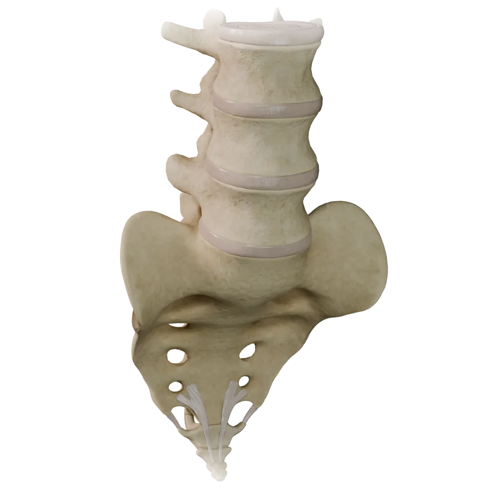
Cartilaginous joints (synchondroses) of the vertebral column
0/20

Fibrous joints of the vertebral column
0/20

Joints of thoracolumbar vertebral column
0/20
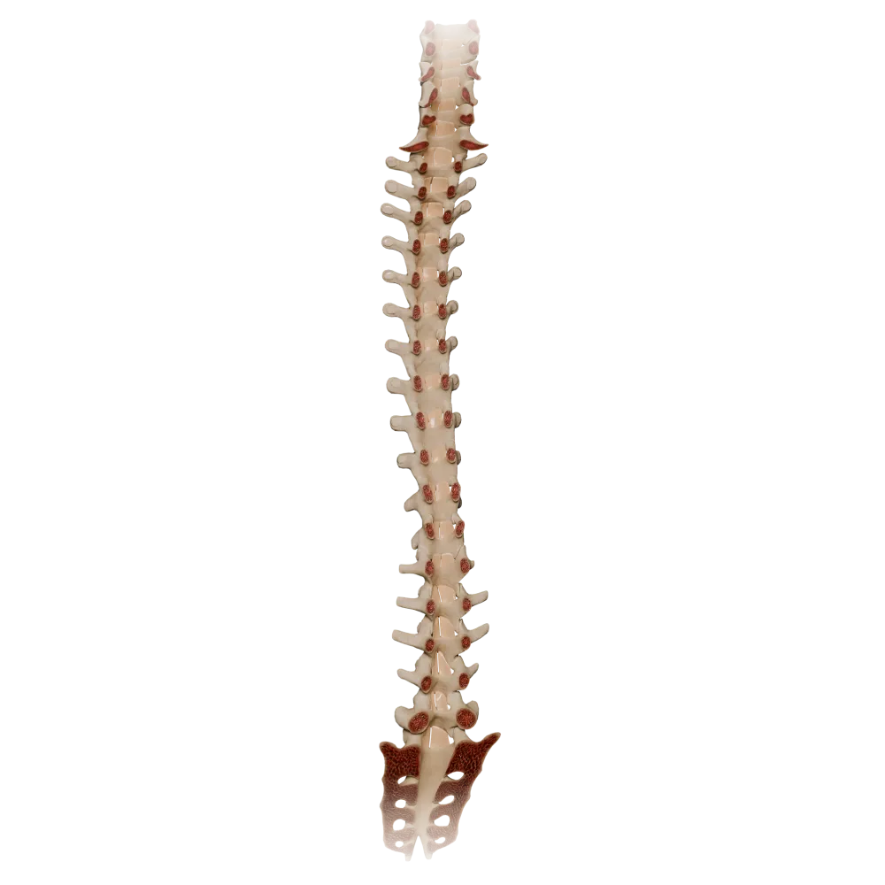
Ligamenta flava
0/20

Synovial joints of vertebral column
0/20
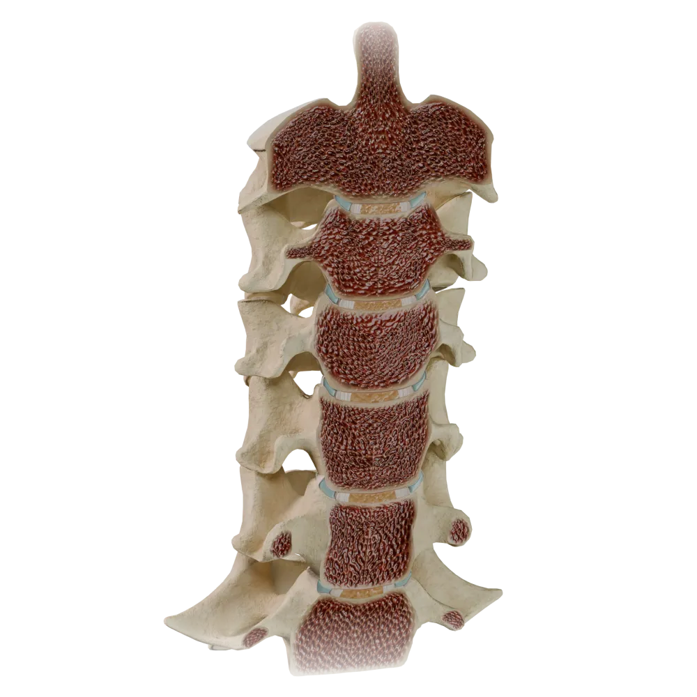
Uncovertebral joints
0/20
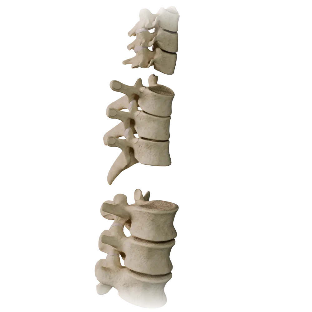
Zygapophyseal joints
0/20
Anatomy quiz on cartilaginous joints of vertebral column
Anatomy quiz on cartilaginous joints of vertebral column: intervertebral discs, articular cartilage, synchondroses.
1/20
bold
text
1. What type of cartilage forms the intervertebral discs?
-
Hyaline cartilage
Intervertebral discs are composed of fibrous (fibrocartilaginous) cartilage, which provides high compressive strength and resistance to loading. Hyaline cartilage covers the articular surfaces, whereas elastic cartilage is found in the auricle and epiglottis. Reticular cartilage is not recognized in the classification of cartilaginous tissues.
-
Elastic cartilage
Intervertebral discs are composed of fibrous (fibrocartilaginous) cartilage, which provides high compressive strength and resistance to loading. Hyaline cartilage covers the articular surfaces, whereas elastic cartilage is found in the auricle and epiglottis. Reticular cartilage is not recognized in the classification of cartilaginous tissues.
-
Fibrous (fibrocartilaginous) cartilage
Intervertebral discs are composed of fibrous (fibrocartilaginous) cartilage, which provides high compressive strength and resistance to loading. Hyaline cartilage covers the articular surfaces, whereas elastic cartilage is found in the auricle and epiglottis. Reticular cartilage is not recognized in the classification of cartilaginous tissues.
-
Reticular cartilage
Intervertebral discs are composed of fibrous (fibrocartilaginous) cartilage, which provides high compressive strength and resistance to loading. Hyaline cartilage covers the articular surfaces, whereas elastic cartilage is found in the auricle and epiglottis. Reticular cartilage is not recognized in the classification of cartilaginous tissues.
-
I find it difficult to answer
Intervertebral discs are composed of fibrous (fibrocartilaginous) cartilage, which provides high compressive strength and resistance to loading. Hyaline cartilage covers the articular surfaces, whereas elastic cartilage is found in the auricle and epiglottis. Reticular cartilage is not recognized in the classification of cartilaginous tissues.
2. What are the two principal structures comprising the intervertebral disc?
-
Nucleus pulposus and anulus fibrosus
The intervertebral disc (discus intervertebralis) consists of the gelatinous nucleus (nucleus pulposus)—a remnant of the notochord—and the fibrous ring (anulus fibrosus), formed by concentric lamellae of fibrous cartilage and connective tissue.
-
Cartilago hyalina and periosteum
The intervertebral disc (discus intervertebralis) consists of the gelatinous nucleus (nucleus pulposus)—a remnant of the notochord—and the fibrous ring (anulus fibrosus), formed by concentric lamellae of fibrous cartilage and connective tissue.
-
Ligamentum longitudinale and membrana tectoria
The intervertebral disc (discus intervertebralis) consists of the gelatinous nucleus (nucleus pulposus)—a remnant of the notochord—and the fibrous ring (anulus fibrosus), formed by concentric lamellae of fibrous cartilage and connective tissue.
-
Nucleus gelatinosus and capsula articularis
The intervertebral disc (discus intervertebralis) consists of the gelatinous nucleus (nucleus pulposus)—a remnant of the notochord—and the fibrous ring (anulus fibrosus), formed by concentric lamellae of fibrous cartilage and connective tissue.
-
I find it difficult to answer
The intervertebral disc (discus intervertebralis) consists of the gelatinous nucleus (nucleus pulposus)—a remnant of the notochord—and the fibrous ring (anulus fibrosus), formed by concentric lamellae of fibrous cartilage and connective tissue.
3. What is the cartilaginous joint between the vertebral bodies called?
-
Synovial joint (junctura synovialis)
The connection of the vertebral bodies by means of an intervertebral disc is a type of synchondrosis with a narrow cleft within the disc. The synovial joint is formed by the zygapophysial articulations. A syndesmosis is a joint formed by fibrous connective tissue.
-
Synchondrosis
The connection of the vertebral bodies by means of an intervertebral disc is a type of synchondrosis with a narrow cleft within the disc. The synovial joint is formed by the zygapophysial articulations. A syndesmosis is a joint formed by fibrous connective tissue.
-
Syndesmosis (syndesmosis)
The connection of the vertebral bodies by means of an intervertebral disc is a type of synchondrosis with a narrow cleft within the disc. The synovial joint is formed by the zygapophysial articulations. A syndesmosis is a joint formed by fibrous connective tissue.
-
Synostosis (synostosis)
The connection of the vertebral bodies by means of an intervertebral disc is a type of synchondrosis with a narrow cleft within the disc. The synovial joint is formed by the zygapophysial articulations. A syndesmosis is a joint formed by fibrous connective tissue.
-
I find it difficult to answer
The connection of the vertebral bodies by means of an intervertebral disc is a type of synchondrosis with a narrow cleft within the disc. The synovial joint is formed by the zygapophysial articulations. A syndesmosis is a joint formed by fibrous connective tissue.
4. How does the height of an intervertebral disc in the lumbar region compare with that in the cervical region?
-
The same in all regions
The height of the intervertebral discs increases in the caudal direction and is greatest in the lumbar region (up to 10–12 mm). This corresponds to the increasing axial load. In the cervical region, the discs are thinner, whereas in the thoracic region, their relative height is the lowest.
-
Greatest in the cervical region
The height of the intervertebral discs increases in the caudal direction and is greatest in the lumbar region (up to 10–12 mm). This corresponds to the increasing axial load. In the cervical region, the discs are thinner, whereas in the thoracic region, their relative height is the lowest.
-
Greatest in the lumbar region
The height of the intervertebral discs increases in the caudal direction and is greatest in the lumbar region (up to 10–12 mm). This corresponds to the increasing axial load. In the cervical region, the discs are thinner, whereas in the thoracic region, their relative height is the lowest.
-
Greatest in the thoracic region
The height of the intervertebral discs increases in the caudal direction and is greatest in the lumbar region (up to 10–12 mm). This corresponds to the increasing axial load. In the cervical region, the discs are thinner, whereas in the thoracic region, their relative height is the lowest.
-
I find it difficult to answer
The height of the intervertebral discs increases in the caudal direction and is greatest in the lumbar region (up to 10–12 mm). This corresponds to the increasing axial load. In the cervical region, the discs are thinner, whereas in the thoracic region, their relative height is the lowest.
5. How many intervertebral discs are present in the vertebral column of an adult?
-
23
An adult has 23 intervertebral discs. There is no disc between the occipital bone and the atlas or between CI–CII (the atlas and axis). The first disc is located between CII and CIII, and the last between LV and the sacrum.
-
24
An adult has 23 intervertebral discs. There is no disc between the occipital bone and the atlas or between CI–CII (the atlas and axis). The first disc is located between CII and CIII, and the last between LV and the sacrum.
-
25
An adult has 23 intervertebral discs. There is no disc between the occipital bone and the atlas or between CI–CII (the atlas and axis). The first disc is located between CII and CIII, and the last between LV and the sacrum.
-
26
An adult has 23 intervertebral discs. There is no disc between the occipital bone and the atlas or between CI–CII (the atlas and axis). The first disc is located between CII and CIII, and the last between LV and the sacrum.
-
I find it difficult to answer
An adult has 23 intervertebral discs. There is no disc between the occipital bone and the atlas or between CI–CII (the atlas and axis). The first disc is located between CII and CIII, and the last between LV and the sacrum.
6. Which structure of the intervertebral disc is derived from the notochord (chorda dorsalis)?
-
Anulus fibrosus
The gelatinous nucleus (nucleus pulposus) is a remnant of the notochord (chorda dorsalis) — the axial skeleton of the embryo. It is rich in water (up to 88% in neonates) and proteoglycans. The anulus fibrosus develops from the sclerotome, not from the notochord.
-
Nucleus pulposus
The gelatinous nucleus (nucleus pulposus) is a remnant of the notochord (chorda dorsalis) — the axial skeleton of the embryo. It is rich in water (up to 88% in neonates) and proteoglycans. The anulus fibrosus develops from the sclerotome, not from the notochord.
-
Cartilago epiphysialis
The gelatinous nucleus (nucleus pulposus) is a remnant of the notochord (chorda dorsalis) — the axial skeleton of the embryo. It is rich in water (up to 88% in neonates) and proteoglycans. The anulus fibrosus develops from the sclerotome, not from the notochord.
-
Ligamentum flavum
The gelatinous nucleus (nucleus pulposus) is a remnant of the notochord (chorda dorsalis) — the axial skeleton of the embryo. It is rich in water (up to 88% in neonates) and proteoglycans. The anulus fibrosus develops from the sclerotome, not from the notochord.
-
I find it difficult to answer
The gelatinous nucleus (nucleus pulposus) is a remnant of the notochord (chorda dorsalis) — the axial skeleton of the embryo. It is rich in water (up to 88% in neonates) and proteoglycans. The anulus fibrosus develops from the sclerotome, not from the notochord.
7. What is the ratio of disc height to vertebral body height in the cervical region?
-
Approximately 1:5
In the cervical region, the ratio of disc height to vertebral body height is approximately 2:5 (about 40%), providing the greatest range of motion. In the thoracic region, this ratio is about 1:5 (the lowest), whereas in the lumbar region it is about 1:3.
-
Approximately 2:5
In the cervical region, the ratio of disc height to vertebral body height is approximately 2:5 (about 40%), providing the greatest range of motion. In the thoracic region, this ratio is about 1:5 (the lowest), whereas in the lumbar region it is about 1:3.
-
Approximately 1:3
In the cervical region, the ratio of disc height to vertebral body height is approximately 2:5 (about 40%), providing the greatest range of motion. In the thoracic region, this ratio is about 1:5 (the lowest), whereas in the lumbar region it is about 1:3.
-
Approximately 1:2
In the cervical region, the ratio of disc height to vertebral body height is approximately 2:5 (about 40%), providing the greatest range of motion. In the thoracic region, this ratio is about 1:5 (the lowest), whereas in the lumbar region it is about 1:3.
-
I find it difficult to answer
In the cervical region, the ratio of disc height to vertebral body height is approximately 2:5 (about 40%), providing the greatest range of motion. In the thoracic region, this ratio is about 1:5 (the lowest), whereas in the lumbar region it is about 1:3.
8. Which synchondrosis of vertebral column connects the sacrum to the coccyx?
-
Symphysis sacroiliaca
The connection between the sacrum and coccyx — synchondrosis sacrococcygea — is a cartilaginous joint that may ossify with age.
-
Synchondrosis sacrococcygea
The connection between the sacrum and coccyx — synchondrosis sacrococcygea — is a cartilaginous joint that may ossify with age.
-
Symphysis pubica
The connection between the sacrum and coccyx — synchondrosis sacrococcygea — is a cartilaginous joint that may ossify with age.
-
Synchondrosis sphenooccipitalis
The connection between the sacrum and coccyx — synchondrosis sacrococcygea — is a cartilaginous joint that may ossify with age.
-
I find it difficult to answer
The connection between the sacrum and coccyx — synchondrosis sacrococcygea — is a cartilaginous joint that may ossify with age.
9. The fibers of the fibrous ring (anulus fibrosus) are oriented relative to those of the adjacent lamellae:
-
Parallel and horizontal
The fibers of the anulus fibrosus are arranged obliquely at an angle of approximately 30° to the horizontal plane; in adjacent concentric lamellae, they cross and are directed in opposite directions. This architecture provides resistance to both compressive and torsional loads.
-
Perpendicular to the axis of the vertebral column
The fibers of the anulus fibrosus are arranged obliquely at an angle of approximately 30° to the horizontal plane; in adjacent concentric lamellae, they cross and are directed in opposite directions. This architecture provides resistance to both compressive and torsional loads.
-
Obliquely, at an angle of ~30° to the horizontal, crossing in adjacent lamellae
The fibers of the anulus fibrosus are arranged obliquely at an angle of approximately 30° to the horizontal plane; in adjacent concentric lamellae, they cross and are directed in opposite directions. This architecture provides resistance to both compressive and torsional loads.
-
Spirally along the vertebral axis
The fibers of the anulus fibrosus are arranged obliquely at an angle of approximately 30° to the horizontal plane; in adjacent concentric lamellae, they cross and are directed in opposite directions. This architecture provides resistance to both compressive and torsional loads.
-
I find it difficult to answer
The fibers of the anulus fibrosus are arranged obliquely at an angle of approximately 30° to the horizontal plane; in adjacent concentric lamellae, they cross and are directed in opposite directions. This architecture provides resistance to both compressive and torsional loads.
10. What are the cartilaginous plates separating the vertebral body from the intervertebral disc called?
-
Cartilago articularis
The cartilaginous endplates — cartilagineae laminae (vertebral endplates) — consist of hyaline cartilage and form the boundary between the vertebral body and the disc. They provide nutrition to the disc by diffusion and absorb loads. Cartilago articularis covers the articular surfaces.
-
Laminae cartilagineae (vertebral endplate)
The cartilaginous endplates — cartilagineae laminae (vertebral endplates) — consist of hyaline cartilage and form the boundary between the vertebral body and the disc. They provide nutrition to the disc by diffusion and absorb loads. Cartilago articularis covers the articular surfaces.
-
Facies intervertebralis
The cartilaginous endplates — cartilagineae laminae (vertebral endplates) — consist of hyaline cartilage and form the boundary between the vertebral body and the disc. They provide nutrition to the disc by diffusion and absorb loads. Cartilago articularis covers the articular surfaces.
-
Periosteum corticale
The cartilaginous endplates — cartilagineae laminae (vertebral endplates) — consist of hyaline cartilage and form the boundary between the vertebral body and the disc. They provide nutrition to the disc by diffusion and absorb loads. Cartilago articularis covers the articular surfaces.
-
I find it difficult to answer
The cartilaginous endplates — cartilagineae laminae (vertebral endplates) — consist of hyaline cartilage and form the boundary between the vertebral body and the disc. They provide nutrition to the disc by diffusion and absorb loads. Cartilago articularis covers the articular surfaces.
11. Between which vertebrae is there no intervertebral disc?
-
CII–CIII
There is no intervertebral disc between CI (the atlas) and CII (the axis). The median atlanto-axial joint is synovial and permits rotation. The first disc is located between CII and CIII. A disc is present between LV and SI.
-
CI–CII (atlas–axis)
There is no intervertebral disc between CI (the atlas) and CII (the axis). The median atlanto-axial joint is synovial and permits rotation. The first disc is located between CII and CIII. A disc is present between LV and SI.
-
CIII–CIV
There is no intervertebral disc between CI (the atlas) and CII (the axis). The median atlanto-axial joint is synovial and permits rotation. The first disc is located between CII and CIII. A disc is present between LV and SI.
-
LV–SI
There is no intervertebral disc between CI (the atlas) and CII (the axis). The median atlanto-axial joint is synovial and permits rotation. The first disc is located between CII and CIII. A disc is present between LV and SI.
-
I find it difficult to answer
There is no intervertebral disc between CI (the atlas) and CII (the axis). The median atlanto-axial joint is synovial and permits rotation. The first disc is located between CII and CIII. A disc is present between LV and SI.
12. How is the intervertebral disc nourished in an adult?
-
By direct blood supply through the disc's own vessels
The intervertebral disc in an adult is an avascular structure and receives nutrients predominantly by diffusion through the cartilaginous endplates from vessels in the cancellous bone of the vertebral bodies. During childhood, blood vessels penetrate the disc and become obliterated by 20–25 years of age.
-
By diffusion from the vessels of the vertebral bodies through the cartilaginous endplates
The intervertebral disc in an adult is an avascular structure and receives nutrients predominantly by diffusion through the cartilaginous endplates from vessels in the cancellous bone of the vertebral bodies. During childhood, blood vessels penetrate the disc and become obliterated by 20–25 years of age.
-
By synovial fluid from the zygapophysial joints
The intervertebral disc in an adult is an avascular structure and receives nutrients predominantly by diffusion through the cartilaginous endplates from vessels in the cancellous bone of the vertebral bodies. During childhood, blood vessels penetrate the disc and become obliterated by 20–25 years of age.
-
By lymphatic capillaries of the anulus fibrosus
The intervertebral disc in an adult is an avascular structure and receives nutrients predominantly by diffusion through the cartilaginous endplates from vessels in the cancellous bone of the vertebral bodies. During childhood, blood vessels penetrate the disc and become obliterated by 20–25 years of age.
-
I find it difficult to answer
The intervertebral disc in an adult is an avascular structure and receives nutrients predominantly by diffusion through the cartilaginous endplates from vessels in the cancellous bone of the vertebral bodies. During childhood, blood vessels penetrate the disc and become obliterated by 20–25 years of age.
13. What is the cartilaginous joint between the first and second sacral segments during growth called?
-
Symphysis sacroiliaca
During growth, the individual sacral segments are connected by cartilaginous layers — synchondroses intersacrales. At the completion of growth, they ossify completely (become synostoses), forming a single sacral bone. Articulatio sacroiliaca is a synovial articulation between the sacrum and the ilium.
-
Synchondrosis intercorporalis
During growth, the individual sacral segments are connected by cartilaginous layers — synchondroses intersacrales. At the completion of growth, they ossify completely (become synostoses), forming a single sacral bone. Articulatio sacroiliaca is a synovial articulation between the sacrum and the ilium.
-
Synchondrosis intersacralis
During growth, the individual sacral segments are connected by cartilaginous layers — synchondroses intersacrales. At the completion of growth, they ossify completely (become synostoses), forming a single sacral bone. Articulatio sacroiliaca is a synovial articulation between the sacrum and the ilium.
-
Articulatio sacroiliaca
During growth, the individual sacral segments are connected by cartilaginous layers — synchondroses intersacrales. At the completion of growth, they ossify completely (become synostoses), forming a single sacral bone. Articulatio sacroiliaca is a synovial articulation between the sacrum and the ilium.
-
I find it difficult to answer
During growth, the individual sacral segments are connected by cartilaginous layers — synchondroses intersacrales. At the completion of growth, they ossify completely (become synostoses), forming a single sacral bone. Articulatio sacroiliaca is a synovial articulation between the sacrum and the ilium.
14. How many intervertebral discs are present in the cervical region of an adult?
-
5
An adult has 5 intervertebral discs. There is no disc between the occipital bone and the atlas or between CI–CII (the atlas and axis). The first disc is located between CII and CIII.
-
7
An adult has 5 intervertebral discs. There is no disc between the occipital bone and the atlas or between CI–CII (the atlas and axis). The first disc is located between CII and CIII.
-
4
An adult has 5 intervertebral discs. There is no disc between the occipital bone and the atlas or between CI–CII (the atlas and axis). The first disc is located between CII and CIII.
-
8
An adult has 5 intervertebral discs. There is no disc between the occipital bone and the atlas or between CI–CII (the atlas and axis). The first disc is located between CII and CIII.
-
I find it difficult to answer
An adult has 5 intervertebral discs. There is no disc between the occipital bone and the atlas or between CI–CII (the atlas and axis). The first disc is located between CII and CIII.
15. What is the shape of the cartilaginous endplates (vertebral endplates) in a transverse section of the vertebral body?
-
Convex superiorly and inferiorly (biconvex)
The cartilaginous endplates have a concave (concave) shape corresponding to the slightly biconcave surface of the vertebral body. This geometry centers the nucleus pulposus and distributes the load evenly across the surface of the disc.
-
Flat
The cartilaginous endplates have a concave (concave) shape corresponding to the slightly biconcave surface of the vertebral body. This geometry centers the nucleus pulposus and distributes the load evenly across the surface of the disc.
-
Concave (concave), conforming to the shape of the vertebral body
The cartilaginous endplates have a concave (concave) shape corresponding to the slightly biconcave surface of the vertebral body. This geometry centers the nucleus pulposus and distributes the load evenly across the surface of the disc.
-
Triangular
The cartilaginous endplates have a concave (concave) shape corresponding to the slightly biconcave surface of the vertebral body. This geometry centers the nucleus pulposus and distributes the load evenly across the surface of the disc.
-
I find it difficult to answer
The cartilaginous endplates have a concave (concave) shape corresponding to the slightly biconcave surface of the vertebral body. This geometry centers the nucleus pulposus and distributes the load evenly across the surface of the disc.
16. What is the thickness of the cartilaginous endplate of the vertebral body?
-
Approximately 0.2–0.5 mm
The cartilaginous endplates of the vertebral bodies are approximately 0.6–1.0 mm thick. They are thinner in the central portion (over the nucleus pulposus) and somewhat thicker peripherally. This thin cartilaginous layer is critically important for nutrition of the disc by diffusion.
-
Approximately 0.6–1.0 mm
The cartilaginous endplates of the vertebral bodies are approximately 0.6–1.0 mm thick. They are thinner in the central portion (over the nucleus pulposus) and somewhat thicker peripherally. This thin cartilaginous layer is critically important for nutrition of the disc by diffusion.
-
Approximately 2–4 mm
The cartilaginous endplates of the vertebral bodies are approximately 0.6–1.0 mm thick. They are thinner in the central portion (over the nucleus pulposus) and somewhat thicker peripherally. This thin cartilaginous layer is critically important for nutrition of the disc by diffusion.
-
Approximately 5–7 mm
The cartilaginous endplates of the vertebral bodies are approximately 0.6–1.0 mm thick. They are thinner in the central portion (over the nucleus pulposus) and somewhat thicker peripherally. This thin cartilaginous layer is critically important for nutrition of the disc by diffusion.
-
I find it difficult to answer
The cartilaginous endplates of the vertebral bodies are approximately 0.6–1.0 mm thick. They are thinner in the central portion (over the nucleus pulposus) and somewhat thicker peripherally. This thin cartilaginous layer is critically important for nutrition of the disc by diffusion.
17. What is the cartilaginous joint between the second and third sacral segments during growth called?
-
Symphysis sacroiliaca
During growth, the individual sacral segments are connected by cartilaginous layers—synchondroses intersacrales. At the completion of growth, they ossify completely (become synostoses), forming a single sacral bone. Articulatio sacroiliaca is a synovial articulation between the sacrum and the ilium.
-
Synchondrosis intercorporalis
During growth, the individual sacral segments are connected by cartilaginous layers—synchondroses intersacrales. At the completion of growth, they ossify completely (become synostoses), forming a single sacral bone. Articulatio sacroiliaca is a synovial articulation between the sacrum and the ilium.
-
Synchondrosis intersacralis
During growth, the individual sacral segments are connected by cartilaginous layers—synchondroses intersacrales. At the completion of growth, they ossify completely (become synostoses), forming a single sacral bone. Articulatio sacroiliaca is a synovial articulation between the sacrum and the ilium.
-
Articulatio sacroiliaca
During growth, the individual sacral segments are connected by cartilaginous layers—synchondroses intersacrales. At the completion of growth, they ossify completely (become synostoses), forming a single sacral bone. Articulatio sacroiliaca is a synovial articulation between the sacrum and the ilium.
-
I find it difficult to answer
During growth, the individual sacral segments are connected by cartilaginous layers—synchondroses intersacrales. At the completion of growth, they ossify completely (become synostoses), forming a single sacral bone. Articulatio sacroiliaca is a synovial articulation between the sacrum and the ilium.
18. How does the water content of the nucleus pulposus change with age?
-
It increases from 70% to 90%
In neonates, the nucleus pulposus contains approximately 88% water; with age, this value gradually decreases to approximately 70% in older adults. Dehydration of the nucleus reduces its capacity for hydrostatic load distribution and decreases disc height.
-
It does not change and remains approximately 80%
In neonates, the nucleus pulposus contains approximately 88% water; with age, this value gradually decreases to approximately 70% in older adults. Dehydration of the nucleus reduces its capacity for hydrostatic load distribution and decreases disc height.
-
It decreases from approximately 88% in neonates to 70% in older adults
In neonates, the nucleus pulposus contains approximately 88% water; with age, this value gradually decreases to approximately 70% in older adults. Dehydration of the nucleus reduces its capacity for hydrostatic load distribution and decreases disc height.
-
It decreases to 40–50% by middle age
In neonates, the nucleus pulposus contains approximately 88% water; with age, this value gradually decreases to approximately 70% in older adults. Dehydration of the nucleus reduces its capacity for hydrostatic load distribution and decreases disc height.
-
I find it difficult to answer
In neonates, the nucleus pulposus contains approximately 88% water; with age, this value gradually decreases to approximately 70% in older adults. Dehydration of the nucleus reduces its capacity for hydrostatic load distribution and decreases disc height.
19. Which region of the vertebral column does NOT have intervertebral discs between all adjacent vertebrae?
-
Lumbar region
In adults, the sacral vertebrae are completely united by synostosis (the intersegmental synchondroses ossify); therefore, there are no true intervertebral discs between them. The cervical region lacks a disc only between CI–CII, not between all adjacent vertebrae. The thoracic and lumbar regions have discs between all adjacent vertebrae.
-
Thoracic region
In adults, the sacral vertebrae are completely united by synostosis (the intersegmental synchondroses ossify); therefore, there are no true intervertebral discs between them. The cervical region lacks a disc only between CI–CII, not between all adjacent vertebrae. The thoracic and lumbar regions have discs between all adjacent vertebrae.
-
Cervical region (between CI–CII)
In adults, the sacral vertebrae are completely united by synostosis (the intersegmental synchondroses ossify); therefore, there are no true intervertebral discs between them. The cervical region lacks a disc only between CI–CII, not between all adjacent vertebrae. The thoracic and lumbar regions have discs between all adjacent vertebrae.
-
Sacral region in adults
In adults, the sacral vertebrae are completely united by synostosis (the intersegmental synchondroses ossify); therefore, there are no true intervertebral discs between them. The cervical region lacks a disc only between CI–CII, not between all adjacent vertebrae. The thoracic and lumbar regions have discs between all adjacent vertebrae.
-
I find it difficult to answer
In adults, the sacral vertebrae are completely united by synostosis (the intersegmental synchondroses ossify); therefore, there are no true intervertebral discs between them. The cervical region lacks a disc only between CI–CII, not between all adjacent vertebrae. The thoracic and lumbar regions have discs between all adjacent vertebrae.
20. What is the function of the nucleus pulposus in the mechanics of the intervertebral disc?
-
It limits rotational movements of the vertebral column
The nucleus pulposus functions as a hydrostatic cushion: as an incompressible gel, it distributes pressure uniformly in all directions to the anulus fibrosus and cartilaginous endplates under axial loading (according to Pascal's principle).
-
It acts as a hydrostatic nucleus, evenly distributing compressive load to the anulus fibrosus
The nucleus pulposus functions as a hydrostatic cushion: as an incompressible gel, it distributes pressure uniformly in all directions to the anulus fibrosus and cartilaginous endplates under axial loading (according to Pascal's principle).
-
It provides the blood supply to the disc
The nucleus pulposus functions as a hydrostatic cushion: as an incompressible gel, it distributes pressure uniformly in all directions to the anulus fibrosus and cartilaginous endplates under axial loading (according to Pascal's principle).
-
It rigidly fixes the vertebral bodies, preventing any movement
The nucleus pulposus functions as a hydrostatic cushion: as an incompressible gel, it distributes pressure uniformly in all directions to the anulus fibrosus and cartilaginous endplates under axial loading (according to Pascal's principle).
-
I find it difficult to answer
The nucleus pulposus functions as a hydrostatic cushion: as an incompressible gel, it distributes pressure uniformly in all directions to the anulus fibrosus and cartilaginous endplates under axial loading (according to Pascal's principle).
Retake this quiz?
Your current progress will be reset.
