
Joints of vertebral column
0/20
By topic

Atlantoaxial joints (median and lateral)
0/20
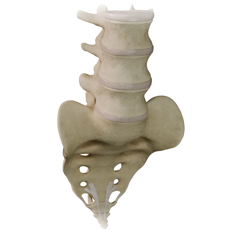
Cartilaginous joints (synchondroses) of the vertebral column
0/20

Fibrous joints of the vertebral column
0/20

Joints of thoracolumbar vertebral column
0/20
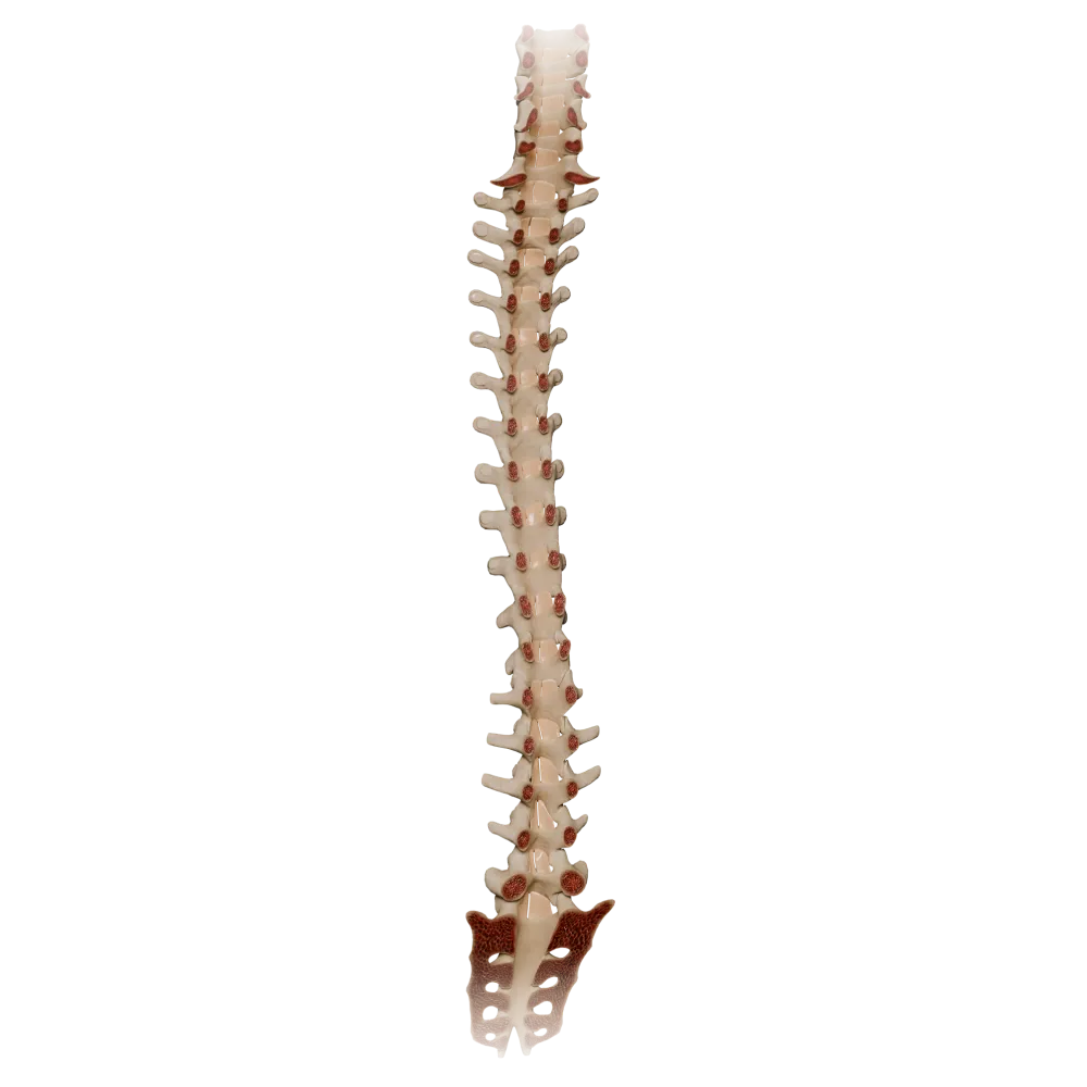
Ligamenta flava
0/20

Synovial joints of vertebral column
0/20
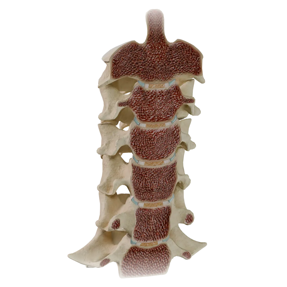
Uncovertebral joints
0/20
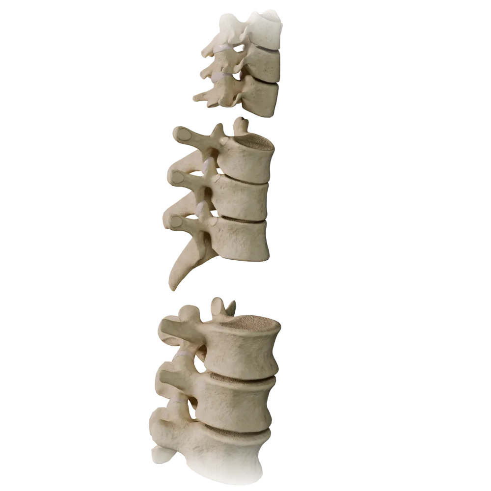
Zygapophyseal joints
0/20
Ligamenta flava anatomy quiz
Quiz on the normal anatomy of the ligamenta flava of the vertebral column: structure, topography, functions, and systemic relationships.
1/20
bold
text
1. Which specific parts of the vertebrae do the ligamenta flava (ligg. flava) connect?
-
Bodies of adjacent vertebrae
The ligamenta flava extend between the laminae of the arches (laminae arcuum) of adjacent vertebrae, filling the spaces between them from the II cervical vertebra to the I sacral vertebra.
-
Arches of adjacent vertebrae
The ligamenta flava extend between the laminae of the arches (laminae arcuum) of adjacent vertebrae, filling the spaces between them from the II cervical vertebra to the I sacral vertebra.
-
Transverse processes of adjacent vertebrae
The ligamenta flava extend between the laminae of the arches (laminae arcuum) of adjacent vertebrae, filling the spaces between them from the II cervical vertebra to the I sacral vertebra.
-
Spinous processes of adjacent vertebrae
The ligamenta flava extend between the laminae of the arches (laminae arcuum) of adjacent vertebrae, filling the spaces between them from the II cervical vertebra to the I sacral vertebra.
-
I find it difficult to answer
The ligamenta flava extend between the laminae of the arches (laminae arcuum) of adjacent vertebrae, filling the spaces between them from the II cervical vertebra to the I sacral vertebra.
2. What tissue forms the basis of the ligamenta flava and determines their characteristic color?
-
Dense fibrous connective tissue with a predominance of collagen fibers
Ligamenta flava consist primarily of elastic fibers (up to 80%), which accounts for their yellow color and high elasticity, allowing the spine to return to its original position after flexion.
-
Reticular connective tissue
Ligamenta flava consist primarily of elastic fibers (up to 80%), which accounts for their yellow color and high elasticity, allowing the spine to return to its original position after flexion.
-
Elastic connective tissue with a predominance of elastic fibers
Ligamenta flava consist primarily of elastic fibers (up to 80%), which accounts for their yellow color and high elasticity, allowing the spine to return to its original position after flexion.
-
Hyaline cartilage
Ligamenta flava consist primarily of elastic fibers (up to 80%), which accounts for their yellow color and high elasticity, allowing the spine to return to its original position after flexion.
-
I find it difficult to answer
Ligamenta flava consist primarily of elastic fibers (up to 80%), which accounts for their yellow color and high elasticity, allowing the spine to return to its original position after flexion.
3. At which vertebra does the superior extent of the ligamenta flava of the vertebral column begin?
-
At the atlas (C I)
The ligamenta flava begin at the axis (C II) and extend inferiorly to the I sacral vertebra. Between the atlas and the axis, the posterior atlanto-axial membrane is present instead of a ligamentum flavum.
-
At the axis (C II)
The ligamenta flava begin at the axis (C II) and extend inferiorly to the I sacral vertebra. Between the atlas and the axis, the posterior atlanto-axial membrane is present instead of a ligamentum flavum.
-
At the III cervical vertebra (C III)
The ligamenta flava begin at the axis (C II) and extend inferiorly to the I sacral vertebra. Between the atlas and the axis, the posterior atlanto-axial membrane is present instead of a ligamentum flavum.
-
At the VII cervical vertebra (C VII)
The ligamenta flava begin at the axis (C II) and extend inferiorly to the I sacral vertebra. Between the atlas and the axis, the posterior atlanto-axial membrane is present instead of a ligamentum flavum.
-
I find it difficult to answer
The ligamenta flava begin at the axis (C II) and extend inferiorly to the I sacral vertebra. Between the atlas and the axis, the posterior atlanto-axial membrane is present instead of a ligamentum flavum.
4. Which movement of the vertebral column is primarily restricted by the ligamenta flava?
-
Extension
The ligamenta flava restrict excessive flexion of the vertebral column: during flexion, they are stretched and passively assist in returning the spine to an upright position, preventing excessive separation of the vertebral arches.
-
Lateral bending
The ligamenta flava restrict excessive flexion of the vertebral column: during flexion, they are stretched and passively assist in returning the spine to an upright position, preventing excessive separation of the vertebral arches.
-
Flexion
The ligamenta flava restrict excessive flexion of the vertebral column: during flexion, they are stretched and passively assist in returning the spine to an upright position, preventing excessive separation of the vertebral arches.
-
Rotation
The ligamenta flava restrict excessive flexion of the vertebral column: during flexion, they are stretched and passively assist in returning the spine to an upright position, preventing excessive separation of the vertebral arches.
-
I find it difficult to answer
The ligamenta flava restrict excessive flexion of the vertebral column: during flexion, they are stretched and passively assist in returning the spine to an upright position, preventing excessive separation of the vertebral arches.
5. The ligamenta flava form part of which wall of the vertebral canal?
-
Anterior wall
The ligamenta flava form the posterior wall of the vertebral canal (canalis vertebralis), lying poster to the contents of the canal. The anterior wall is formed by the vertebral bodies and intervertebral discs.
-
Lateral wall
The ligamenta flava form the posterior wall of the vertebral canal (canalis vertebralis), lying poster to the contents of the canal. The anterior wall is formed by the vertebral bodies and intervertebral discs.
-
Posterior wall
The ligamenta flava form the posterior wall of the vertebral canal (canalis vertebralis), lying poster to the contents of the canal. The anterior wall is formed by the vertebral bodies and intervertebral discs.
-
Inferior wall
The ligamenta flava form the posterior wall of the vertebral canal (canalis vertebralis), lying poster to the contents of the canal. The anterior wall is formed by the vertebral bodies and intervertebral discs.
-
I find it difficult to answer
The ligamenta flava form the posterior wall of the vertebral canal (canalis vertebralis), lying poster to the contents of the canal. The anterior wall is formed by the vertebral bodies and intervertebral discs.
6. What is the functional role of the ligamenta flava in maintaining body position?
-
Actively contract like muscle tissue during extension
The high content of elastic fibers provides passive resistance to gravity: the ligamenta flava are constantly pretensioned and help maintain an upright posture without additional muscular effort.
-
Passively maintain the upright position of the spine through the resilience of elastic fibers
The high content of elastic fibers provides passive resistance to gravity: the ligamenta flava are constantly pretensioned and help maintain an upright posture without additional muscular effort.
-
Produce synovial fluid to lubricate the intervertebral joints
The high content of elastic fibers provides passive resistance to gravity: the ligamenta flava are constantly pretensioned and help maintain an upright posture without additional muscular effort.
-
Transmit axial load from the vertebral bodies to the intervertebral discs
The high content of elastic fibers provides passive resistance to gravity: the ligamenta flava are constantly pretensioned and help maintain an upright posture without additional muscular effort.
-
I find it difficult to answer
The high content of elastic fibers provides passive resistance to gravity: the ligamenta flava are constantly pretensioned and help maintain an upright posture without additional muscular effort.
7. Between which structures are the ligamenta flava located in the transverse plane?
-
Between the vertebral body and the intervertebral disc
In the transverse plane, the Ligamenta flava occupy the space between the laminae (laminae) of the arches of adjacent vertebrae, extending laterally to the capsules of the zygapophyseal joints.
-
Between the articular processes
In the transverse plane, the Ligamenta flava occupy the space between the laminae (laminae) of the arches of adjacent vertebrae, extending laterally to the capsules of the zygapophyseal joints.
-
Between the adjacent laminae of the vertebral arches (laminae)
In the transverse plane, the Ligamenta flava occupy the space between the laminae (laminae) of the arches of adjacent vertebrae, extending laterally to the capsules of the zygapophyseal joints.
-
Between the transverse processes and costal facets
In the transverse plane, the Ligamenta flava occupy the space between the laminae (laminae) of the arches of adjacent vertebrae, extending laterally to the capsules of the zygapophyseal joints.
-
I find it difficult to answer
In the transverse plane, the Ligamenta flava occupy the space between the laminae (laminae) of the arches of adjacent vertebrae, extending laterally to the capsules of the zygapophyseal joints.
8. What is the inferior extent of the ligamenta flava along the vertebral column?
-
V lumbar vertebra (L V)
The ligamenta flava extend from C II to S I, inclusive. Inferiorly, in the sacral and coccygeal regions, the vertebral arches fuse, and separate ligaments are not formed.
-
I sacral vertebra (S I)
The ligamenta flava extend from C II to S I, inclusive. Inferiorly, in the sacral and coccygeal regions, the vertebral arches fuse, and separate ligaments are not formed.
-
II sacral vertebra (S II)
The ligamenta flava extend from C II to S I, inclusive. Inferiorly, in the sacral and coccygeal regions, the vertebral arches fuse, and separate ligaments are not formed.
-
Coccyx (os coccygis)
The ligamenta flava extend from C II to S I, inclusive. Inferiorly, in the sacral and coccygeal regions, the vertebral arches fuse, and separate ligaments are not formed.
-
I find it difficult to answer
The ligamenta flava extend from C II to S I, inclusive. Inferiorly, in the sacral and coccygeal regions, the vertebral arches fuse, and separate ligaments are not formed.
9. What ratio of elastic to collagen fibers is characteristic of the ligamenta flava?
-
Collagen fibers constitute approximately 80%, and elastic fibers approximately 20%
According to histological studies, the ligamenta flava contain approximately 80% elastic fibers and 20% collagen fibers—the highest proportion of elastic fibers among all ligaments of the human body.
-
Elastic and collagen fibers are present in approximately equal proportions
According to histological studies, the ligamenta flava contain approximately 80% elastic fibers and 20% collagen fibers—the highest proportion of elastic fibers among all ligaments of the human body.
-
Elastic fibers constitute approximately 80%, and collagen fibers approximately 20%
According to histological studies, the ligamenta flava contain approximately 80% elastic fibers and 20% collagen fibers—the highest proportion of elastic fibers among all ligaments of the human body.
-
Reticular fibers constitute more than 50%, with the remainder being elastic fibers
According to histological studies, the ligamenta flava contain approximately 80% elastic fibers and 20% collagen fibers—the highest proportion of elastic fibers among all ligaments of the human body.
-
I find it difficult to answer
According to histological studies, the ligamenta flava contain approximately 80% elastic fibers and 20% collagen fibers—the highest proportion of elastic fibers among all ligaments of the human body.
10. At which level of the vertebral column are the ligamenta flava the thickest?
-
In the cervical region
In the lumbar region, the ligamenta flava are the thickest (up to 5–6 mm), corresponding to the maximum load on this region of the spine. In the cervical region, they are thinner and broader.
-
In the thoracic region
In the lumbar region, the ligamenta flava are the thickest (up to 5–6 mm), corresponding to the maximum load on this region of the spine. In the cervical region, they are thinner and broader.
-
In the lumbar region
In the lumbar region, the ligamenta flava are the thickest (up to 5–6 mm), corresponding to the maximum load on this region of the spine. In the cervical region, they are thinner and broader.
-
At the lumbosacral junction (L V – S I)
In the lumbar region, the ligamenta flava are the thickest (up to 5–6 mm), corresponding to the maximum load on this region of the spine. In the cervical region, they are thinner and broader.
-
I find it difficult to answer
In the lumbar region, the ligamenta flava are the thickest (up to 5–6 mm), corresponding to the maximum load on this region of the spine. In the cervical region, they are thinner and broader.
11. Which synovial joints of the vertebral column do the ligamenta flava adjoin medially, supplementing their capsules?
-
Atlanto-occipital joints
The lateral margin of each ligamentum flavum merges with the medial part of the capsule of the zygapophyseal (facet) joint, forming a single functional complex of posterior stabilizing structures of the vertebral column.
-
Zygapophyseal (facet) joints
The lateral margin of each ligamentum flavum merges with the medial part of the capsule of the zygapophyseal (facet) joint, forming a single functional complex of posterior stabilizing structures of the vertebral column.
-
Costovertebral joints
The lateral margin of each ligamentum flavum merges with the medial part of the capsule of the zygapophyseal (facet) joint, forming a single functional complex of posterior stabilizing structures of the vertebral column.
-
Sacroiliac joints
The lateral margin of each ligamentum flavum merges with the medial part of the capsule of the zygapophyseal (facet) joint, forming a single functional complex of posterior stabilizing structures of the vertebral column.
-
I find it difficult to answer
The lateral margin of each ligamentum flavum merges with the medial part of the capsule of the zygapophyseal (facet) joint, forming a single functional complex of posterior stabilizing structures of the vertebral column.
12. What role do the ligamenta flava play in relation to the epidural space of the vertebral canal?
-
Form the anterior wall of the epidural space
The ligamenta flava form the posterior wall of the epidural space (spatium epidurale). The epidural fat body and venous plexuses are anteverted relative to the ligamenta flava within the vertebral canal.
-
Completely separate the epidural space from the subarachnoid space
The ligamenta flava form the posterior wall of the epidural space (spatium epidurale). The epidural fat body and venous plexuses are anteverted relative to the ligamenta flava within the vertebral canal.
-
Form the posterior wall of the epidural space
The ligamenta flava form the posterior wall of the epidural space (spatium epidurale). The epidural fat body and venous plexuses are anteverted relative to the ligamenta flava within the vertebral canal.
-
Divide the epidural space into right and left halves
The ligamenta flava form the posterior wall of the epidural space (spatium epidurale). The epidural fat body and venous plexuses are anteverted relative to the ligamenta flava within the vertebral canal.
-
I find it difficult to answer
The ligamenta flava form the posterior wall of the epidural space (spatium epidurale). The epidural fat body and venous plexuses are anteverted relative to the ligamenta flava within the vertebral canal.
13. How does the width of the ligamenta flava in the cervical region of the spine compare with that in the lumbar region?
-
In the cervical region, they are broader and thinner; in the lumbar region, they are narrower and thicker
In the cervical region, the ligamenta flava are relatively broad but thin, corresponding to a greater range of motion (particularly rotation). In the lumbar region, they are significantly thicker but occupy a smaller area, providing firm stabilization under axial loading.
-
In the cervical region, they are narrower and thicker; in the lumbar region, they are broader and thinner
In the cervical region, the ligamenta flava are relatively broad but thin, corresponding to a greater range of motion (particularly rotation). In the lumbar region, they are significantly thicker but occupy a smaller area, providing firm stabilization under axial loading.
-
Their width and thickness are uniform throughout
In the cervical region, the ligamenta flava are relatively broad but thin, corresponding to a greater range of motion (particularly rotation). In the lumbar region, they are significantly thicker but occupy a smaller area, providing firm stabilization under axial loading.
-
The ligamenta flava are absent in the cervical region
In the cervical region, the ligamenta flava are relatively broad but thin, corresponding to a greater range of motion (particularly rotation). In the lumbar region, they are significantly thicker but occupy a smaller area, providing firm stabilization under axial loading.
-
I find it difficult to answer
In the cervical region, the ligamenta flava are relatively broad but thin, corresponding to a greater range of motion (particularly rotation). In the lumbar region, they are significantly thicker but occupy a smaller area, providing firm stabilization under axial loading.
14. To which group of bone articulations (according to arthrological classification) do the Ligamenta flava belong?
-
Synchondroses
The ligamenta flava are syndesmoses (syndesmoses)—continuous articulations in which bones are connected by connective-tissue structures (in this case, an elastic ligament) without a joint cavity.
-
Synostoses
The ligamenta flava are syndesmoses (syndesmoses)—continuous articulations in which bones are connected by connective-tissue structures (in this case, an elastic ligament) without a joint cavity.
-
Syndesmoses
The ligamenta flava are syndesmoses (syndesmoses)—continuous articulations in which bones are connected by connective-tissue structures (in this case, an elastic ligament) without a joint cavity.
-
Symphyses
The ligamenta flava are syndesmoses (syndesmoses)—continuous articulations in which bones are connected by connective-tissue structures (in this case, an elastic ligament) without a joint cavity.
-
I find it difficult to answer
The ligamenta flava are syndesmoses (syndesmoses)—continuous articulations in which bones are connected by connective-tissue structures (in this case, an elastic ligament) without a joint cavity.
15. Between which vertebrae are the ligamenta flava separated most widely during maximal flexion of the spine?
-
Between C IV and C V
At the L V–S I level, the range of flexion is maximal in the lumbar region, and the ligamentum flavum between these vertebrae undergoes the greatest stretching during flexion. It is at this level that the distance between the vertebral arches is greatest during flexion.
-
Between T IV and T V
At the L V–S I level, the range of flexion is maximal in the lumbar region, and the ligamentum flavum between these vertebrae undergoes the greatest stretching during flexion. It is at this level that the distance between the vertebral arches is greatest during flexion.
-
Between L III and L IV
At the L V–S I level, the range of flexion is maximal in the lumbar region, and the ligamentum flavum between these vertebrae undergoes the greatest stretching during flexion. It is at this level that the distance between the vertebral arches is greatest during flexion.
-
Between L V and S I
At the L V–S I level, the range of flexion is maximal in the lumbar region, and the ligamentum flavum between these vertebrae undergoes the greatest stretching during flexion. It is at this level that the distance between the vertebral arches is greatest during flexion.
-
I find it difficult to answer
At the L V–S I level, the range of flexion is maximal in the lumbar region, and the ligamentum flavum between these vertebrae undergoes the greatest stretching during flexion. It is at this level that the distance between the vertebral arches is greatest during flexion.
16. Which structures are located immediately anteverted relative to the ligamenta flava in the vertebral canal?
-
Posterior longitudinal ligament and vertebral bodies
Immediately anteverted relative to the ligamenta flava are the epidural fat body, internal vertebral venous plexuses, and dura mater (dura mater spinalis), with the meninges of the spinal cord enclosed within it.
-
Epidural adipose tissue with venous plexuses and the spinal dura
Immediately anteverted relative to the ligamenta flava are the epidural fat body, internal vertebral venous plexuses, and dura mater (dura mater spinalis), with the meninges of the spinal cord enclosed within it.
-
Spinal nerves and their roots without coverings
Immediately anteverted relative to the ligamenta flava are the epidural fat body, internal vertebral venous plexuses, and dura mater (dura mater spinalis), with the meninges of the spinal cord enclosed within it.
-
Spinal pia mater
Immediately anteverted relative to the ligamenta flava are the epidural fat body, internal vertebral venous plexuses, and dura mater (dura mater spinalis), with the meninges of the spinal cord enclosed within it.
-
I find it difficult to answer
Immediately anteverted relative to the ligamenta flava are the epidural fat body, internal vertebral venous plexuses, and dura mater (dura mater spinalis), with the meninges of the spinal cord enclosed within it.
17. The ligamenta flava connect:
-
Arches of adjacent vertebrae
The ligamenta flava fill the spaces between the arches of adjacent vertebrae, bounding the vertebral canal posteriorly. They consist of elastic tissue (hence their yellow color) and prevent excessive flexion of the spine.
-
The bodies of adjacent vertebrae along their lateral surfaces
The ligamenta flava fill the spaces between the arches of adjacent vertebrae, bounding the vertebral canal posteriorly. They consist of elastic tissue (hence their yellow color) and prevent excessive flexion of the spine.
-
Spinous processes of adjacent vertebrae
The ligamenta flava fill the spaces between the arches of adjacent vertebrae, bounding the vertebral canal posteriorly. They consist of elastic tissue (hence their yellow color) and prevent excessive flexion of the spine.
-
Articular processes of adjacent vertebrae
The ligamenta flava fill the spaces between the arches of adjacent vertebrae, bounding the vertebral canal posteriorly. They consist of elastic tissue (hence their yellow color) and prevent excessive flexion of the spine.
-
I find it difficult to answer
The ligamenta flava fill the spaces between the arches of adjacent vertebrae, bounding the vertebral canal posteriorly. They consist of elastic tissue (hence their yellow color) and prevent excessive flexion of the spine.
18. How do the ligamenta flava differ from the posterior longitudinal ligament of the vertebral column in composition and location?
-
The ligamenta flava contain predominantly collagen and are located on the posterior surfaces of the vertebral bodies
The ligamenta flava are elastic, connect the vertebral arches, and form the posterior wall of the vertebral canal. The posterior longitudinal ligament (lig. longitudinale posterius) is collagenous, runs along the posterior surfaces of the vertebral bodies, and forms the anterior wall of the vertebral canal.
-
The ligamenta flava contain predominantly elastic fibers and connect the vertebral arches, whereas the posterior longitudinal ligament is collagenous and runs along the posterior surfaces of the vertebral bodies
The ligamenta flava are elastic, connect the vertebral arches, and form the posterior wall of the vertebral canal. The posterior longitudinal ligament (lig. longitudinale posterius) is collagenous, runs along the posterior surfaces of the vertebral bodies, and forms the anterior wall of the vertebral canal.
-
Both types of ligaments have the same composition and differ only topographically
The ligamenta flava are elastic, connect the vertebral arches, and form the posterior wall of the vertebral canal. The posterior longitudinal ligament (lig. longitudinale posterius) is collagenous, runs along the posterior surfaces of the vertebral bodies, and forms the anterior wall of the vertebral canal.
-
The posterior longitudinal ligament is elastic, whereas the ligamenta flava are collagenous
The ligamenta flava are elastic, connect the vertebral arches, and form the posterior wall of the vertebral canal. The posterior longitudinal ligament (lig. longitudinale posterius) is collagenous, runs along the posterior surfaces of the vertebral bodies, and forms the anterior wall of the vertebral canal.
-
I find it difficult to answer
The ligamenta flava are elastic, connect the vertebral arches, and form the posterior wall of the vertebral canal. The posterior longitudinal ligament (lig. longitudinale posterius) is collagenous, runs along the posterior surfaces of the vertebral bodies, and forms the anterior wall of the vertebral canal.
19. What is the innervation of the ligamenta flava of the vertebral column?
-
The ligamenta flava lack nerve endings and have no innervation
The ligamenta flava are innervated by recurrent meningeal branches (rami meningei recurrentes, sinuvertebral nerve of Luschka) of the spinal nerves, which return to the vertebral canal through the intervertebral foramen and supply the structures of its posterior wall.
-
Innervated by branches of the intercostal nerves exclusively in the thoracic region
The ligamenta flava are innervated by recurrent meningeal branches (rami meningei recurrentes, sinuvertebral nerve of Luschka) of the spinal nerves, which return to the vertebral canal through the intervertebral foramen and supply the structures of its posterior wall.
-
Innervated by recurrent meningeal branches (rami meningei) of the spinal nerves
The ligamenta flava are innervated by recurrent meningeal branches (rami meningei recurrentes, sinuvertebral nerve of Luschka) of the spinal nerves, which return to the vertebral canal through the intervertebral foramen and supply the structures of its posterior wall.
-
Innervated only by sympathetic fibers from the paravertebral ganglia
The ligamenta flava are innervated by recurrent meningeal branches (rami meningei recurrentes, sinuvertebral nerve of Luschka) of the spinal nerves, which return to the vertebral canal through the intervertebral foramen and supply the structures of its posterior wall.
-
I find it difficult to answer
The ligamenta flava are innervated by recurrent meningeal branches (rami meningei recurrentes, sinuvertebral nerve of Luschka) of the spinal nerves, which return to the vertebral canal through the intervertebral foramen and supply the structures of its posterior wall.
20. Which of the following ligamentous structures of the vertebral column interacts most closely with the ligamenta flava, both functionally and topographically, as a single posterior stabilizing complex?
-
Anterior longitudinal ligament (lig. longitudinale anterius)
The ligamenta flava, interspinous ligaments (ligg. interspinalia), supraspinous ligament (lig. supraspinale), and capsules of the zygapophyseal joints form a single posterior ligamentous complex (posterior ligamentous complex), jointly restricting flexion and providing posterior stability of the vertebral column.
-
Interspinous and supraspinous ligaments together with the capsules of the zygapophyseal joints
The ligamenta flava, interspinous ligaments (ligg. interspinalia), supraspinous ligament (lig. supraspinale), and capsules of the zygapophyseal joints form a single posterior ligamentous complex (posterior ligamentous complex), jointly restricting flexion and providing posterior stability of the vertebral column.
-
Ligaments of the uncovertebral joints
The ligamenta flava, interspinous ligaments (ligg. interspinalia), supraspinous ligament (lig. supraspinale), and capsules of the zygapophyseal joints form a single posterior ligamentous complex (posterior ligamentous complex), jointly restricting flexion and providing posterior stability of the vertebral column.
-
Alar ligaments (ligg. alaria)
The ligamenta flava, interspinous ligaments (ligg. interspinalia), supraspinous ligament (lig. supraspinale), and capsules of the zygapophyseal joints form a single posterior ligamentous complex (posterior ligamentous complex), jointly restricting flexion and providing posterior stability of the vertebral column.
-
I find it difficult to answer
The ligamenta flava, interspinous ligaments (ligg. interspinalia), supraspinous ligament (lig. supraspinale), and capsules of the zygapophyseal joints form a single posterior ligamentous complex (posterior ligamentous complex), jointly restricting flexion and providing posterior stability of the vertebral column.
Retake this quiz?
Your current progress will be reset.
