
Joints of vertebral column
0/20
By topic

Atlantoaxial joints (median and lateral)
0/20
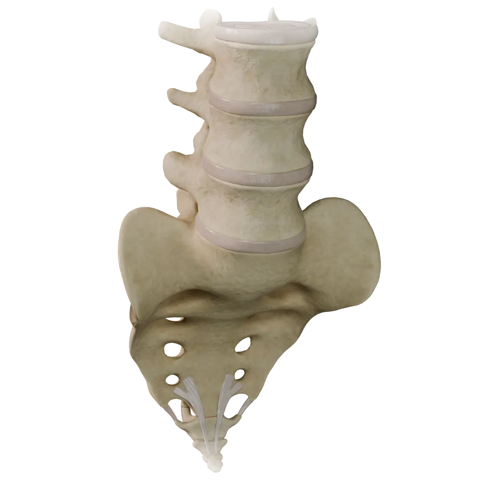
Cartilaginous joints (synchondroses) of the vertebral column
0/20

Fibrous joints of the vertebral column
0/20

Joints of thoracolumbar vertebral column
0/20
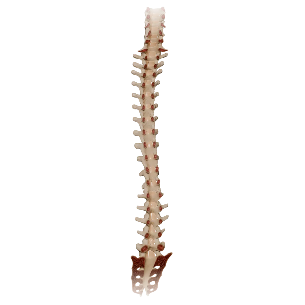
Ligamenta flava
0/20

Synovial joints of vertebral column
0/20
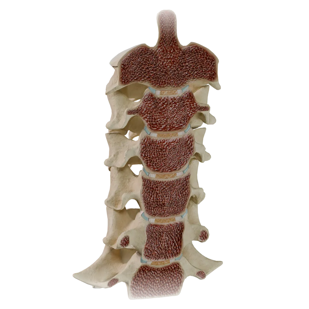
Uncovertebral joints
0/20
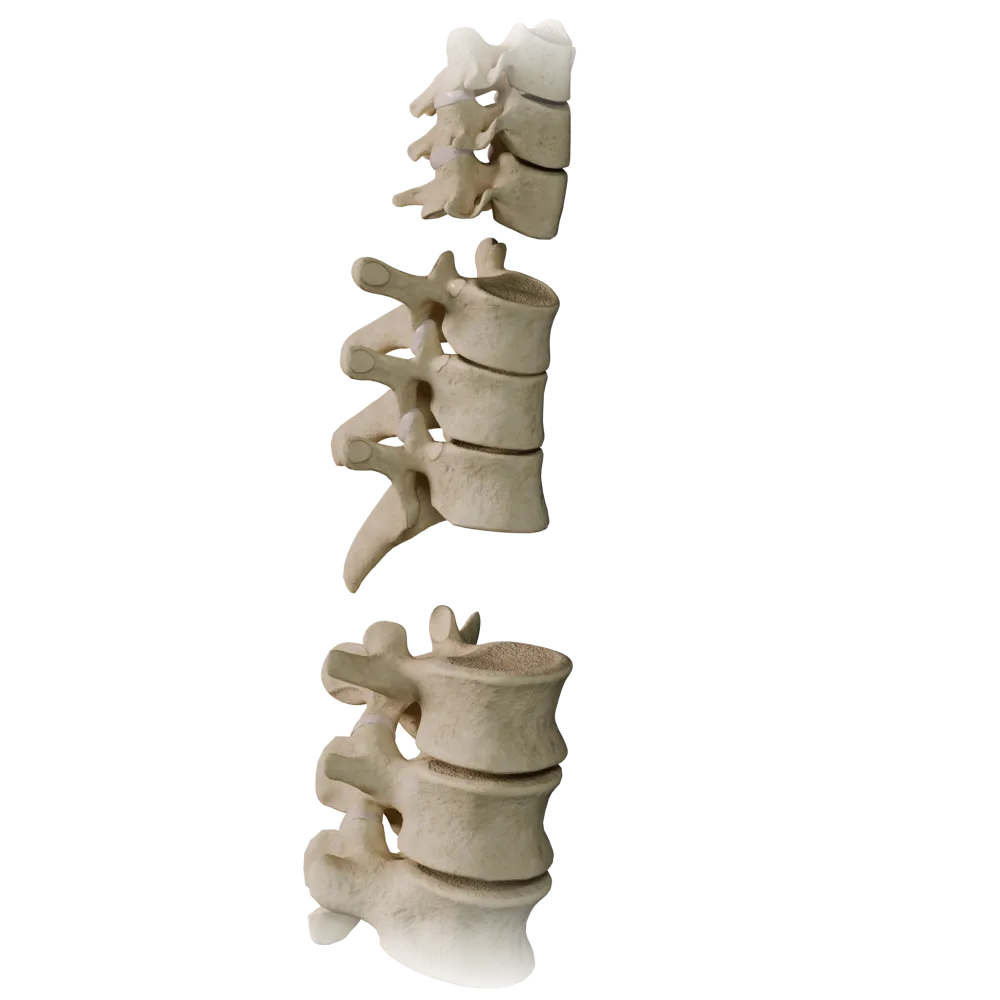
Zygapophyseal joints
0/20
Anatomy quiz on the atlantoaxial joints (median and lateral)
Anatomy quiz on the atlantoaxial joints: median and lateral—structure, ligaments, movements, blood supply, and innervation.
1/20
bold
text
1. What type of joint, based on the shape of its articular surfaces, is the median atlantoaxial joint (articulatio atlantoaxialis mediana)?
-
Ellipsoid (condylaris)
The median atlantoaxial joint is a trochoid (pivot) joint: the dens of the axis rotates within an osteoligamentous ring formed by the anterior arch of the atlas and the transverse ligament of the atlas.
-
Trochoid (trochoidea)
The median atlantoaxial joint is a trochoid (pivot) joint: the dens of the axis rotates within an osteoligamentous ring formed by the anterior arch of the atlas and the transverse ligament of the atlas.
-
Saddle (sellaris)
The median atlantoaxial joint is a trochoid (pivot) joint: the dens of the axis rotates within an osteoligamentous ring formed by the anterior arch of the atlas and the transverse ligament of the atlas.
-
Plane (plana)
The median atlantoaxial joint is a trochoid (pivot) joint: the dens of the axis rotates within an osteoligamentous ring formed by the anterior arch of the atlas and the transverse ligament of the atlas.
-
I find it difficult to answer
The median atlantoaxial joint is a trochoid (pivot) joint: the dens of the axis rotates within an osteoligamentous ring formed by the anterior arch of the atlas and the transverse ligament of the atlas.
2. The transverse ligament of the atlas (ligamentum transversum atlantis) attaches to:
-
The lateral masses of the atlas on both sides
The transverse ligament of the atlas extends between the medial surfaces of the lateral masses of the atlas, holding the dens of the axis against the anterior arch of the atlas and preventing its dorsal displacement.
-
The anterior arch of the atlas and the body of the axis
The transverse ligament of the atlas extends between the medial surfaces of the lateral masses of the atlas, holding the dens of the axis against the anterior arch of the atlas and preventing its dorsal displacement.
-
The occipital bone and the posterior arch of the atlas
The transverse ligament of the atlas extends between the medial surfaces of the lateral masses of the atlas, holding the dens of the axis against the anterior arch of the atlas and preventing its dorsal displacement.
-
The dens of the axis and the anterior arch of the atlas
The transverse ligament of the atlas extends between the medial surfaces of the lateral masses of the atlas, holding the dens of the axis against the anterior arch of the atlas and preventing its dorsal displacement.
-
I find it difficult to answer
The transverse ligament of the atlas extends between the medial surfaces of the lateral masses of the atlas, holding the dens of the axis against the anterior arch of the atlas and preventing its dorsal displacement.
3. What is the primary movement at the lateral atlantoaxial joints (articulationes atlantoaxiales laterales)?
-
Flexion and extension
The lateral atlantoaxial joints participate in head rotation together with the median joint. Their plane articular surfaces permit gliding during rotation of the atlas around the dens of the axis.
-
Lateral flexion
The lateral atlantoaxial joints participate in head rotation together with the median joint. Their plane articular surfaces permit gliding during rotation of the atlas around the dens of the axis.
-
Rotation around the vertical axis
The lateral atlantoaxial joints participate in head rotation together with the median joint. Their plane articular surfaces permit gliding during rotation of the atlas around the dens of the axis.
-
Gliding (translation)
The lateral atlantoaxial joints participate in head rotation together with the median joint. Their plane articular surfaces permit gliding during rotation of the atlas around the dens of the axis.
-
I find it difficult to answer
The lateral atlantoaxial joints participate in head rotation together with the median joint. Their plane articular surfaces permit gliding during rotation of the atlas around the dens of the axis.
4. What forms the anterior wall of the median atlantoaxial joint?
-
Anterior arch of the atlas (arcus anterior atlantis)
The anterior wall of the median atlantoaxial joint is formed anteriorly by the anterior arch of the atlas (facet for the dens) and posteriorly by the transverse ligament of the atlas.
-
Posterior arch of the atlas (arcus posterior atlantis)
The anterior wall of the median atlantoaxial joint is formed anteriorly by the anterior arch of the atlas (facet for the dens) and posteriorly by the transverse ligament of the atlas.
-
Body of the axis (corpus axis)
The anterior wall of the median atlantoaxial joint is formed anteriorly by the anterior arch of the atlas (facet for the dens) and posteriorly by the transverse ligament of the atlas.
-
Occipital bone (os occipitale)
The anterior wall of the median atlantoaxial joint is formed anteriorly by the anterior arch of the atlas (facet for the dens) and posteriorly by the transverse ligament of the atlas.
-
I find it difficult to answer
The anterior wall of the median atlantoaxial joint is formed anteriorly by the anterior arch of the atlas (facet for the dens) and posteriorly by the transverse ligament of the atlas.
5. The cruciform ligament of the atlas (ligamentum cruciforme atlantis) consists of the following components:
-
Transverse ligament of the atlas and longitudinal bands (fasciculi longitudinales)
The cruciform ligament of the atlas consists of the transverse ligament of the atlas and two longitudinal bands: the superior band (to the anterior semicircumference of the foramen magnum) and the inferior band (to the body of the axis).
-
Apical ligament of the dens and alar ligaments
The cruciform ligament of the atlas consists of the transverse ligament of the atlas and two longitudinal bands: the superior band (to the anterior semicircumference of the foramen magnum) and the inferior band (to the body of the axis).
-
Anterior atlantoaxial membrane and posterior atlantoaxial membrane
The cruciform ligament of the atlas consists of the transverse ligament of the atlas and two longitudinal bands: the superior band (to the anterior semicircumference of the foramen magnum) and the inferior band (to the body of the axis).
-
Nuchal ligament and anterior longitudinal ligament
The cruciform ligament of the atlas consists of the transverse ligament of the atlas and two longitudinal bands: the superior band (to the anterior semicircumference of the foramen magnum) and the inferior band (to the body of the axis).
-
I find it difficult to answer
The cruciform ligament of the atlas consists of the transverse ligament of the atlas and two longitudinal bands: the superior band (to the anterior semicircumference of the foramen magnum) and the inferior band (to the body of the axis).
6. The apical ligament of the dens (ligamentum apicis dentis) connects:
-
The lateral surfaces of the dens to the occipital condyles
Ligamentum apicis dentis is a weak rudimentary ligament extending from the apex of the dens (apex dentis) to the anterior margin of the foramen magnum (foramen magnum). It is considered a remnant of the notochord.
-
The apex of the dens of the axis to the anterior margin of the foramen magnum
Ligamentum apicis dentis is a weak rudimentary ligament extending from the apex of the dens (apex dentis) to the anterior margin of the foramen magnum (foramen magnum). It is considered a remnant of the notochord.
-
The dens of the axis to the transverse ligament of the atlas
Ligamentum apicis dentis is a weak rudimentary ligament extending from the apex of the dens (apex dentis) to the anterior margin of the foramen magnum (foramen magnum). It is considered a remnant of the notochord.
-
The lateral masses of the atlas to the occipital bone
Ligamentum apicis dentis is a weak rudimentary ligament extending from the apex of the dens (apex dentis) to the anterior margin of the foramen magnum (foramen magnum). It is considered a remnant of the notochord.
-
I find it difficult to answer
Ligamentum apicis dentis is a weak rudimentary ligament extending from the apex of the dens (apex dentis) to the anterior margin of the foramen magnum (foramen magnum). It is considered a remnant of the notochord.
7. The alar ligaments (ligamenta alaria) attach laterally to:
-
The transverse processes of the atlas
The alar ligaments run from the lateral surfaces of the apex of the dens of the axis obliquely superiorly and laterally to the medial surfaces of the occipital condyles, limiting excessive rotation and lateral flexion.
-
The medial surfaces of the occipital condyles
The alar ligaments run from the lateral surfaces of the apex of the dens of the axis obliquely superiorly and laterally to the medial surfaces of the occipital condyles, limiting excessive rotation and lateral flexion.
-
The posterior arch of the atlas
The alar ligaments run from the lateral surfaces of the apex of the dens of the axis obliquely superiorly and laterally to the medial surfaces of the occipital condyles, limiting excessive rotation and lateral flexion.
-
The body of the axis
The alar ligaments run from the lateral surfaces of the apex of the dens of the axis obliquely superiorly and laterally to the medial surfaces of the occipital condyles, limiting excessive rotation and lateral flexion.
-
I find it difficult to answer
The alar ligaments run from the lateral surfaces of the apex of the dens of the axis obliquely superiorly and laterally to the medial surfaces of the occipital condyles, limiting excessive rotation and lateral flexion.
8. The tectorial membrane (membrana tectoria) is a continuation of which structure?
-
Anterior longitudinal ligament (ligamentum longitudinale anterius)
The tectorial membrane (membrana tectoria) is a broad cranial expansion of the posterior longitudinal ligament. It covers the cruciform ligament of the atlas and the alar ligaments dorsally, attaching to the basilar part of the occipital bone.
-
Posterior longitudinal ligament (ligamentum longitudinale posterius)
The tectorial membrane (membrana tectoria) is a broad cranial expansion of the posterior longitudinal ligament. It covers the cruciform ligament of the atlas and the alar ligaments dorsally, attaching to the basilar part of the occipital bone.
-
Nuchal ligament (ligamentum nuchae)
The tectorial membrane (membrana tectoria) is a broad cranial expansion of the posterior longitudinal ligament. It covers the cruciform ligament of the atlas and the alar ligaments dorsally, attaching to the basilar part of the occipital bone.
-
Posterior atlantoaxial membrane (membrana atlantoaxialis posterior)
The tectorial membrane (membrana tectoria) is a broad cranial expansion of the posterior longitudinal ligament. It covers the cruciform ligament of the atlas and the alar ligaments dorsally, attaching to the basilar part of the occipital bone.
-
I find it difficult to answer
The tectorial membrane (membrana tectoria) is a broad cranial expansion of the posterior longitudinal ligament. It covers the cruciform ligament of the atlas and the alar ligaments dorsally, attaching to the basilar part of the occipital bone.
9. Classification of the lateral atlantoaxial joints by number of axes of rotation:
-
Triaxial
The lateral atlantoaxial joints are plane joints (articulationes planae) that are functionally uniaxial: they participate in rotation together with the median atlantoaxial joint, forming a single combined joint with it.
-
Biaxial
The lateral atlantoaxial joints are plane joints (articulationes planae) that are functionally uniaxial: they participate in rotation together with the median atlantoaxial joint, forming a single combined joint with it.
-
Uniaxial
The lateral atlantoaxial joints are plane joints (articulationes planae) that are functionally uniaxial: they participate in rotation together with the median atlantoaxial joint, forming a single combined joint with it.
-
Nonaxial (amphiarthrosis)
The lateral atlantoaxial joints are plane joints (articulationes planae) that are functionally uniaxial: they participate in rotation together with the median atlantoaxial joint, forming a single combined joint with it.
-
I find it difficult to answer
The lateral atlantoaxial joints are plane joints (articulationes planae) that are functionally uniaxial: they participate in rotation together with the median atlantoaxial joint, forming a single combined joint with it.
10. The articular surfaces of the lateral atlantoaxial joints are formed by:
-
The inferior articular surfaces of the atlas and the superior articular surfaces of the axis
The lateral atlantoaxial joints are formed between the inferior articular surfaces (foveae articulares inferiores) of the lateral masses of the atlas and the superior articular surfaces (facies articulares superiores) of the axis.
-
The superior articular surfaces of the atlas and the occipital condyles
The lateral atlantoaxial joints are formed between the inferior articular surfaces (foveae articulares inferiores) of the lateral masses of the atlas and the superior articular surfaces (facies articulares superiores) of the axis.
-
The transverse processes of the atlas and the axis
The lateral atlantoaxial joints are formed between the inferior articular surfaces (foveae articulares inferiores) of the lateral masses of the atlas and the superior articular surfaces (facies articulares superiores) of the axis.
-
The lateral masses of the atlas and the transverse processes of the axis
The lateral atlantoaxial joints are formed between the inferior articular surfaces (foveae articulares inferiores) of the lateral masses of the atlas and the superior articular surfaces (facies articulares superiores) of the axis.
-
I find it difficult to answer
The lateral atlantoaxial joints are formed between the inferior articular surfaces (foveae articulares inferiores) of the lateral masses of the atlas and the superior articular surfaces (facies articulares superiores) of the axis.
11. What is the cross-sectional shape of the dens of the axis (dens axis) that is important for its function in the median joint?
-
Triangular
The dens of the axis has an oval cross-section with flattening in the anteroposterior direction. The anterior articular surface (facies articularis anterior) contacts the facet for the dens of the atlas, while the posterior surface contacts the transverse ligament of the atlas.
-
Oval, flattened anteriorly and posteriorly
The dens of the axis has an oval cross-section with flattening in the anteroposterior direction. The anterior articular surface (facies articularis anterior) contacts the facet for the dens of the atlas, while the posterior surface contacts the transverse ligament of the atlas.
-
Square
The dens of the axis has an oval cross-section with flattening in the anteroposterior direction. The anterior articular surface (facies articularis anterior) contacts the facet for the dens of the atlas, while the posterior surface contacts the transverse ligament of the atlas.
-
Cylindrical with a uniformly rounded cross-section
The dens of the axis has an oval cross-section with flattening in the anteroposterior direction. The anterior articular surface (facies articularis anterior) contacts the facet for the dens of the atlas, while the posterior surface contacts the transverse ligament of the atlas.
-
I find it difficult to answer
The dens of the axis has an oval cross-section with flattening in the anteroposterior direction. The anterior articular surface (facies articularis anterior) contacts the facet for the dens of the atlas, while the posterior surface contacts the transverse ligament of the atlas.
12. The anterior atlantoaxial membrane (membrana atlantoaxialis anterior) extends between:
-
The posterior arch of the atlas and the arch of the axis
Membrana atlantoaxialis anterior is a dense fibrous sheet connecting the inferior margin of the anterior arch of the atlas to the anterior surface of the body of the axis. It is reinforced in the midline by the anterior longitudinal ligament.
-
The inferior margin of the anterior arch of the atlas and the anterior surface of the body of the axis
Membrana atlantoaxialis anterior is a dense fibrous sheet connecting the inferior margin of the anterior arch of the atlas to the anterior surface of the body of the axis. It is reinforced in the midline by the anterior longitudinal ligament.
-
The anterior arch of the atlas and the occipital bone
Membrana atlantoaxialis anterior is a dense fibrous sheet connecting the inferior margin of the anterior arch of the atlas to the anterior surface of the body of the axis. It is reinforced in the midline by the anterior longitudinal ligament.
-
The lateral masses of the atlas and the body of the axis
Membrana atlantoaxialis anterior is a dense fibrous sheet connecting the inferior margin of the anterior arch of the atlas to the anterior surface of the body of the axis. It is reinforced in the midline by the anterior longitudinal ligament.
-
I find it difficult to answer
Membrana atlantoaxialis anterior is a dense fibrous sheet connecting the inferior margin of the anterior arch of the atlas to the anterior surface of the body of the axis. It is reinforced in the midline by the anterior longitudinal ligament.
13. The primary function of the transverse ligament of the atlas in the median atlantoaxial joint is:
-
Limiting rotation of the atlas
The transverse ligament of the atlas is the principal stabilizer preventing posterior displacement of the dens of the axis into the vertebral canal and protecting the spinal cord.
-
Preventing posterior displacement of the dens of the axis into the vertebral canal
The transverse ligament of the atlas is the principal stabilizer preventing posterior displacement of the dens of the axis into the vertebral canal and protecting the spinal cord.
-
Connecting the atlas to the occipital bone
The transverse ligament of the atlas is the principal stabilizer preventing posterior displacement of the dens of the axis into the vertebral canal and protecting the spinal cord.
-
Facilitating flexion at the atlanto-occipital joint
The transverse ligament of the atlas is the principal stabilizer preventing posterior displacement of the dens of the axis into the vertebral canal and protecting the spinal cord.
-
I find it difficult to answer
The transverse ligament of the atlas is the principal stabilizer preventing posterior displacement of the dens of the axis into the vertebral canal and protecting the spinal cord.
14. Which nerve provides innervation to the atlantoaxial joints?
-
Fourth cervical spinal nerve (C4)
The atlantoaxial joints are innervated predominantly by branches of the second cervical spinal nerve (C2). Articular branches from C2 reach both the median and lateral atlantoaxial joints.
-
Second cervical spinal nerve (C2)
The atlantoaxial joints are innervated predominantly by branches of the second cervical spinal nerve (C2). Articular branches from C2 reach both the median and lateral atlantoaxial joints.
-
Accessory nerve (n. accessorius, XI)
The atlantoaxial joints are innervated predominantly by branches of the second cervical spinal nerve (C2). Articular branches from C2 reach both the median and lateral atlantoaxial joints.
-
Greater occipital nerve (n. occipitalis major)
The atlantoaxial joints are innervated predominantly by branches of the second cervical spinal nerve (C2). Articular branches from C2 reach both the median and lateral atlantoaxial joints.
-
I find it difficult to answer
The atlantoaxial joints are innervated predominantly by branches of the second cervical spinal nerve (C2). Articular branches from C2 reach both the median and lateral atlantoaxial joints.
15. The blood supply to the atlantoaxial joints is provided predominantly by branches of the:
-
External carotid artery (a. carotis externa)
The blood supply to the atlantoaxial complex is provided by muscular and spinal branches of the vertebral artery (a. vertebralis), which passes through the transverse foramina of the cervical vertebrae and curves around the lateral masses of the atlas.
-
Vertebral artery (a. vertebralis)
The blood supply to the atlantoaxial complex is provided by muscular and spinal branches of the vertebral artery (a. vertebralis), which passes through the transverse foramina of the cervical vertebrae and curves around the lateral masses of the atlas.
-
Posterior inferior cerebellar artery (a. cerebelli inferior posterior)
The blood supply to the atlantoaxial complex is provided by muscular and spinal branches of the vertebral artery (a. vertebralis), which passes through the transverse foramina of the cervical vertebrae and curves around the lateral masses of the atlas.
-
Ascending cervical artery (a. cervicalis ascendens)
The blood supply to the atlantoaxial complex is provided by muscular and spinal branches of the vertebral artery (a. vertebralis), which passes through the transverse foramina of the cervical vertebrae and curves around the lateral masses of the atlas.
-
I find it difficult to answer
The blood supply to the atlantoaxial complex is provided by muscular and spinal branches of the vertebral artery (a. vertebralis), which passes through the transverse foramina of the cervical vertebrae and curves around the lateral masses of the atlas.
16. The range of rotation (rotational movement) in the atlanto-axial complex to each side is approximately:
-
15–20°
The atlantoaxial complex provides approximately 45–50° of rotation to each side, accounting for approximately 50% of the total rotational range of the cervical spine. The movement is limited by the alar ligaments.
-
30–35°
The atlantoaxial complex provides approximately 45–50° of rotation to each side, accounting for approximately 50% of the total rotational range of the cervical spine. The movement is limited by the alar ligaments.
-
45–50°
The atlantoaxial complex provides approximately 45–50° of rotation to each side, accounting for approximately 50% of the total rotational range of the cervical spine. The movement is limited by the alar ligaments.
-
60–70°
The atlantoaxial complex provides approximately 45–50° of rotation to each side, accounting for approximately 50% of the total rotational range of the cervical spine. The movement is limited by the alar ligaments.
-
I find it difficult to answer
The atlantoaxial complex provides approximately 45–50° of rotation to each side, accounting for approximately 50% of the total rotational range of the cervical spine. The movement is limited by the alar ligaments.
17. The posterior atlantoaxial membrane (membrana atlantoaxialis posterior) corresponds to which structure in the underlying vertebral segments?
-
Anterior longitudinal ligament
The posterior atlanto-axial membrane (membrana atlantoaxialis posterior) is homologous to the Ligamenta flava (ligamenta flava) of the lower intervertebral spaces. It connects the inferior margin of the posterior arch of the atlas to the superior margin of the arch of the axis.
-
Ligamentum flavum (ligamentum flavum)
The posterior atlanto-axial membrane (membrana atlantoaxialis posterior) is homologous to the Ligamenta flava (ligamenta flava) of the lower intervertebral spaces. It connects the inferior margin of the posterior arch of the atlas to the superior margin of the arch of the axis.
-
Interspinous ligament (ligamentum interspinale)
The posterior atlanto-axial membrane (membrana atlantoaxialis posterior) is homologous to the Ligamenta flava (ligamenta flava) of the lower intervertebral spaces. It connects the inferior margin of the posterior arch of the atlas to the superior margin of the arch of the axis.
-
Supraspinous ligament (ligamentum supraspinale)
The posterior atlanto-axial membrane (membrana atlantoaxialis posterior) is homologous to the Ligamenta flava (ligamenta flava) of the lower intervertebral spaces. It connects the inferior margin of the posterior arch of the atlas to the superior margin of the arch of the axis.
-
I find it difficult to answer
The posterior atlanto-axial membrane (membrana atlantoaxialis posterior) is homologous to the Ligamenta flava (ligamenta flava) of the lower intervertebral spaces. It connects the inferior margin of the posterior arch of the atlas to the superior margin of the arch of the axis.
18. In terms of articular capsule structure, the median atlantoaxial joint is correctly characterized as:
-
A two-chambered joint with an intra-articular disc
The median atlantoaxial joint actually contains two synovial cavities: one between the dens and the anterior arch of the atlas (anterior joint of the dens) and one between the dens and the transverse ligament of the atlas (posterior joint of the dens), each with its own capsule.
-
A joint with two separate synovial cavities—anterior and posterior
The median atlantoaxial joint actually contains two synovial cavities: one between the dens and the anterior arch of the atlas (anterior joint of the dens) and one between the dens and the transverse ligament of the atlas (posterior joint of the dens), each with its own capsule.
-
A single-chambered joint with a single articular cavity
The median atlantoaxial joint actually contains two synovial cavities: one between the dens and the anterior arch of the atlas (anterior joint of the dens) and one between the dens and the transverse ligament of the atlas (posterior joint of the dens), each with its own capsule.
-
A synchondrosis without an articular capsule
The median atlantoaxial joint actually contains two synovial cavities: one between the dens and the anterior arch of the atlas (anterior joint of the dens) and one between the dens and the transverse ligament of the atlas (posterior joint of the dens), each with its own capsule.
-
I find it difficult to answer
The median atlantoaxial joint actually contains two synovial cavities: one between the dens and the anterior arch of the atlas (anterior joint of the dens) and one between the dens and the transverse ligament of the atlas (posterior joint of the dens), each with its own capsule.
19. The alar ligaments (ligamenta alaria) perform the following primary function:
-
Guide flexion at the atlanto-occipital joint
The alar ligaments are strong paired structures located on either side of the dens of the axis. They serve as the principal restraints against excessive rotation (the contralateral alar ligament becomes taut during rotation) and lateral flexion of the head.
-
Limit excessive rotation and lateral flexion of the head
The alar ligaments are strong paired structures located on either side of the dens of the axis. They serve as the principal restraints against excessive rotation (the contralateral alar ligament becomes taut during rotation) and lateral flexion of the head.
-
Prevent anterior displacement of the dens
The alar ligaments are strong paired structures located on either side of the dens of the axis. They serve as the principal restraints against excessive rotation (the contralateral alar ligament becomes taut during rotation) and lateral flexion of the head.
-
Connect the transverse processes of the atlas to the occipital bone
The alar ligaments are strong paired structures located on either side of the dens of the axis. They serve as the principal restraints against excessive rotation (the contralateral alar ligament becomes taut during rotation) and lateral flexion of the head.
-
I find it difficult to answer
The alar ligaments are strong paired structures located on either side of the dens of the axis. They serve as the principal restraints against excessive rotation (the contralateral alar ligament becomes taut during rotation) and lateral flexion of the head.
20. All three atlantoaxial joints (the median and two lateral joints) are functionally integrated into:
-
A simple joint with a single articular cavity
The median and two lateral atlantoaxial joints are anatomically separate but functionally unified as a combined joint (articulatio combinata). Movement (rotation) is possible only with the simultaneous participation of all three joints.
-
A combined joint functioning simultaneously
The median and two lateral atlantoaxial joints are anatomically separate but functionally unified as a combined joint (articulatio combinata). Movement (rotation) is possible only with the simultaneous participation of all three joints.
-
A syndesmosis with limited mobility
The median and two lateral atlantoaxial joints are anatomically separate but functionally unified as a combined joint (articulatio combinata). Movement (rotation) is possible only with the simultaneous participation of all three joints.
-
An amphiarthrosis with minimal movement
The median and two lateral atlantoaxial joints are anatomically separate but functionally unified as a combined joint (articulatio combinata). Movement (rotation) is possible only with the simultaneous participation of all three joints.
-
I find it difficult to answer
The median and two lateral atlantoaxial joints are anatomically separate but functionally unified as a combined joint (articulatio combinata). Movement (rotation) is possible only with the simultaneous participation of all three joints.
Retake this quiz?
Your current progress will be reset.
