
Joints of vertebral column
0/20
By topic

Atlantoaxial joints (median and lateral)
0/20
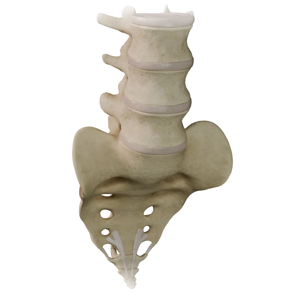
Cartilaginous joints (synchondroses) of the vertebral column
0/20

Fibrous joints of the vertebral column
0/20

Joints of thoracolumbar vertebral column
0/20
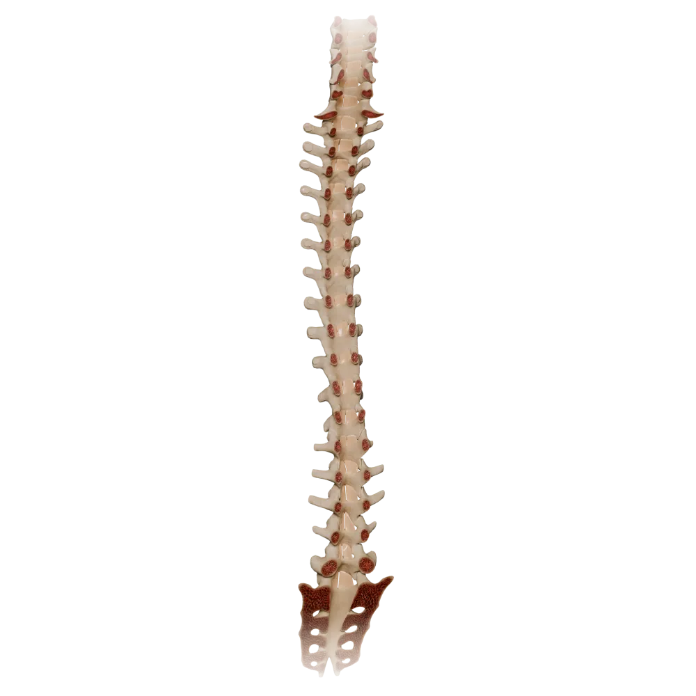
Ligamenta flava
0/20

Synovial joints of vertebral column
0/20
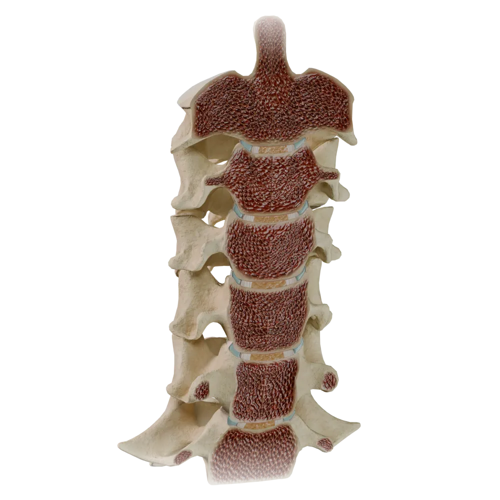
Uncovertebral joints
0/20
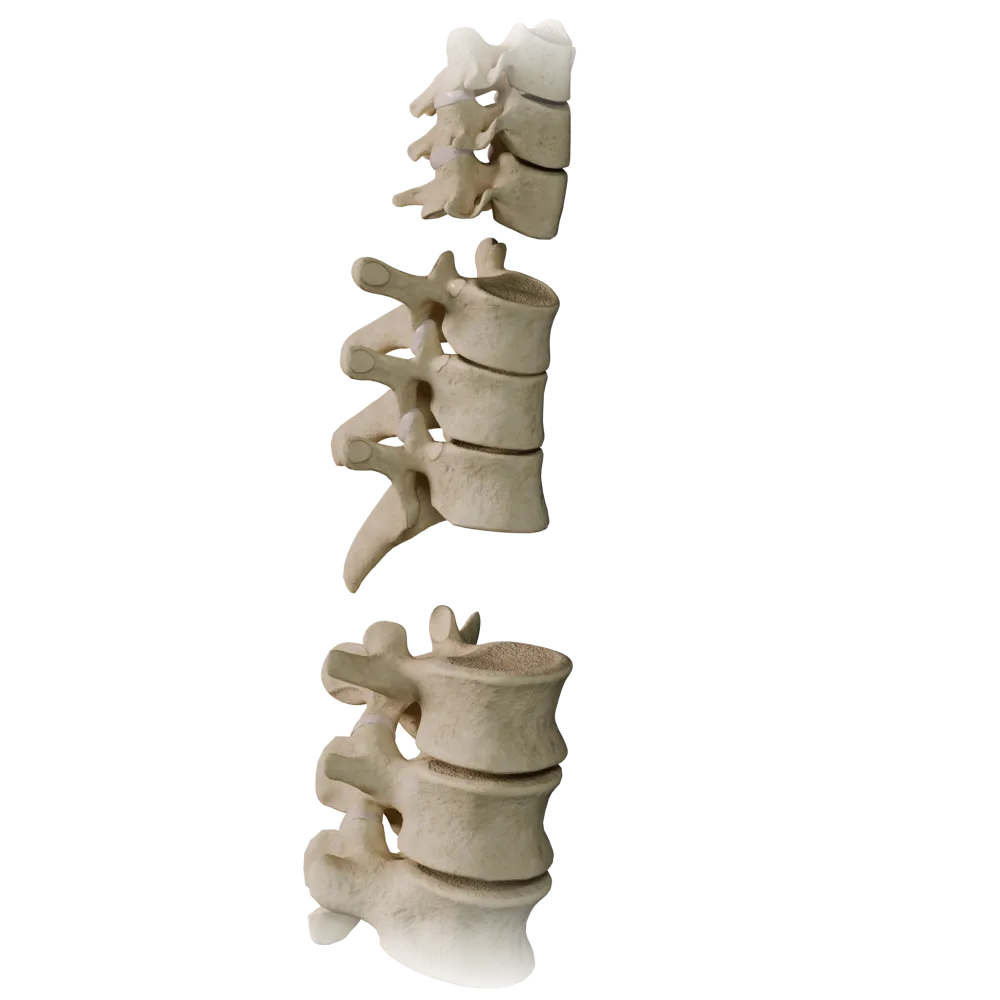
Zygapophyseal joints
0/20
Anatomy quiz on the joints of the thoracolumbar vertebral column
Normal anatomy test on the joints of the thoracolumbar vertebral column: joints, ligaments, cartilage, functional axes.
1/20
bold
text
1. What type of joint is formed by the bodies of adjacent thoracic vertebrae?
-
Syndesmosis
The bodies of adjacent vertebrae are connected by intervertebral discs—fibrocartilaginous symphyses (symphysis intervertebralis). The disc consists of the nucleus pulposus and annulus fibrosus, which classifies the joint as a type of synchondrosis with fibrocartilage.
-
Synchondrosis(intervertebral disc)
The bodies of adjacent vertebrae are connected by intervertebral discs—fibrocartilaginous symphyses (symphysis intervertebralis). The disc consists of the nucleus pulposus and annulus fibrosus, which classifies the joint as a type of synchondrosis with fibrocartilage.
-
Synostosis
The bodies of adjacent vertebrae are connected by intervertebral discs—fibrocartilaginous symphyses (symphysis intervertebralis). The disc consists of the nucleus pulposus and annulus fibrosus, which classifies the joint as a type of synchondrosis with fibrocartilage.
-
Synchondrosis with hyaline cartilage
The bodies of adjacent vertebrae are connected by intervertebral discs—fibrocartilaginous symphyses (symphysis intervertebralis). The disc consists of the nucleus pulposus and annulus fibrosus, which classifies the joint as a type of synchondrosis with fibrocartilage.
-
I find it difficult to answer
The bodies of adjacent vertebrae are connected by intervertebral discs—fibrocartilaginous symphyses (symphysis intervertebralis). The disc consists of the nucleus pulposus and annulus fibrosus, which classifies the joint as a type of synchondrosis with fibrocartilage.
2. Which ligament attaches to the tips of the spinous processes of the thoracic and lumbar vertebrae, forming a median structure?
-
Lig. supraspinale
The supraspinous ligament (lig. supraspinale) extends as a continuous band along the tips of the spinous processes from the seventh cervical vertebra to the sacrum. In the cervical region, it continues as the ligamentum nuchae. The interspinous ligaments (lig. interspinale) connect the lateral surfaces of the spinous processes, but not their tips.
-
Lig. interspinale
The supraspinous ligament (lig. supraspinale) extends as a continuous band along the tips of the spinous processes from the seventh cervical vertebra to the sacrum. In the cervical region, it continues as the ligamentum nuchae. The interspinous ligaments (lig. interspinale) connect the lateral surfaces of the spinous processes, but not their tips.
-
Lig. flavum
The supraspinous ligament (lig. supraspinale) extends as a continuous band along the tips of the spinous processes from the seventh cervical vertebra to the sacrum. In the cervical region, it continues as the ligamentum nuchae. The interspinous ligaments (lig. interspinale) connect the lateral surfaces of the spinous processes, but not their tips.
-
Lig. longitudinale posterius
The supraspinous ligament (lig. supraspinale) extends as a continuous band along the tips of the spinous processes from the seventh cervical vertebra to the sacrum. In the cervical region, it continues as the ligamentum nuchae. The interspinous ligaments (lig. interspinale) connect the lateral surfaces of the spinous processes, but not their tips.
-
I find it difficult to answer
The supraspinous ligament (lig. supraspinale) extends as a continuous band along the tips of the spinous processes from the seventh cervical vertebra to the sacrum. In the cervical region, it continues as the ligamentum nuchae. The interspinous ligaments (lig. interspinale) connect the lateral surfaces of the spinous processes, but not their tips.
3. To which type do the intervertebral (zygapophyseal) joints of the thoracic and lumbar regions belong according to the shape of their articular surfaces?
-
Ball-and-socket joints
Articulationes zygapophysiales (zygapophyseal joints) are formed by the flat articular surfaces of the superior and inferior articular processes. These are planar joints (articulatio plana) permitting small-amplitude gliding movements. The shape of the articular surfaces determines the plane and range of motion in each region.
-
Planar joints
Articulationes zygapophysiales (zygapophyseal joints) are formed by the flat articular surfaces of the superior and inferior articular processes. These are planar joints (articulatio plana) permitting small-amplitude gliding movements. The shape of the articular surfaces determines the plane and range of motion in each region.
-
Ellipsoid joints
Articulationes zygapophysiales (zygapophyseal joints) are formed by the flat articular surfaces of the superior and inferior articular processes. These are planar joints (articulatio plana) permitting small-amplitude gliding movements. The shape of the articular surfaces determines the plane and range of motion in each region.
-
Hinge joints
Articulationes zygapophysiales (zygapophyseal joints) are formed by the flat articular surfaces of the superior and inferior articular processes. These are planar joints (articulatio plana) permitting small-amplitude gliding movements. The shape of the articular surfaces determines the plane and range of motion in each region.
-
I find it difficult to answer
Articulationes zygapophysiales (zygapophyseal joints) are formed by the flat articular surfaces of the superior and inferior articular processes. These are planar joints (articulatio plana) permitting small-amplitude gliding movements. The shape of the articular surfaces determines the plane and range of motion in each region.
4. In which direction are the articular surfaces of the zygapophyseal joints oriented in the lumbar spine?
-
In the frontal plane
In the lumbar region, the articular surfaces of the zygapophyseal joints are oriented predominantly in the sagittal plane, which limits rotation and results in the predominance of flexion-extension. In the thoracic region, they are oriented closer to the frontal plane, permitting rotation.
-
In the horizontal plane
In the lumbar region, the articular surfaces of the zygapophyseal joints are oriented predominantly in the sagittal plane, which limits rotation and results in the predominance of flexion-extension. In the thoracic region, they are oriented closer to the frontal plane, permitting rotation.
-
In the sagittal (parasagittal) plane
In the lumbar region, the articular surfaces of the zygapophyseal joints are oriented predominantly in the sagittal plane, which limits rotation and results in the predominance of flexion-extension. In the thoracic region, they are oriented closer to the frontal plane, permitting rotation.
-
At an angle of 45° to the horizontal
In the lumbar region, the articular surfaces of the zygapophyseal joints are oriented predominantly in the sagittal plane, which limits rotation and results in the predominance of flexion-extension. In the thoracic region, they are oriented closer to the frontal plane, permitting rotation.
-
I find it difficult to answer
In the lumbar region, the articular surfaces of the zygapophyseal joints are oriented predominantly in the sagittal plane, which limits rotation and results in the predominance of flexion-extension. In the thoracic region, they are oriented closer to the frontal plane, permitting rotation.
5. Which structure of the intervertebral disc is located at its center and functions as a hydrostatic shock absorber?
-
Annulus fibrosus
Nucleus pulposus — the gelatinous nucleus in the center of the disc, a remnant of the notochord (notochord). It contains a large amount of water and proteoglycans, providing hydrostatic absorption of axial loads. The annulus fibrosus is the outer fibrous ring that contains the nucleus.
-
Nucleus pulposus
Nucleus pulposus — the gelatinous nucleus in the center of the disc, a remnant of the notochord (notochord). It contains a large amount of water and proteoglycans, providing hydrostatic absorption of axial loads. The annulus fibrosus is the outer fibrous ring that contains the nucleus.
-
Ligamentum flavum
Nucleus pulposus — the gelatinous nucleus in the center of the disc, a remnant of the notochord (notochord). It contains a large amount of water and proteoglycans, providing hydrostatic absorption of axial loads. The annulus fibrosus is the outer fibrous ring that contains the nucleus.
-
Cartilago hyalina
Nucleus pulposus — the gelatinous nucleus in the center of the disc, a remnant of the notochord (notochord). It contains a large amount of water and proteoglycans, providing hydrostatic absorption of axial loads. The annulus fibrosus is the outer fibrous ring that contains the nucleus.
-
I find it difficult to answer
Nucleus pulposus — the gelatinous nucleus in the center of the disc, a remnant of the notochord (notochord). It contains a large amount of water and proteoglycans, providing hydrostatic absorption of axial loads. The annulus fibrosus is the outer fibrous ring that contains the nucleus.
6. Ligamentum longitudinale anterius attaches to the vertebral bodies and intervertebral discs. What are its superior and inferior boundaries?
-
From the occipital bone to the coccyx
The anterior longitudinal ligament (lig. longitudinale anterius) extends from the basilar part of the occipital bone (os occipitale) to the anterior surface of the sacrum. It is the longest ligament in the body and prevents excessive extension of the spine.
-
From the atlas to the sacrum
The anterior longitudinal ligament (lig. longitudinale anterius) extends from the basilar part of the occipital bone (os occipitale) to the anterior surface of the sacrum. It is the longest ligament in the body and prevents excessive extension of the spine.
-
From the occipital bone to the sacrum
The anterior longitudinal ligament (lig. longitudinale anterius) extends from the basilar part of the occipital bone (os occipitale) to the anterior surface of the sacrum. It is the longest ligament in the body and prevents excessive extension of the spine.
-
From the epistropheus to the coccyx
The anterior longitudinal ligament (lig. longitudinale anterius) extends from the basilar part of the occipital bone (os occipitale) to the anterior surface of the sacrum. It is the longest ligament in the body and prevents excessive extension of the spine.
-
I find it difficult to answer
The anterior longitudinal ligament (lig. longitudinale anterius) extends from the basilar part of the occipital bone (os occipitale) to the anterior surface of the sacrum. It is the longest ligament in the body and prevents excessive extension of the spine.
7. Ligamentum longitudinale posterius is located on the posterior surface of the vertebral bodies. What configuration does it have in the thoracic and lumbar regions?
-
Uniformly wide throughout its entire length
The posterior longitudinal ligament in the thoracic and lumbar regions has a characteristic serrated appearance: it widens at the level of the intervertebral discs and narrows at the level of the vertebral bodies. This is anatomically significant because the lateral portions of the discs are relatively less protected by the ligament.
-
Widens at the discs and narrows at the vertebral bodies
The posterior longitudinal ligament in the thoracic and lumbar regions has a characteristic serrated appearance: it widens at the level of the intervertebral discs and narrows at the level of the vertebral bodies. This is anatomically significant because the lateral portions of the discs are relatively less protected by the ligament.
-
Narrows at the discs and widens at the vertebral bodies
The posterior longitudinal ligament in the thoracic and lumbar regions has a characteristic serrated appearance: it widens at the level of the intervertebral discs and narrows at the level of the vertebral bodies. This is anatomically significant because the lateral portions of the discs are relatively less protected by the ligament.
-
Is interrupted in the lumbar region
The posterior longitudinal ligament in the thoracic and lumbar regions has a characteristic serrated appearance: it widens at the level of the intervertebral discs and narrows at the level of the vertebral bodies. This is anatomically significant because the lateral portions of the discs are relatively less protected by the ligament.
-
I find it difficult to answer
The posterior longitudinal ligament in the thoracic and lumbar regions has a characteristic serrated appearance: it widens at the level of the intervertebral discs and narrows at the level of the vertebral bodies. This is anatomically significant because the lateral portions of the discs are relatively less protected by the ligament.
8. Ligamenta flava (ligg. flava) connect the arches of adjacent vertebrae. Of which tissue are they predominantly composed?
-
Dense irregular connective tissue
Ligg. flava have a high content of elastic fibers (up to 80%), which gives them their yellow color and the ability to return to their original position after flexion. This elasticity maintains the vertical position of the spine and contributes to trunk extension.
-
Hyaline cartilage
Ligg. flava have a high content of elastic fibers (up to 80%), which gives them their yellow color and the ability to return to their original position after flexion. This elasticity maintains the vertical position of the spine and contributes to trunk extension.
-
Elastic connective tissue
Ligg. flava have a high content of elastic fibers (up to 80%), which gives them their yellow color and the ability to return to their original position after flexion. This elasticity maintains the vertical position of the spine and contributes to trunk extension.
-
Fibrocartilage
Ligg. flava have a high content of elastic fibers (up to 80%), which gives them their yellow color and the ability to return to their original position after flexion. This elasticity maintains the vertical position of the spine and contributes to trunk extension.
-
I find it difficult to answer
Ligg. flava have a high content of elastic fibers (up to 80%), which gives them their yellow color and the ability to return to their original position after flexion. This elasticity maintains the vertical position of the spine and contributes to trunk extension.
9. Which ligaments connect the transverse processes in the thoracic and lumbar regions of the spine?
-
Ligg. interspinalia
The intertransverse ligaments (ligg. intertransversaria) connect the apices of the transverse processes of adjacent vertebrae. They are well developed in the lumbar region. Their function is to limit lateral flexion. Ligg. costotransversaria are separate ligaments of the costotransverse joints.
-
Ligg. intertransversaria
The intertransverse ligaments (ligg. intertransversaria) connect the apices of the transverse processes of adjacent vertebrae. They are well developed in the lumbar region. Their function is to limit lateral flexion. Ligg. costotransversaria are separate ligaments of the costotransverse joints.
-
Ligg. flava
The intertransverse ligaments (ligg. intertransversaria) connect the apices of the transverse processes of adjacent vertebrae. They are well developed in the lumbar region. Their function is to limit lateral flexion. Ligg. costotransversaria are separate ligaments of the costotransverse joints.
-
Ligg. costotransversaria
The intertransverse ligaments (ligg. intertransversaria) connect the apices of the transverse processes of adjacent vertebrae. They are well developed in the lumbar region. Their function is to limit lateral flexion. Ligg. costotransversaria are separate ligaments of the costotransverse joints.
-
I find it difficult to answer
The intertransverse ligaments (ligg. intertransversaria) connect the apices of the transverse processes of adjacent vertebrae. They are well developed in the lumbar region. Their function is to limit lateral flexion. Ligg. costotransversaria are separate ligaments of the costotransverse joints.
10. How many costovertebral joints does a typical rib (III–IX) form?
-
One—only art. capitis costae
A typical rib forms two synovial joints with a vertebra: articulatio capitis costae (the head of the rib with the bodies of two adjacent vertebrae) and articulatio costotransversaria (the tubercle of the rib with the transverse process of the corresponding vertebra). Together, they function as a single “compound” joint during respiratory movements.
-
Two—art. capitis costae and art. costotransversaria
A typical rib forms two synovial joints with a vertebra: articulatio capitis costae (the head of the rib with the bodies of two adjacent vertebrae) and articulatio costotransversaria (the tubercle of the rib with the transverse process of the corresponding vertebra). Together, they function as a single “compound” joint during respiratory movements.
-
Three—art. capitis costae, art. costotransversaria and art. sternocostalis
A typical rib forms two synovial joints with a vertebra: articulatio capitis costae (the head of the rib with the bodies of two adjacent vertebrae) and articulatio costotransversaria (the tubercle of the rib with the transverse process of the corresponding vertebra). Together, they function as a single “compound” joint during respiratory movements.
-
One—only art. costotransversaria
A typical rib forms two synovial joints with a vertebra: articulatio capitis costae (the head of the rib with the bodies of two adjacent vertebrae) and articulatio costotransversaria (the tubercle of the rib with the transverse process of the corresponding vertebra). Together, they function as a single “compound” joint during respiratory movements.
-
I find it difficult to answer
A typical rib forms two synovial joints with a vertebra: articulatio capitis costae (the head of the rib with the bodies of two adjacent vertebrae) and articulatio costotransversaria (the tubercle of the rib with the transverse process of the corresponding vertebra). Together, they function as a single “compound” joint during respiratory movements.
11. Articulatio capitis costae of typical ribs (II–X) is a compound joint. What divides its cavity?
-
Articular labrum (labrum)
In the articulatio capitis costae of typical ribs, the cavity is divided by the intra-articular ligament of the head of the rib (lig. capitis costae intraarticulare), which extends from the crest of the head of the rib to the intervertebral disc. This makes it a compound joint (articulatio composita). The intra-articular ligament is absent in ribs I, XI, and XII.
-
Intra-articular ligament (lig. capitis costae intraarticulare)
In the articulatio capitis costae of typical ribs, the cavity is divided by the intra-articular ligament of the head of the rib (lig. capitis costae intraarticulare), which extends from the crest of the head of the rib to the intervertebral disc. This makes it a compound joint (articulatio composita). The intra-articular ligament is absent in ribs I, XI, and XII.
-
Articular meniscus
In the articulatio capitis costae of typical ribs, the cavity is divided by the intra-articular ligament of the head of the rib (lig. capitis costae intraarticulare), which extends from the crest of the head of the rib to the intervertebral disc. This makes it a compound joint (articulatio composita). The intra-articular ligament is absent in ribs I, XI, and XII.
-
Ligamenta flava
In the articulatio capitis costae of typical ribs, the cavity is divided by the intra-articular ligament of the head of the rib (lig. capitis costae intraarticulare), which extends from the crest of the head of the rib to the intervertebral disc. This makes it a compound joint (articulatio composita). The intra-articular ligament is absent in ribs I, XI, and XII.
-
I find it difficult to answer
In the articulatio capitis costae of typical ribs, the cavity is divided by the intra-articular ligament of the head of the rib (lig. capitis costae intraarticulare), which extends from the crest of the head of the rib to the intervertebral disc. This makes it a compound joint (articulatio composita). The intra-articular ligament is absent in ribs I, XI, and XII.
12. Which type of movement predominates in the thoracic spine because of the frontal orientation of the articular surfaces of the zygapophyseal joints?
-
Flexion and extension
The articular surfaces of the thoracic zygapophyseal joints, oriented in the frontal plane, permit rotation as the primary movement. Flexion-extension is also limited by the presence of the ribs and the overlapping arrangement of the spinous processes.
-
Rotation (turning)
The articular surfaces of the thoracic zygapophyseal joints, oriented in the frontal plane, permit rotation as the primary movement. Flexion-extension is also limited by the presence of the ribs and the overlapping arrangement of the spinous processes.
-
Lateral bending
The articular surfaces of the thoracic zygapophyseal joints, oriented in the frontal plane, permit rotation as the primary movement. Flexion-extension is also limited by the presence of the ribs and the overlapping arrangement of the spinous processes.
-
All types of movement equally
The articular surfaces of the thoracic zygapophyseal joints, oriented in the frontal plane, permit rotation as the primary movement. Flexion-extension is also limited by the presence of the ribs and the overlapping arrangement of the spinous processes.
-
I find it difficult to answer
The articular surfaces of the thoracic zygapophyseal joints, oriented in the frontal plane, permit rotation as the primary movement. Flexion-extension is also limited by the presence of the ribs and the overlapping arrangement of the spinous processes.
13. Lig. costotransversarium laterale is stretched between the tip of the transverse process and the tubercle of the rib. To which joint does it belong?
-
Art. capitis costae
Lig. costotransversarium laterale is a reinforcing ligament of the articulatio costotransversaria, connecting the transverse process of the vertebra to the tubercle of the corresponding rib. Alongside it, there are lig. costotransversarium (between the neck of the rib and the transverse process) and lig. costotransversarium superius.
-
Art. costotransversaria
Lig. costotransversarium laterale is a reinforcing ligament of the articulatio costotransversaria, connecting the transverse process of the vertebra to the tubercle of the corresponding rib. Alongside it, there are lig. costotransversarium (between the neck of the rib and the transverse process) and lig. costotransversarium superius.
-
Art. sternocostalis
Lig. costotransversarium laterale is a reinforcing ligament of the articulatio costotransversaria, connecting the transverse process of the vertebra to the tubercle of the corresponding rib. Alongside it, there are lig. costotransversarium (between the neck of the rib and the transverse process) and lig. costotransversarium superius.
-
Art. zygapophysialis
Lig. costotransversarium laterale is a reinforcing ligament of the articulatio costotransversaria, connecting the transverse process of the vertebra to the tubercle of the corresponding rib. Alongside it, there are lig. costotransversarium (between the neck of the rib and the transverse process) and lig. costotransversarium superius.
-
I find it difficult to answer
Lig. costotransversarium laterale is a reinforcing ligament of the articulatio costotransversaria, connecting the transverse process of the vertebra to the tubercle of the corresponding rib. Alongside it, there are lig. costotransversarium (between the neck of the rib and the transverse process) and lig. costotransversarium superius.
14. How many intervertebral discs are there in the thoracic region of the vertebral column?
-
11
The thoracic region (Th1–Th12) comprises 12 thoracic vertebrae. Intervertebral discs are located between adjacent vertebrae, beginning at the Th1–Th2 interspace. Thus, 11 intervertebral discs are formed between the 12 thoracic vertebrae (Th1–Th2 … Th11–Th12 interspaces).
-
12
The thoracic region (Th1–Th12) comprises 12 thoracic vertebrae. Intervertebral discs are located between adjacent vertebrae, beginning at the Th1–Th2 interspace. Thus, 11 intervertebral discs are formed between the 12 thoracic vertebrae (Th1–Th2 … Th11–Th12 interspaces).
-
10
The thoracic region (Th1–Th12) comprises 12 thoracic vertebrae. Intervertebral discs are located between adjacent vertebrae, beginning at the Th1–Th2 interspace. Thus, 11 intervertebral discs are formed between the 12 thoracic vertebrae (Th1–Th2 … Th11–Th12 interspaces).
-
13
The thoracic region (Th1–Th12) comprises 12 thoracic vertebrae. Intervertebral discs are located between adjacent vertebrae, beginning at the Th1–Th2 interspace. Thus, 11 intervertebral discs are formed between the 12 thoracic vertebrae (Th1–Th2 … Th11–Th12 interspaces).
-
I find it difficult to answer
The thoracic region (Th1–Th12) comprises 12 thoracic vertebrae. Intervertebral discs are located between adjacent vertebrae, beginning at the Th1–Th2 interspace. Thus, 11 intervertebral discs are formed between the 12 thoracic vertebrae (Th1–Th2 … Th11–Th12 interspaces).
15. Articulatio lumbosacralis—the articulation between L5 and S1—includes which components?
-
Only the symphysis between the bodies of L5 and S1
The lumbosacral joint (articulatio lumbosacralis) is structured according to the same principle as the intervertebral joints: it includes the L5–S1 intervertebral disc (symphysis) and paired zygapophyseal joints (articulationes zygapophysiales L5–S1). These components function jointly.
-
An intervertebral disc between the bodies and paired zygapophyseal joints
The lumbosacral joint (articulatio lumbosacralis) is structured according to the same principle as the intervertebral joints: it includes the L5–S1 intervertebral disc (symphysis) and paired zygapophyseal joints (articulationes zygapophysiales L5–S1). These components function jointly.
-
Only paired zygapophyseal joints without a disc
The lumbosacral joint (articulatio lumbosacralis) is structured according to the same principle as the intervertebral joints: it includes the L5–S1 intervertebral disc (symphysis) and paired zygapophyseal joints (articulationes zygapophysiales L5–S1). These components function jointly.
-
Synostosis of the vertebral bodies and zygapophyseal joints
The lumbosacral joint (articulatio lumbosacralis) is structured according to the same principle as the intervertebral joints: it includes the L5–S1 intervertebral disc (symphysis) and paired zygapophyseal joints (articulationes zygapophysiales L5–S1). These components function jointly.
-
I find it difficult to answer
The lumbosacral joint (articulatio lumbosacralis) is structured according to the same principle as the intervertebral joints: it includes the L5–S1 intervertebral disc (symphysis) and paired zygapophyseal joints (articulationes zygapophysiales L5–S1). These components function jointly.
16. Which ligament, extending between the transverse processes of L5 and the wings of the ilium, reinforces the lumbosacral joint?
-
Lig. iliolumbale
The iliolumbar ligament (lig. iliolumbale) originates from the transverse processes of LIV–LV and attaches to the crista iliaca and the wing of the ilium, stabilizing the lumbosacral junction. Lig. sacrospinale and sacrotuberale are sacropelvic ligaments.
-
Lig. sacrospinale
The iliolumbar ligament (lig. iliolumbale) originates from the transverse processes of LIV–LV and attaches to the crista iliaca and the wing of the ilium, stabilizing the lumbosacral junction. Lig. sacrospinale and sacrotuberale are sacropelvic ligaments.
-
Lig. sacrotuberale
The iliolumbar ligament (lig. iliolumbale) originates from the transverse processes of LIV–LV and attaches to the crista iliaca and the wing of the ilium, stabilizing the lumbosacral junction. Lig. sacrospinale and sacrotuberale are sacropelvic ligaments.
-
Lig. inguinale
The iliolumbar ligament (lig. iliolumbale) originates from the transverse processes of LIV–LV and attaches to the crista iliaca and the wing of the ilium, stabilizing the lumbosacral junction. Lig. sacrospinale and sacrotuberale are sacropelvic ligaments.
-
I find it difficult to answer
The iliolumbar ligament (lig. iliolumbale) originates from the transverse processes of LIV–LV and attaches to the crista iliaca and the wing of the ilium, stabilizing the lumbosacral junction. Lig. sacrospinale and sacrotuberale are sacropelvic ligaments.
17. The annulus fibrosus of the intervertebral disc is formed predominantly by which type of connective tissue?
-
Loose irregular connective tissue
The fibrous ring (annulus fibrosus) consists of 15–25 concentrically arranged lamellae formed by type I and II collagen fibers and fibrocartilage. The fibers of adjacent lamellae cross at an angle of ~30° to the horizontal, providing strength under multiaxial loads.
-
Concentrically arranged lamellae of fibrocartilage and collagen fibers
The fibrous ring (annulus fibrosus) consists of 15–25 concentrically arranged lamellae formed by type I and II collagen fibers and fibrocartilage. The fibers of adjacent lamellae cross at an angle of ~30° to the horizontal, providing strength under multiaxial loads.
-
Elastic tissue with a predominance of elastic fibers
The fibrous ring (annulus fibrosus) consists of 15–25 concentrically arranged lamellae formed by type I and II collagen fibers and fibrocartilage. The fibers of adjacent lamellae cross at an angle of ~30° to the horizontal, providing strength under multiaxial loads.
-
Hyaline cartilage
The fibrous ring (annulus fibrosus) consists of 15–25 concentrically arranged lamellae formed by type I and II collagen fibers and fibrocartilage. The fibers of adjacent lamellae cross at an angle of ~30° to the horizontal, providing strength under multiaxial loads.
-
I find it difficult to answer
The fibrous ring (annulus fibrosus) consists of 15–25 concentrically arranged lamellae formed by type I and II collagen fibers and fibrocartilage. The fibers of adjacent lamellae cross at an angle of ~30° to the horizontal, providing strength under multiaxial loads.
18. How does the articulatio capitis costae of rib I differ from the articulatio capitis costae of typical ribs?
-
It has an intra-articular ligament and a cartilaginous meniscus
The head of rib I has a single articular surface and articulates exclusively with the body of ThI (unlike typical ribs). The intra-articular ligament is absent, and the joint cavity is undivided—the joint is simple (articulatio simplex). The joints of ribs XI and XII are structured similarly.
-
It is a simple joint without an intra-articular ligament; the head articulates only with the body of ThI
The head of rib I has a single articular surface and articulates exclusively with the body of ThI (unlike typical ribs). The intra-articular ligament is absent, and the joint cavity is undivided—the joint is simple (articulatio simplex). The joints of ribs XI and XII are structured similarly.
-
It is a saddle joint
The head of rib I has a single articular surface and articulates exclusively with the body of ThI (unlike typical ribs). The intra-articular ligament is absent, and the joint cavity is undivided—the joint is simple (articulatio simplex). The joints of ribs XI and XII are structured similarly.
-
It has additional articular cartilage in the form of a labrum
The head of rib I has a single articular surface and articulates exclusively with the body of ThI (unlike typical ribs). The intra-articular ligament is absent, and the joint cavity is undivided—the joint is simple (articulatio simplex). The joints of ribs XI and XII are structured similarly.
-
I find it difficult to answer
The head of rib I has a single articular surface and articulates exclusively with the body of ThI (unlike typical ribs). The intra-articular ligament is absent, and the joint cavity is undivided—the joint is simple (articulatio simplex). The joints of ribs XI and XII are structured similarly.
19. What functional role do the ligg. flava perform during extension of the vertebral column?
-
They actively contract, producing extension
During flexion of the spine, the ligg. flava are stretched, storing elastic potential energy. When returning to the vertical position (extension), they passively contract, releasing the stored energy and assisting extension. At full extension, they are relaxed.
-
They are passively tensioned, preventing excessive extension
During flexion of the spine, the ligg. flava are stretched, storing elastic potential energy. When returning to the vertical position (extension), they passively contract, releasing the stored energy and assisting extension. At full extension, they are relaxed.
-
They relax and store potential energy for subsequent flexion
During flexion of the spine, the ligg. flava are stretched, storing elastic potential energy. When returning to the vertical position (extension), they passively contract, releasing the stored energy and assisting extension. At full extension, they are relaxed.
-
They do not participate in extension and function only during flexion
During flexion of the spine, the ligg. flava are stretched, storing elastic potential energy. When returning to the vertical position (extension), they passively contract, releasing the stored energy and assisting extension. At full extension, they are relaxed.
-
I find it difficult to answer
During flexion of the spine, the ligg. flava are stretched, storing elastic potential energy. When returning to the vertical position (extension), they passively contract, releasing the stored energy and assisting extension. At full extension, they are relaxed.
20. Which movement in the lumbar spine has the greatest range compared with the other regions?
-
Rotation
The sagittal orientation of the articular surfaces of the lumbar zygapophyseal joints provides the greatest range of flexion and extension among all regions of the spine. Rotation is substantially limited—the articular surfaces mechanically block it. Lateral flexion is moderate.
-
Lateral bending
The sagittal orientation of the articular surfaces of the lumbar zygapophyseal joints provides the greatest range of flexion and extension among all regions of the spine. Rotation is substantially limited—the articular surfaces mechanically block it. Lateral flexion is moderate.
-
Flexion and extension
The sagittal orientation of the articular surfaces of the lumbar zygapophyseal joints provides the greatest range of flexion and extension among all regions of the spine. Rotation is substantially limited—the articular surfaces mechanically block it. Lateral flexion is moderate.
-
All movements are limited equally
The sagittal orientation of the articular surfaces of the lumbar zygapophyseal joints provides the greatest range of flexion and extension among all regions of the spine. Rotation is substantially limited—the articular surfaces mechanically block it. Lateral flexion is moderate.
-
I find it difficult to answer
The sagittal orientation of the articular surfaces of the lumbar zygapophyseal joints provides the greatest range of flexion and extension among all regions of the spine. Rotation is substantially limited—the articular surfaces mechanically block it. Lateral flexion is moderate.
Retake this quiz?
Your current progress will be reset.
