
Joints of vertebral column
0/20
By topic

Atlantoaxial joints (median and lateral)
0/20
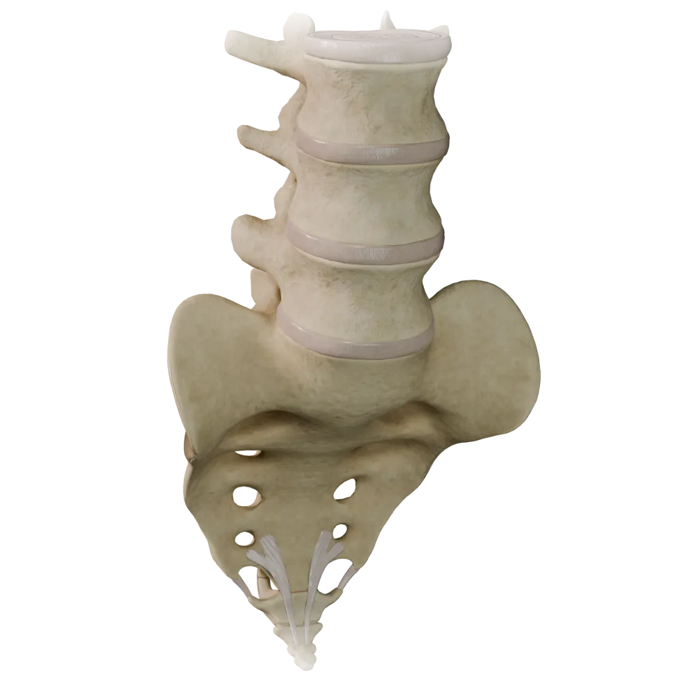
Cartilaginous joints (synchondroses) of the vertebral column
0/20

Fibrous joints of the vertebral column
0/20

Joints of thoracolumbar vertebral column
0/20
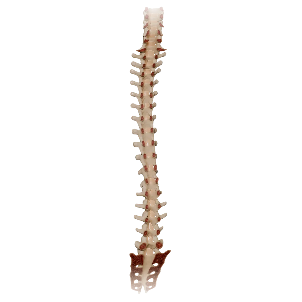
Ligamenta flava
0/20

Synovial joints of vertebral column
0/20
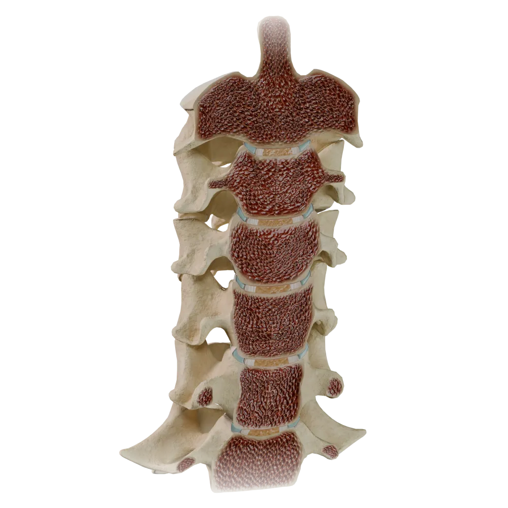
Uncovertebral joints
0/20
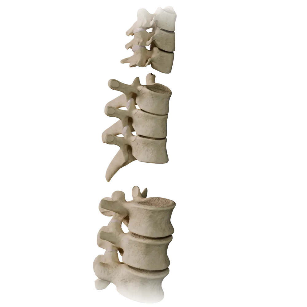
Zygapophyseal joints
0/20
Quiz on zygapophyseal joints in the system of articulations
Anatomy quiz on the zygapophyseal joints of the vertebral column: structure, classification, ligaments, and biomechanics.
1/20
bold
text
1. To which type of joints based on the shape of their articular surfaces do the zygapophyseal joints in the cervical region of the vertebral column belong?
-
Cylindrical
The cervical zygapophyseal (facet) joints are formed by the flat articular surfaces of the superior and inferior articular processes, which classifies them as plane joints (articulationes planae).
-
Plane
The cervical zygapophyseal (facet) joints are formed by the flat articular surfaces of the superior and inferior articular processes, which classifies them as plane joints (articulationes planae).
-
Ellipsoid
The cervical zygapophyseal (facet) joints are formed by the flat articular surfaces of the superior and inferior articular processes, which classifies them as plane joints (articulationes planae).
-
Saddle-shaped
The cervical zygapophyseal (facet) joints are formed by the flat articular surfaces of the superior and inferior articular processes, which classifies them as plane joints (articulationes planae).
-
I find it difficult to answer
The cervical zygapophyseal (facet) joints are formed by the flat articular surfaces of the superior and inferior articular processes, which classifies them as plane joints (articulationes planae).
2. Which region of the vertebral column is characterized by articular surfaces of the zygapophyseal joints oriented in the frontal plane?
-
Cervical
In the thoracic region of the vertebral column, the articular surfaces of the zygapophyseal joints are oriented predominantly in the frontal plane, which limits flexion and extension but permits considerable rotation.
-
Thoracic
In the thoracic region of the vertebral column, the articular surfaces of the zygapophyseal joints are oriented predominantly in the frontal plane, which limits flexion and extension but permits considerable rotation.
-
Lumbar
In the thoracic region of the vertebral column, the articular surfaces of the zygapophyseal joints are oriented predominantly in the frontal plane, which limits flexion and extension but permits considerable rotation.
-
Sacral
In the thoracic region of the vertebral column, the articular surfaces of the zygapophyseal joints are oriented predominantly in the frontal plane, which limits flexion and extension but permits considerable rotation.
-
I find it difficult to answer
In the thoracic region of the vertebral column, the articular surfaces of the zygapophyseal joints are oriented predominantly in the frontal plane, which limits flexion and extension but permits considerable rotation.
3. The articular capsule of the zygapophyseal joints attaches:
-
At the margin of the articular cartilage
The capsule of the zygapophyseal joint attaches directly at the margin of the articular cartilage (along the perimeter of the articular surface), which is typical of joints with flat or slightly concave surfaces.
-
5–7 mm from the margin of the articular cartilage
The capsule of the zygapophyseal joint attaches directly at the margin of the articular cartilage (along the perimeter of the articular surface), which is typical of joints with flat or slightly concave surfaces.
-
To the vertebral body
The capsule of the zygapophyseal joint attaches directly at the margin of the articular cartilage (along the perimeter of the articular surface), which is typical of joints with flat or slightly concave surfaces.
-
To the vertebral arch, bypassing the articular processes
The capsule of the zygapophyseal joint attaches directly at the margin of the articular cartilage (along the perimeter of the articular surface), which is typical of joints with flat or slightly concave surfaces.
-
I find it difficult to answer
The capsule of the zygapophyseal joint attaches directly at the margin of the articular cartilage (along the perimeter of the articular surface), which is typical of joints with flat or slightly concave surfaces.
4. Which ligament is the strongest and directly reinforces the zygapophyseal joints posteriorly?
-
Anterior longitudinal ligament
The ligamentum flavum (ligamentum flavum), composed predominantly of elastic fibers, is located between the arches of adjacent vertebrae and lies directly on the capsules of the zygapophysial joints, reinforcing them posteriorly.
-
Ligamentum flavum
The ligamentum flavum (ligamentum flavum), composed predominantly of elastic fibers, is located between the arches of adjacent vertebrae and lies directly on the capsules of the zygapophysial joints, reinforcing them posteriorly.
-
Posterior longitudinal ligament
The ligamentum flavum (ligamentum flavum), composed predominantly of elastic fibers, is located between the arches of adjacent vertebrae and lies directly on the capsules of the zygapophysial joints, reinforcing them posteriorly.
-
Interspinal ligament
The ligamentum flavum (ligamentum flavum), composed predominantly of elastic fibers, is located between the arches of adjacent vertebrae and lies directly on the capsules of the zygapophysial joints, reinforcing them posteriorly.
-
I find it difficult to answer
The ligamentum flavum (ligamentum flavum), composed predominantly of elastic fibers, is located between the arches of adjacent vertebrae and lies directly on the capsules of the zygapophysial joints, reinforcing them posteriorly.
5. The articular processes forming the zygapophyseal joints are derived from the:
-
Vertebral body
The articular processes (processus articulares superiores et inferiores) arise from the vertebral arch at the junction of its pedicle and lamina and are direct derivatives of the arch.
-
Vertebral arch
The articular processes (processus articulares superiores et inferiores) arise from the vertebral arch at the junction of its pedicle and lamina and are direct derivatives of the arch.
-
Transverse process
The articular processes (processus articulares superiores et inferiores) arise from the vertebral arch at the junction of its pedicle and lamina and are direct derivatives of the arch.
-
Spinous process
The articular processes (processus articulares superiores et inferiores) arise from the vertebral arch at the junction of its pedicle and lamina and are direct derivatives of the arch.
-
I find it difficult to answer
The articular processes (processus articulares superiores et inferiores) arise from the vertebral arch at the junction of its pedicle and lamina and are direct derivatives of the arch.
6. What forms the joint space of the zygapophyseal joint?
-
The inferior articular process of the superior vertebra and the superior articular process of the inferior vertebra
The zygapophyseal joint is formed by the inferior articular process (processus articularis inferior) of the superior vertebra and the superior articular process (processus articularis superior) of the inferior vertebra.
-
Two superior articular processes of adjacent vertebrae
The zygapophyseal joint is formed by the inferior articular process (processus articularis inferior) of the superior vertebra and the superior articular process (processus articularis superior) of the inferior vertebra.
-
The body of the superior vertebra and the arch of the inferior vertebra
The zygapophyseal joint is formed by the inferior articular process (processus articularis inferior) of the superior vertebra and the superior articular process (processus articularis superior) of the inferior vertebra.
-
The transverse process and body of a single vertebra
The zygapophyseal joint is formed by the inferior articular process (processus articularis inferior) of the superior vertebra and the superior articular process (processus articularis superior) of the inferior vertebra.
-
I find it difficult to answer
The zygapophyseal joint is formed by the inferior articular process (processus articularis inferior) of the superior vertebra and the superior articular process (processus articularis superior) of the inferior vertebra.
7. Based on the number of axes of movement, the zygapophyseal joints are:
-
Triaxial
Movements occur around three principal axes (frontal, sagittal, and vertical).
-
Biaxial
Movements occur around three principal axes (frontal, sagittal, and vertical).
-
Uniaxial
Movements occur around three principal axes (frontal, sagittal, and vertical).
-
Multiaxial
Movements occur around three principal axes (frontal, sagittal, and vertical).
-
I find it difficult to answer
Movements occur around three principal axes (frontal, sagittal, and vertical).
8. The synovial membrane of the zygapophyseal joint:
-
Is absent because the joint is a symphysis
The synovial membrane lines the internal surface of the joint capsule and frequently forms folds (meniscoid structures) that project into the joint cavity.
-
Lines the internal surface
The synovial membrane lines the internal surface of the joint capsule and frequently forms folds (meniscoid structures) that project into the joint cavity.
-
Covers the entire surface of the intervertebral disc
The synovial membrane lines the internal surface of the joint capsule and frequently forms folds (meniscoid structures) that project into the joint cavity.
-
Is fused with the posterior longitudinal ligament
The synovial membrane lines the internal surface of the joint capsule and frequently forms folds (meniscoid structures) that project into the joint cavity.
-
I find it difficult to answer
The synovial membrane lines the internal surface of the joint capsule and frequently forms folds (meniscoid structures) that project into the joint cavity.
9. In which region of the vertebral column are the articular surfaces of the zygapophyseal joints oriented predominantly in the sagittal plane?
-
Cervical
In the lumbar region, the articular surfaces of the zygapophyseal joints are oriented in the sagittal plane (or close to it), which determines the biomechanics of this region, with flexion/extension predominating and rotation being limited.
-
Thoracic
In the lumbar region, the articular surfaces of the zygapophyseal joints are oriented in the sagittal plane (or close to it), which determines the biomechanics of this region, with flexion/extension predominating and rotation being limited.
-
Lumbar
In the lumbar region, the articular surfaces of the zygapophyseal joints are oriented in the sagittal plane (or close to it), which determines the biomechanics of this region, with flexion/extension predominating and rotation being limited.
-
Thoracic and cervical simultaneously
In the lumbar region, the articular surfaces of the zygapophyseal joints are oriented in the sagittal plane (or close to it), which determines the biomechanics of this region, with flexion/extension predominating and rotation being limited.
-
I find it difficult to answer
In the lumbar region, the articular surfaces of the zygapophyseal joints are oriented in the sagittal plane (or close to it), which determines the biomechanics of this region, with flexion/extension predominating and rotation being limited.
10. According to structural classification, what type of articulation between the vertebrae do the zygapophyseal joints represent?
-
Syndesmosis
Zygapophyseal joints are diarthroses (juncturae synoviales)—true synovial joints comprising a joint cavity, articular cartilage, a synovial membrane, and a capsule.
-
Synchondrosis
Zygapophyseal joints are diarthroses (juncturae synoviales)—true synovial joints comprising a joint cavity, articular cartilage, a synovial membrane, and a capsule.
-
Synostosis
Zygapophyseal joints are diarthroses (juncturae synoviales)—true synovial joints comprising a joint cavity, articular cartilage, a synovial membrane, and a capsule.
-
Diarthrosis (true joint)
Zygapophyseal joints are diarthroses (juncturae synoviales)—true synovial joints comprising a joint cavity, articular cartilage, a synovial membrane, and a capsule.
-
I find it difficult to answer
Zygapophyseal joints are diarthroses (juncturae synoviales)—true synovial joints comprising a joint cavity, articular cartilage, a synovial membrane, and a capsule.
11. What type of cartilage covers the articular surfaces of the zygapophyseal joints?
-
Fibrocartilage
The articular surfaces of the zygapophyseal joints are covered by hyaline cartilage, as are those of most synovial joints in the human body.
-
Hyaline cartilage
The articular surfaces of the zygapophyseal joints are covered by hyaline cartilage, as are those of most synovial joints in the human body.
-
Elastic cartilage
The articular surfaces of the zygapophyseal joints are covered by hyaline cartilage, as are those of most synovial joints in the human body.
-
The articular surfaces are not covered by cartilage
The articular surfaces of the zygapophyseal joints are covered by hyaline cartilage, as are those of most synovial joints in the human body.
-
I find it difficult to answer
The articular surfaces of the zygapophyseal joints are covered by hyaline cartilage, as are those of most synovial joints in the human body.
12. The posterior ramus of a spinal nerve innervates the zygapophyseal joint at:
-
Only one level
Each zygapophyseal joint receives innervation from the posterior rami of spinal nerves at two adjacent levels—the nerve at the same level and the nerve one level above—providing overlapping innervation.
-
Two adjacent levels
Each zygapophyseal joint receives innervation from the posterior rami of spinal nerves at two adjacent levels—the nerve at the same level and the nerve one level above—providing overlapping innervation.
-
Three levels of the vertebral column
Each zygapophyseal joint receives innervation from the posterior rami of spinal nerves at two adjacent levels—the nerve at the same level and the nerve one level above—providing overlapping innervation.
-
Only part of the capsule
Each zygapophyseal joint receives innervation from the posterior rami of spinal nerves at two adjacent levels—the nerve at the same level and the nerve one level above—providing overlapping innervation.
-
I find it difficult to answer
Each zygapophyseal joint receives innervation from the posterior rami of spinal nerves at two adjacent levels—the nerve at the same level and the nerve one level above—providing overlapping innervation.
13. What are the branches that directly innervate the capsule of the zygapophyseal joint called?
-
Lateral branches of the posterior branches of the spinal nerves
The capsule of the zygapophyseal joint is innervated by the medial branches (rami mediales) of the posterior rami of the spinal nerves of the corresponding and superior segments.
-
Medial branches of the posterior branches of the spinal nerves
The capsule of the zygapophyseal joint is innervated by the medial branches (rami mediales) of the posterior rami of the spinal nerves of the corresponding and superior segments.
-
Anterior branches of the spinal nerves
The capsule of the zygapophyseal joint is innervated by the medial branches (rami mediales) of the posterior rami of the spinal nerves of the corresponding and superior segments.
-
Sinuvertebral nerve of Luschka
The capsule of the zygapophyseal joint is innervated by the medial branches (rami mediales) of the posterior rami of the spinal nerves of the corresponding and superior segments.
-
I find it difficult to answer
The capsule of the zygapophyseal joint is innervated by the medial branches (rami mediales) of the posterior rami of the spinal nerves of the corresponding and superior segments.
14. What is the principal biomechanical function of the zygapophyseal joints together with the intervertebral disc?
-
Exclusive absorption of axial loading
The zygapophyseal joints, together with the intervertebral disc, form a “three-joint complex” (functional spinal unit): they guide and restrict movements depending on the orientation of the articular surfaces and participate in the transmission of axial load (particularly during extension).
-
Guidance and restriction of movements of the vertebral column, as well as transmission of part of the load
The zygapophyseal joints, together with the intervertebral disc, form a “three-joint complex” (functional spinal unit): they guide and restrict movements depending on the orientation of the articular surfaces and participate in the transmission of axial load (particularly during extension).
-
Rotational movements only
The zygapophyseal joints, together with the intervertebral disc, form a “three-joint complex” (functional spinal unit): they guide and restrict movements depending on the orientation of the articular surfaces and participate in the transmission of axial load (particularly during extension).
-
Only fixation in the neutral position without movement
The zygapophyseal joints, together with the intervertebral disc, form a “three-joint complex” (functional spinal unit): they guide and restrict movements depending on the orientation of the articular surfaces and participate in the transmission of axial load (particularly during extension).
-
I find it difficult to answer
The zygapophyseal joints, together with the intervertebral disc, form a “three-joint complex” (functional spinal unit): they guide and restrict movements depending on the orientation of the articular surfaces and participate in the transmission of axial load (particularly during extension).
15. How many pairs of zygapophyseal joints are present in the cervical region of the vertebral column (C2–C7)?
-
4 pairs
In the cervical region of the vertebral column, zygapophyseal joints are formed between C2–C3, C3–C4, C4–C5, C5–C6, and C6–C7, constituting 5 pairs of joints. The C1–C2 joint (atlanto-axial joint) is a separate anatomical structure.
-
5 pairs
In the cervical region of the vertebral column, zygapophyseal joints are formed between C2–C3, C3–C4, C4–C5, C5–C6, and C6–C7, constituting 5 pairs of joints. The C1–C2 joint (atlanto-axial joint) is a separate anatomical structure.
-
6 pairs
In the cervical region of the vertebral column, zygapophyseal joints are formed between C2–C3, C3–C4, C4–C5, C5–C6, and C6–C7, constituting 5 pairs of joints. The C1–C2 joint (atlanto-axial joint) is a separate anatomical structure.
-
7 pairs
In the cervical region of the vertebral column, zygapophyseal joints are formed between C2–C3, C3–C4, C4–C5, C5–C6, and C6–C7, constituting 5 pairs of joints. The C1–C2 joint (atlanto-axial joint) is a separate anatomical structure.
-
I find it difficult to answer
In the cervical region of the vertebral column, zygapophyseal joints are formed between C2–C3, C3–C4, C4–C5, C5–C6, and C6–C7, constituting 5 pairs of joints. The C1–C2 joint (atlanto-axial joint) is a separate anatomical structure.
16. Which vascular structure supplies blood to the zygapophyseal joints?
-
Anterior intercostal arteries
The zygapophyseal joints are supplied by the posterior branches of segmental arteries (posterior branches of the intercostal, lumbar, and sacral arteries), which accompany the posterior rami of the spinal nerves.
-
Posterior branches of the intercostal and lumbar arteries
The zygapophyseal joints are supplied by the posterior branches of segmental arteries (posterior branches of the intercostal, lumbar, and sacral arteries), which accompany the posterior rami of the spinal nerves.
-
Vertebral artery (cervical region only)
The zygapophyseal joints are supplied by the posterior branches of segmental arteries (posterior branches of the intercostal, lumbar, and sacral arteries), which accompany the posterior rami of the spinal nerves.
-
Internal vertebral venous plexus
The zygapophyseal joints are supplied by the posterior branches of segmental arteries (posterior branches of the intercostal, lumbar, and sacral arteries), which accompany the posterior rami of the spinal nerves.
-
I find it difficult to answer
The zygapophyseal joints are supplied by the posterior branches of segmental arteries (posterior branches of the intercostal, lumbar, and sacral arteries), which accompany the posterior rami of the spinal nerves.
17. How many pairs of zygapophyseal joints are present in the lumbar region of the vertebral column?
-
3 pairs
The lumbar region contains 5 vertebrae (L1–L5). Zygapophyseal joints are formed between the articular processes of adjacent vertebrae: L1–L2, L2–L3, L3–L4, L4–L5, and L5–S1 (lumbosacral articulation). However, if only joints strictly within the lumbar region are considered (between L1–L2, L2–L3, L3–L4, and L4–L5), there are 4 pairs. However, in clinical anatomy and radiology, “lumbar zygapophyseal joints” often refers to all joints involving lumbar vertebrae, including the L5–S1 articulation (this is also a zygapophyseal joint, although the sacrum is not part of the lumbar region).
-
5 pairs
The lumbar region contains 5 vertebrae (L1–L5). Zygapophyseal joints are formed between the articular processes of adjacent vertebrae: L1–L2, L2–L3, L3–L4, L4–L5, and L5–S1 (lumbosacral articulation). However, if only joints strictly within the lumbar region are considered (between L1–L2, L2–L3, L3–L4, and L4–L5), there are 4 pairs. However, in clinical anatomy and radiology, “lumbar zygapophyseal joints” often refers to all joints involving lumbar vertebrae, including the L5–S1 articulation (this is also a zygapophyseal joint, although the sacrum is not part of the lumbar region).
-
6 pairs
The lumbar region contains 5 vertebrae (L1–L5). Zygapophyseal joints are formed between the articular processes of adjacent vertebrae: L1–L2, L2–L3, L3–L4, L4–L5, and L5–S1 (lumbosacral articulation). However, if only joints strictly within the lumbar region are considered (between L1–L2, L2–L3, L3–L4, and L4–L5), there are 4 pairs. However, in clinical anatomy and radiology, “lumbar zygapophyseal joints” often refers to all joints involving lumbar vertebrae, including the L5–S1 articulation (this is also a zygapophyseal joint, although the sacrum is not part of the lumbar region).
-
7 pairs
The lumbar region contains 5 vertebrae (L1–L5). Zygapophyseal joints are formed between the articular processes of adjacent vertebrae: L1–L2, L2–L3, L3–L4, L4–L5, and L5–S1 (lumbosacral articulation). However, if only joints strictly within the lumbar region are considered (between L1–L2, L2–L3, L3–L4, and L4–L5), there are 4 pairs. However, in clinical anatomy and radiology, “lumbar zygapophyseal joints” often refers to all joints involving lumbar vertebrae, including the L5–S1 articulation (this is also a zygapophyseal joint, although the sacrum is not part of the lumbar region).
-
I find it difficult to answer
The lumbar region contains 5 vertebrae (L1–L5). Zygapophyseal joints are formed between the articular processes of adjacent vertebrae: L1–L2, L2–L3, L3–L4, L4–L5, and L5–S1 (lumbosacral articulation). However, if only joints strictly within the lumbar region are considered (between L1–L2, L2–L3, L3–L4, and L4–L5), there are 4 pairs. However, in clinical anatomy and radiology, “lumbar zygapophyseal joints” often refers to all joints involving lumbar vertebrae, including the L5–S1 articulation (this is also a zygapophyseal joint, although the sacrum is not part of the lumbar region).
18. How does the orientation of the articular surfaces of the zygapophyseal joints in the upper cervical region (C2–C4) differ from that in the lumbar region?
-
Sagittal in the cervical region and horizontal in the lumbar region
In the cervical region, the articular surfaces of the zygapophyseal joints are oriented at an angle of approximately 45° to the horizontal plane (nearly horizontal), permitting considerable rotation. In the lumbar region, they are oriented predominantly in the sagittal plane, limiting rotation.
-
Nearly horizontal in the cervical region (at an angle of ~45° to the horizontal) and sagittal in the lumbar region
In the cervical region, the articular surfaces of the zygapophyseal joints are oriented at an angle of approximately 45° to the horizontal plane (nearly horizontal), permitting considerable rotation. In the lumbar region, they are oriented predominantly in the sagittal plane, limiting rotation.
-
The orientation is identical in both regions—frontal
In the cervical region, the articular surfaces of the zygapophyseal joints are oriented at an angle of approximately 45° to the horizontal plane (nearly horizontal), permitting considerable rotation. In the lumbar region, they are oriented predominantly in the sagittal plane, limiting rotation.
-
Frontal in the cervical region and oblique in the lumbar region
In the cervical region, the articular surfaces of the zygapophyseal joints are oriented at an angle of approximately 45° to the horizontal plane (nearly horizontal), permitting considerable rotation. In the lumbar region, they are oriented predominantly in the sagittal plane, limiting rotation.
-
I find it difficult to answer
In the cervical region, the articular surfaces of the zygapophyseal joints are oriented at an angle of approximately 45° to the horizontal plane (nearly horizontal), permitting considerable rotation. In the lumbar region, they are oriented predominantly in the sagittal plane, limiting rotation.
19. Which ligament, located above the spinous processes, indirectly contributes to stabilization of the zygapophyseal joints in the cervical region?
-
Anterior longitudinal ligament
The nuchal ligament (ligamentum nuchae) is a strong median fibrous plate of the cervical region that is homologous to the supraspinous ligament. It limits flexion of the cervical region and thereby indirectly reduces the load on the capsules of the zygapophyseal joints during flexion.
-
Nuchal ligament (ligamentum nuchae)
The nuchal ligament (ligamentum nuchae) is a strong median fibrous plate of the cervical region that is homologous to the supraspinous ligament. It limits flexion of the cervical region and thereby indirectly reduces the load on the capsules of the zygapophyseal joints during flexion.
-
Intertransverse ligament
The nuchal ligament (ligamentum nuchae) is a strong median fibrous plate of the cervical region that is homologous to the supraspinous ligament. It limits flexion of the cervical region and thereby indirectly reduces the load on the capsules of the zygapophyseal joints during flexion.
-
Alar ligament
The nuchal ligament (ligamentum nuchae) is a strong median fibrous plate of the cervical region that is homologous to the supraspinous ligament. It limits flexion of the cervical region and thereby indirectly reduces the load on the capsules of the zygapophyseal joints during flexion.
-
I find it difficult to answer
The nuchal ligament (ligamentum nuchae) is a strong median fibrous plate of the cervical region that is homologous to the supraspinous ligament. It limits flexion of the cervical region and thereby indirectly reduces the load on the capsules of the zygapophyseal joints during flexion.
20. Which movement is restricted most by the zygapophyseal joints of the lumbar region due to the sagittal orientation of their articular surfaces?
-
Flexion (flexion)
The sagittal orientation of the articular surfaces of the zygapophysial joints in the lumbar region mechanically blocks axial rotation (rotation), because when rotation is attempted, the articular surfaces abut one another. This is a fundamental biomechanical distinction between the lumbar region and the thoracic and cervical regions.
-
Extension (extension)
The sagittal orientation of the articular surfaces of the zygapophysial joints in the lumbar region mechanically blocks axial rotation (rotation), because when rotation is attempted, the articular surfaces abut one another. This is a fundamental biomechanical distinction between the lumbar region and the thoracic and cervical regions.
-
Axial rotation (rotation)
The sagittal orientation of the articular surfaces of the zygapophysial joints in the lumbar region mechanically blocks axial rotation (rotation), because when rotation is attempted, the articular surfaces abut one another. This is a fundamental biomechanical distinction between the lumbar region and the thoracic and cervical regions.
-
Lateral flexion
The sagittal orientation of the articular surfaces of the zygapophysial joints in the lumbar region mechanically blocks axial rotation (rotation), because when rotation is attempted, the articular surfaces abut one another. This is a fundamental biomechanical distinction between the lumbar region and the thoracic and cervical regions.
-
I find it difficult to answer
The sagittal orientation of the articular surfaces of the zygapophysial joints in the lumbar region mechanically blocks axial rotation (rotation), because when rotation is attempted, the articular surfaces abut one another. This is a fundamental biomechanical distinction between the lumbar region and the thoracic and cervical regions.
Retake this quiz?
Your current progress will be reset.
