
Joints of vertebral column
0/20
By topic

Atlantoaxial joints (median and lateral)
0/20
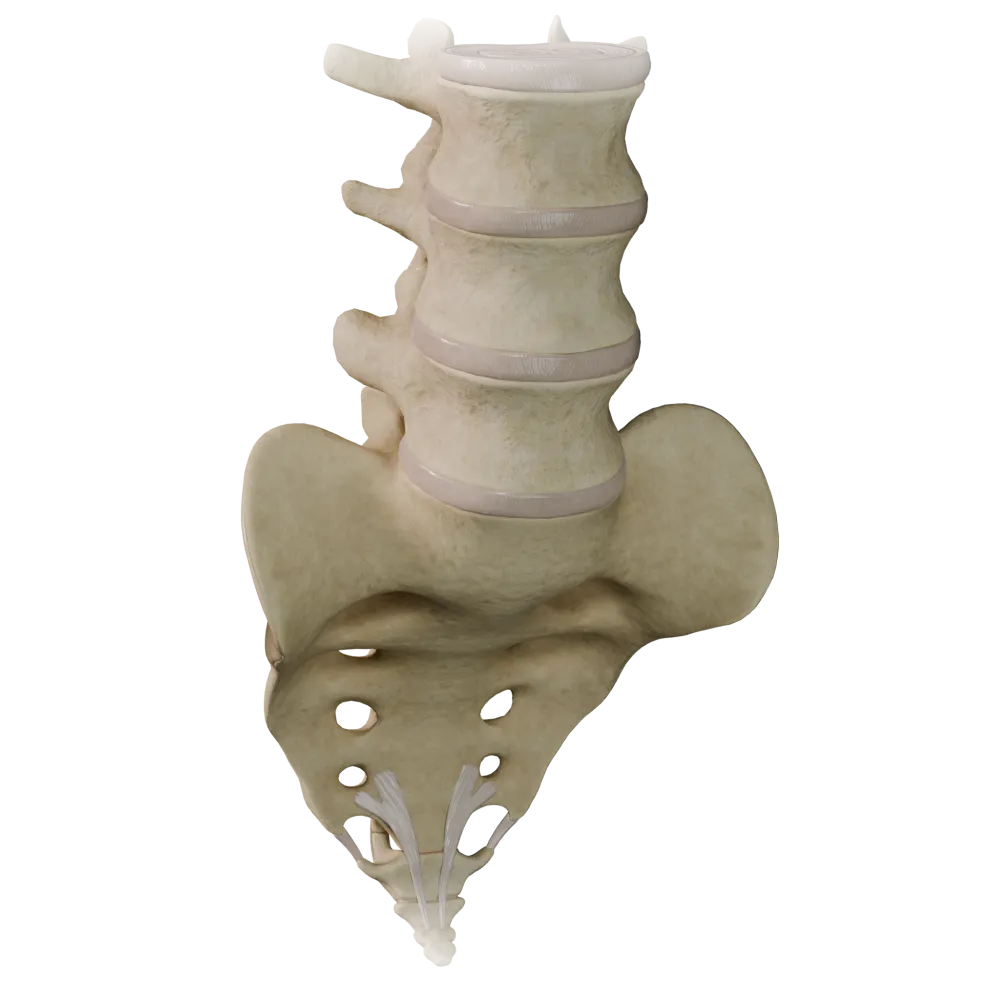
Cartilaginous joints (synchondroses) of the vertebral column
0/20

Fibrous joints of the vertebral column
0/20

Joints of thoracolumbar vertebral column
0/20
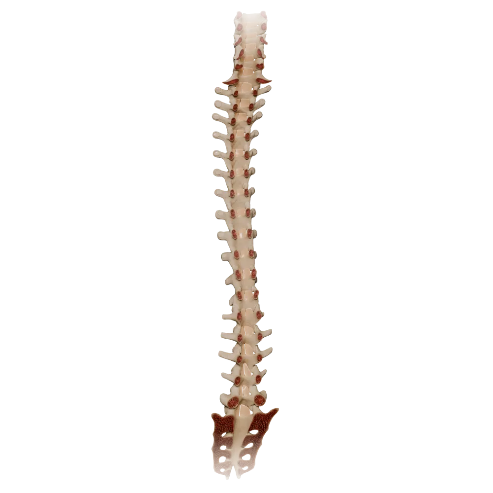
Ligamenta flava
0/20

Synovial joints of vertebral column
0/20
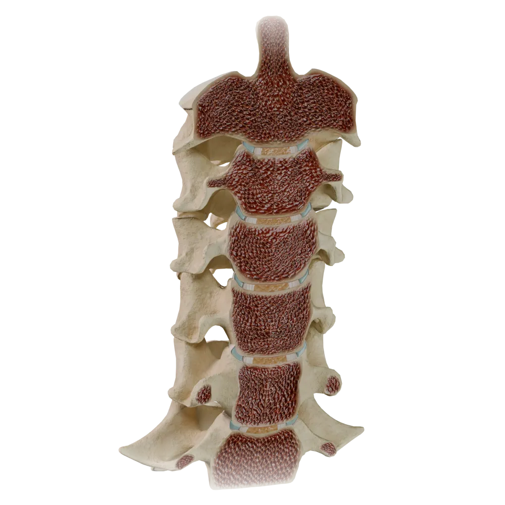
Uncovertebral joints
0/20
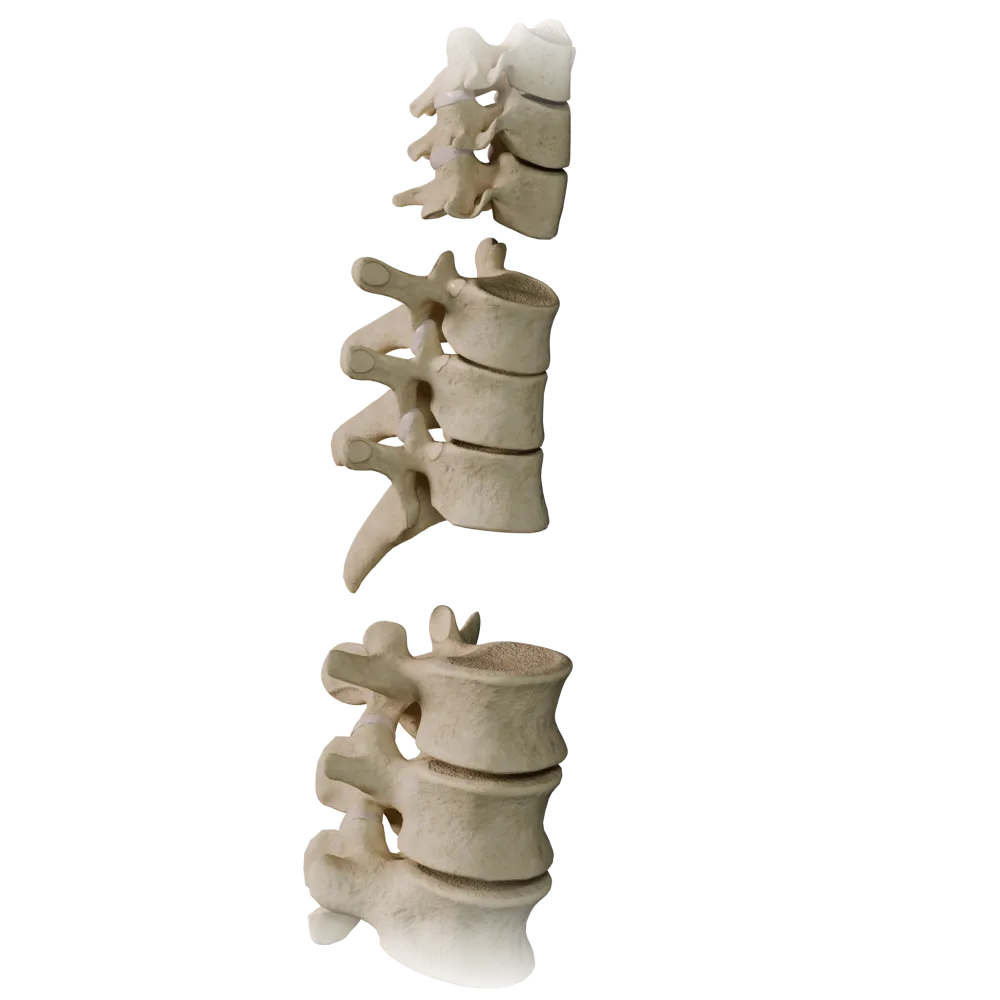
Zygapophyseal joints
0/20
Anatomy quiz on the synovial joints of vertebral column
The quiz covers the normal structure, biomechanics, and functional anatomy of the synovial joints of vertebral column.
1/20
bold
text
1. What type of joint is the median atlantoaxial joint (articulatio atlantoaxialis mediana)?
-
Ellipsoid joint
The median atlantoaxial joint is formed by the dens of the axis and the anterior arch of the atlas; the articular surfaces are cylindrical, permitting rotation around a vertical axis—a characteristic feature of a trochoid (pivot) joint.
-
Trochoid (pivot) joint
The median atlantoaxial joint is formed by the dens of the axis and the anterior arch of the atlas; the articular surfaces are cylindrical, permitting rotation around a vertical axis—a characteristic feature of a trochoid (pivot) joint.
-
Saddle joint
The median atlantoaxial joint is formed by the dens of the axis and the anterior arch of the atlas; the articular surfaces are cylindrical, permitting rotation around a vertical axis—a characteristic feature of a trochoid (pivot) joint.
-
Ball-and-socket joint
The median atlantoaxial joint is formed by the dens of the axis and the anterior arch of the atlas; the articular surfaces are cylindrical, permitting rotation around a vertical axis—a characteristic feature of a trochoid (pivot) joint.
-
I find it difficult to answer
The median atlantoaxial joint is formed by the dens of the axis and the anterior arch of the atlas; the articular surfaces are cylindrical, permitting rotation around a vertical axis—a characteristic feature of a trochoid (pivot) joint.
2. Which ligament restricts excessive extension of the vertebral column?
-
Ligamentum supraspinale.
The anterior longitudinal ligament (lig. longitudinale anterius) runs along the anterior surface of the vertebral bodies and is the principal structure restricting extension.
-
Ligamentum longitudinale anterius
The anterior longitudinal ligament (lig. longitudinale anterius) runs along the anterior surface of the vertebral bodies and is the principal structure restricting extension.
-
Ligamentum longitudinale posterius
The anterior longitudinal ligament (lig. longitudinale anterius) runs along the anterior surface of the vertebral bodies and is the principal structure restricting extension.
-
Ligamentum nuchae
The anterior longitudinal ligament (lig. longitudinale anterius) runs along the anterior surface of the vertebral bodies and is the principal structure restricting extension.
-
I find it difficult to answer
The anterior longitudinal ligament (lig. longitudinale anterius) runs along the anterior surface of the vertebral bodies and is the principal structure restricting extension.
3. What is the shape of the articular surfaces in the atlantooccipital joint (articulatio atlantooccipitalis)?
-
Flat articular surfaces
The atlantooccipital joint is condylar (ellipsoid): the articular facets of the superior articular surfaces of the atlas articulate with the ellipsoid condyles of the occipital bone, permitting flexion, extension, and slight lateral flexion.
-
Ellipsoid condylar surfaces
The atlantooccipital joint is condylar (ellipsoid): the articular facets of the superior articular surfaces of the atlas articulate with the ellipsoid condyles of the occipital bone, permitting flexion, extension, and slight lateral flexion.
-
Saddle-shaped articular surfaces
The atlantooccipital joint is condylar (ellipsoid): the articular facets of the superior articular surfaces of the atlas articulate with the ellipsoid condyles of the occipital bone, permitting flexion, extension, and slight lateral flexion.
-
Cylindrical articular surfaces
The atlantooccipital joint is condylar (ellipsoid): the articular facets of the superior articular surfaces of the atlas articulate with the ellipsoid condyles of the occipital bone, permitting flexion, extension, and slight lateral flexion.
-
I find it difficult to answer
The atlantooccipital joint is condylar (ellipsoid): the articular facets of the superior articular surfaces of the atlas articulate with the ellipsoid condyles of the occipital bone, permitting flexion, extension, and slight lateral flexion.
4. Between which vertebrae are intervertebral discs (disci intervertebrales) normally absent?
-
Between C2 and C3
An intervertebral disc is absent between the atlas (C1) and the axis (C2), because they are connected by the atlantoaxial joints. A disc is also absent between the occipital bone and the atlas.
-
Between C1 and C2 (the atlas and axis)
An intervertebral disc is absent between the atlas (C1) and the axis (C2), because they are connected by the atlantoaxial joints. A disc is also absent between the occipital bone and the atlas.
-
Between L4 and L5
An intervertebral disc is absent between the atlas (C1) and the axis (C2), because they are connected by the atlantoaxial joints. A disc is also absent between the occipital bone and the atlas.
-
Between T12 and L1
An intervertebral disc is absent between the atlas (C1) and the axis (C2), because they are connected by the atlantoaxial joints. A disc is also absent between the occipital bone and the atlas.
-
I find it difficult to answer
An intervertebral disc is absent between the atlas (C1) and the axis (C2), because they are connected by the atlantoaxial joints. A disc is also absent between the occipital bone and the atlas.
5. The gelatinous nucleus (nucleus pulposus) of the intervertebral disc is derived from which embryonic structure?
-
Sclerotome of the somite
The nucleus pulposus is a remnant of the notochord (chorda dorsalis). During embryogenesis, the notochord regresses within the vertebral bodies, persisting only in the centers of the discs as the nucleus pulposus.
-
Notochord (chorda dorsalis)
The nucleus pulposus is a remnant of the notochord (chorda dorsalis). During embryogenesis, the notochord regresses within the vertebral bodies, persisting only in the centers of the discs as the nucleus pulposus.
-
Dermatome of the somite
The nucleus pulposus is a remnant of the notochord (chorda dorsalis). During embryogenesis, the notochord regresses within the vertebral bodies, persisting only in the centers of the discs as the nucleus pulposus.
-
Splanchnic mesoderm
The nucleus pulposus is a remnant of the notochord (chorda dorsalis). During embryogenesis, the notochord regresses within the vertebral bodies, persisting only in the centers of the discs as the nucleus pulposus.
-
I find it difficult to answer
The nucleus pulposus is a remnant of the notochord (chorda dorsalis). During embryogenesis, the notochord regresses within the vertebral bodies, persisting only in the centers of the discs as the nucleus pulposus.
6. What are the primary movements at the lateral atlantoaxial joint (articulatio atlantoaxialis lateralis)?
-
Flexion and extension
The median and paired lateral atlantoaxial joints function together, primarily permitting rotation of the head around the vertical axis (axial rotation). This range of motion is approximately 30–45° in each direction.
-
Lateral flexion
The median and paired lateral atlantoaxial joints function together, primarily permitting rotation of the head around the vertical axis (axial rotation). This range of motion is approximately 30–45° in each direction.
-
Rotation around the vertical axis
The median and paired lateral atlantoaxial joints function together, primarily permitting rotation of the head around the vertical axis (axial rotation). This range of motion is approximately 30–45° in each direction.
-
Circumduction (circumductio)
The median and paired lateral atlantoaxial joints function together, primarily permitting rotation of the head around the vertical axis (axial rotation). This range of motion is approximately 30–45° in each direction.
-
I find it difficult to answer
The median and paired lateral atlantoaxial joints function together, primarily permitting rotation of the head around the vertical axis (axial rotation). This range of motion is approximately 30–45° in each direction.
7. Ligamentum flavum connects:
-
Spinous processes of adjacent vertebrae
The ligamenta flava (ligg. flava) connect the laminae (laminae) of the arches of adjacent vertebrae. They contain numerous elastic fibers, which give them their yellow color and permit elastic recoil of the vertebral column from flexion.
-
Laminae of the arches of adjacent vertebrae (laminae arcuum)
The ligamenta flava (ligg. flava) connect the laminae (laminae) of the arches of adjacent vertebrae. They contain numerous elastic fibers, which give them their yellow color and permit elastic recoil of the vertebral column from flexion.
-
Transverse processes of adjacent vertebrae
The ligamenta flava (ligg. flava) connect the laminae (laminae) of the arches of adjacent vertebrae. They contain numerous elastic fibers, which give them their yellow color and permit elastic recoil of the vertebral column from flexion.
-
Bodies of adjacent vertebrae
The ligamenta flava (ligg. flava) connect the laminae (laminae) of the arches of adjacent vertebrae. They contain numerous elastic fibers, which give them their yellow color and permit elastic recoil of the vertebral column from flexion.
-
I find it difficult to answer
The ligamenta flava (ligg. flava) connect the laminae (laminae) of the arches of adjacent vertebrae. They contain numerous elastic fibers, which give them their yellow color and permit elastic recoil of the vertebral column from flexion.
8. The cruciform ligament of the atlas (lig. cruciforme atlantis) consists of:
-
The anterior and posterior longitudinal ligaments
Lig. cruciforme atlantis is formed by the transverse atlantal ligament (lig. transversum atlantis), to which superior and inferior longitudinal bundles are attached, extending to the occipital bone and the body of the axis, respectively. It retains the dens of the axis.
-
The transverse atlantal ligament and longitudinal bundles
Lig. cruciforme atlantis is formed by the transverse atlantal ligament (lig. transversum atlantis), to which superior and inferior longitudinal bundles are attached, extending to the occipital bone and the body of the axis, respectively. It retains the dens of the axis.
-
The alar ligaments and apical ligament of the dens
Lig. cruciforme atlantis is formed by the transverse atlantal ligament (lig. transversum atlantis), to which superior and inferior longitudinal bundles are attached, extending to the occipital bone and the body of the axis, respectively. It retains the dens of the axis.
-
The articular capsule and lig. transversum
Lig. cruciforme atlantis is formed by the transverse atlantal ligament (lig. transversum atlantis), to which superior and inferior longitudinal bundles are attached, extending to the occipital bone and the body of the axis, respectively. It retains the dens of the axis.
-
I find it difficult to answer
Lig. cruciforme atlantis is formed by the transverse atlantal ligament (lig. transversum atlantis), to which superior and inferior longitudinal bundles are attached, extending to the occipital bone and the body of the axis, respectively. It retains the dens of the axis.
9. The transverse atlantal ligament (lig. transversum atlantis) is part of the cruciform ligament and is stretched between:
-
The dens of the axis and the anterior arch of the atlas
The transverse atlantal ligament is the strongest part of the cruciform ligament (lig. cruciforme atlantis). It is stretched between the medial surfaces of the lateral masses of the atlas, passing posterior to the dens of the axis and holding it against the anterior arch. It prevents posterior displacement of the dens toward the spinal cord.
-
The lateral masses of the atlas
The transverse atlantal ligament is the strongest part of the cruciform ligament (lig. cruciforme atlantis). It is stretched between the medial surfaces of the lateral masses of the atlas, passing posterior to the dens of the axis and holding it against the anterior arch. It prevents posterior displacement of the dens toward the spinal cord.
-
The dens of the axis and the transverse atlantal ligament
The transverse atlantal ligament is the strongest part of the cruciform ligament (lig. cruciforme atlantis). It is stretched between the medial surfaces of the lateral masses of the atlas, passing posterior to the dens of the axis and holding it against the anterior arch. It prevents posterior displacement of the dens toward the spinal cord.
-
The apex of the dens and the posterior arch of the atlas
The transverse atlantal ligament is the strongest part of the cruciform ligament (lig. cruciforme atlantis). It is stretched between the medial surfaces of the lateral masses of the atlas, passing posterior to the dens of the axis and holding it against the anterior arch. It prevents posterior displacement of the dens toward the spinal cord.
-
I find it difficult to answer
The transverse atlantal ligament is the strongest part of the cruciform ligament (lig. cruciforme atlantis). It is stretched between the medial surfaces of the lateral masses of the atlas, passing posterior to the dens of the axis and holding it against the anterior arch. It prevents posterior displacement of the dens toward the spinal cord.
10. The alar ligaments (ligg. alaria) are stretched between:
-
The dens of the axis and the occipital bone
The alar ligaments are paired, strong ligaments extending from the superior part of the dens of the axis (from its lateral surfaces) to the lateral margins of the foramen magnum (occipital bone). They restrict rotation and lateral flexion of the head. Their rupture results in dangerous displacement of the dens.
-
The lateral masses of the atlas and the occipital bone
The alar ligaments are paired, strong ligaments extending from the superior part of the dens of the axis (from its lateral surfaces) to the lateral margins of the foramen magnum (occipital bone). They restrict rotation and lateral flexion of the head. Their rupture results in dangerous displacement of the dens.
-
The dens of the axis and the transverse atlantal ligament
The alar ligaments are paired, strong ligaments extending from the superior part of the dens of the axis (from its lateral surfaces) to the lateral margins of the foramen magnum (occipital bone). They restrict rotation and lateral flexion of the head. Their rupture results in dangerous displacement of the dens.
-
The occipital condyles and the atlas
The alar ligaments are paired, strong ligaments extending from the superior part of the dens of the axis (from its lateral surfaces) to the lateral margins of the foramen magnum (occipital bone). They restrict rotation and lateral flexion of the head. Their rupture results in dangerous displacement of the dens.
-
I find it difficult to answer
The alar ligaments are paired, strong ligaments extending from the superior part of the dens of the axis (from its lateral surfaces) to the lateral margins of the foramen magnum (occipital bone). They restrict rotation and lateral flexion of the head. Their rupture results in dangerous displacement of the dens.
11. The fibrous ring (anulus fibrosus) of the intervertebral disc is composed predominantly of:
-
Hyaline cartilage
The anulus fibrosus consists of concentric lamellae of fibrocartilage and dense regular connective tissue with obliquely oriented collagen fibers (predominantly type I collagen). This architecture provides resistance to compression, rotation, and shear.
-
Obliquely arranged lamellae of fibrocartilage and dense connective tissue
The anulus fibrosus consists of concentric lamellae of fibrocartilage and dense regular connective tissue with obliquely oriented collagen fibers (predominantly type I collagen). This architecture provides resistance to compression, rotation, and shear.
-
Loose connective tissue with type III collagen
The anulus fibrosus consists of concentric lamellae of fibrocartilage and dense regular connective tissue with obliquely oriented collagen fibers (predominantly type I collagen). This architecture provides resistance to compression, rotation, and shear.
-
Elastic fibers without a cartilaginous matrix
The anulus fibrosus consists of concentric lamellae of fibrocartilage and dense regular connective tissue with obliquely oriented collagen fibers (predominantly type I collagen). This architecture provides resistance to compression, rotation, and shear.
-
I find it difficult to answer
The anulus fibrosus consists of concentric lamellae of fibrocartilage and dense regular connective tissue with obliquely oriented collagen fibers (predominantly type I collagen). This architecture provides resistance to compression, rotation, and shear.
12. Unlike the alar ligaments (ligg. alaria), the apical ligament of the dens (lig. apicis dentis) is stretched:
-
Between the dens and the anterior arch of the atlas
This is a rudimentary ligament extending strictly along the median line from the apex of the dens (dens axis) to the anterior margin of the foramen magnum (basilar part of the occipital bone). Unlike the alar ligaments (which are paired and extend laterally), the apical ligament is unpaired and weak
-
Between the dens and the transverse atlantal ligament
This is a rudimentary ligament extending strictly along the median line from the apex of the dens (dens axis) to the anterior margin of the foramen magnum (basilar part of the occipital bone). Unlike the alar ligaments (which are paired and extend laterally), the apical ligament is unpaired and weak
-
From the apex of the dens to the basilar part of the occipital bone
This is a rudimentary ligament extending strictly along the median line from the apex of the dens (dens axis) to the anterior margin of the foramen magnum (basilar part of the occipital bone). Unlike the alar ligaments (which are paired and extend laterally), the apical ligament is unpaired and weak
-
Between the dens and the lateral masses of the atlas
This is a rudimentary ligament extending strictly along the median line from the apex of the dens (dens axis) to the anterior margin of the foramen magnum (basilar part of the occipital bone). Unlike the alar ligaments (which are paired and extend laterally), the apical ligament is unpaired and weak
-
I find it difficult to answer
This is a rudimentary ligament extending strictly along the median line from the apex of the dens (dens axis) to the anterior margin of the foramen magnum (basilar part of the occipital bone). Unlike the alar ligaments (which are paired and extend laterally), the apical ligament is unpaired and weak
13. The alar ligaments (ligg. alaria) attach to:
-
The anterior arch of the atlas and the dens of the axis
Ligg. alaria extend from the lateral surfaces of the dens of the axis to the medial surfaces of the occipital condyles. They restrict excessive rotation and lateral flexion of the head.
-
The lateral surfaces of the dens of the axis and the occipital condyles
Ligg. alaria extend from the lateral surfaces of the dens of the axis to the medial surfaces of the occipital condyles. They restrict excessive rotation and lateral flexion of the head.
-
The transverse processes of the atlas and the body of the axis
Ligg. alaria extend from the lateral surfaces of the dens of the axis to the medial surfaces of the occipital condyles. They restrict excessive rotation and lateral flexion of the head.
-
The posterior arch of the atlas and the basilar part of the occipital bone
Ligg. alaria extend from the lateral surfaces of the dens of the axis to the medial surfaces of the occipital condyles. They restrict excessive rotation and lateral flexion of the head.
-
I find it difficult to answer
Ligg. alaria extend from the lateral surfaces of the dens of the axis to the medial surfaces of the occipital condyles. They restrict excessive rotation and lateral flexion of the head.
14. To which type of synovial joint do the zygapophysial (facet) joints of the vertebral column (articulationes zygapophysiales) belong?
-
Ball-and-socket joints
Articulationes zygapophysiales are plane (gliding) joints with nearly flat articular surfaces. The range of motion at each individual joint is minimal; however, collectively, they permit considerable mobility throughout the vertebral column.
-
Plane joints (articulationes planae)
Articulationes zygapophysiales are plane (gliding) joints with nearly flat articular surfaces. The range of motion at each individual joint is minimal; however, collectively, they permit considerable mobility throughout the vertebral column.
-
Hinge joints
Articulationes zygapophysiales are plane (gliding) joints with nearly flat articular surfaces. The range of motion at each individual joint is minimal; however, collectively, they permit considerable mobility throughout the vertebral column.
-
Ellipsoid joints
Articulationes zygapophysiales are plane (gliding) joints with nearly flat articular surfaces. The range of motion at each individual joint is minimal; however, collectively, they permit considerable mobility throughout the vertebral column.
-
I find it difficult to answer
Articulationes zygapophysiales are plane (gliding) joints with nearly flat articular surfaces. The range of motion at each individual joint is minimal; however, collectively, they permit considerable mobility throughout the vertebral column.
15. The ligament that connects the apices of the spinous processes from C7 to the sacrum and is a direct continuation of the nuchal ligament is called:
-
Lig. interspinale
Lig. supraspinale is a band of dense connective tissue connecting the apices of the spinous processes from C7 to the sacrum. In the cervical region, it continues as the strong nuchal ligament (lig. nuchae). It restricts flexion of the vertebral column.
-
Lig. supraspinale
Lig. supraspinale is a band of dense connective tissue connecting the apices of the spinous processes from C7 to the sacrum. In the cervical region, it continues as the strong nuchal ligament (lig. nuchae). It restricts flexion of the vertebral column.
-
Lig. longitudinale posterius
Lig. supraspinale is a band of dense connective tissue connecting the apices of the spinous processes from C7 to the sacrum. In the cervical region, it continues as the strong nuchal ligament (lig. nuchae). It restricts flexion of the vertebral column.
-
Lig. flavum
Lig. supraspinale is a band of dense connective tissue connecting the apices of the spinous processes from C7 to the sacrum. In the cervical region, it continues as the strong nuchal ligament (lig. nuchae). It restricts flexion of the vertebral column.
-
I find it difficult to answer
Lig. supraspinale is a band of dense connective tissue connecting the apices of the spinous processes from C7 to the sacrum. In the cervical region, it continues as the strong nuchal ligament (lig. nuchae). It restricts flexion of the vertebral column.
16. Anatomically, the Cruveilhier joint is:
-
Articulatio atlantooccipitalis
The Cruveilhier joint is the eponymous name for the median atlantoaxial joint (articulatio atlantoaxialis mediana), formed by the dens of the axis, the facet for the dens on the anterior arch of the atlas, and the transverse atlantal ligament.
-
Articulatio atlantoaxialis mediana
The Cruveilhier joint is the eponymous name for the median atlantoaxial joint (articulatio atlantoaxialis mediana), formed by the dens of the axis, the facet for the dens on the anterior arch of the atlas, and the transverse atlantal ligament.
-
Articulatio atlantoaxialis lateralis
The Cruveilhier joint is the eponymous name for the median atlantoaxial joint (articulatio atlantoaxialis mediana), formed by the dens of the axis, the facet for the dens on the anterior arch of the atlas, and the transverse atlantal ligament.
-
Articulatio lumbosacralis
The Cruveilhier joint is the eponymous name for the median atlantoaxial joint (articulatio atlantoaxialis mediana), formed by the dens of the axis, the facet for the dens on the anterior arch of the atlas, and the transverse atlantal ligament.
-
I find it difficult to answer
The Cruveilhier joint is the eponymous name for the median atlantoaxial joint (articulatio atlantoaxialis mediana), formed by the dens of the axis, the facet for the dens on the anterior arch of the atlas, and the transverse atlantal ligament.
17. The intervertebral (vertebral) synovial joints in the cervical region, located between the uncinate processes of the vertebral bodies and the beveled lateral surfaces of the overlying vertebral bodies, are called:
-
Articulationes zygapophysiales cervicis
The uncovertebral joints (articulationes uncovertebrales, joints of Luschka) form between the processus uncinati (uncinate processes) of the bodies of the underlying cervical vertebrae (C3–C7) and the beveled margins of the bodies of the overlying vertebrae. They restrict lateral flexion in the cervical region.
-
Articulationes uncovertebrales (joints of Luschka)
The uncovertebral joints (articulationes uncovertebrales, joints of Luschka) form between the processus uncinati (uncinate processes) of the bodies of the underlying cervical vertebrae (C3–C7) and the beveled margins of the bodies of the overlying vertebrae. They restrict lateral flexion in the cervical region.
-
Articulationes costovertebrates
The uncovertebral joints (articulationes uncovertebrales, joints of Luschka) form between the processus uncinati (uncinate processes) of the bodies of the underlying cervical vertebrae (C3–C7) and the beveled margins of the bodies of the overlying vertebrae. They restrict lateral flexion in the cervical region.
-
Articulationes atlantoaxiales laterales
The uncovertebral joints (articulationes uncovertebrales, joints of Luschka) form between the processus uncinati (uncinate processes) of the bodies of the underlying cervical vertebrae (C3–C7) and the beveled margins of the bodies of the overlying vertebrae. They restrict lateral flexion in the cervical region.
-
I find it difficult to answer
The uncovertebral joints (articulationes uncovertebrales, joints of Luschka) form between the processus uncinati (uncinate processes) of the bodies of the underlying cervical vertebrae (C3–C7) and the beveled margins of the bodies of the overlying vertebrae. They restrict lateral flexion in the cervical region.
18. The intertransverse ligaments (ligg. intertransversaria) connect:
-
Articular processes of adjacent vertebrae
Ligg. intertransversaria are stretched between the transverse processes of adjacent vertebrae. They restrict lateral flexion to the contralateral side and are most highly developed in the thoracic region.
-
Transverse processes of adjacent vertebrae
Ligg. intertransversaria are stretched between the transverse processes of adjacent vertebrae. They restrict lateral flexion to the contralateral side and are most highly developed in the thoracic region.
-
Arches of adjacent vertebrae
Ligg. intertransversaria are stretched between the transverse processes of adjacent vertebrae. They restrict lateral flexion to the contralateral side and are most highly developed in the thoracic region.
-
Bodies of adjacent vertebrae
Ligg. intertransversaria are stretched between the transverse processes of adjacent vertebrae. They restrict lateral flexion to the contralateral side and are most highly developed in the thoracic region.
-
I find it difficult to answer
Ligg. intertransversaria are stretched between the transverse processes of adjacent vertebrae. They restrict lateral flexion to the contralateral side and are most highly developed in the thoracic region.
19. Membrana tectoria is the cranial continuation of which ligament?
-
Lig. longitudinale anterius
Membrana tectoria is a broad, strong sheet that is the cranial continuation of the posterior longitudinal ligament (lig. longitudinale posterius). It covers the cruciform ligament of the atlas and the alar ligaments, attaching to the basilar part of the occipital bone.
-
Lig. longitudinale posterius
Membrana tectoria is a broad, strong sheet that is the cranial continuation of the posterior longitudinal ligament (lig. longitudinale posterius). It covers the cruciform ligament of the atlas and the alar ligaments, attaching to the basilar part of the occipital bone.
-
Lig. nuchae
Membrana tectoria is a broad, strong sheet that is the cranial continuation of the posterior longitudinal ligament (lig. longitudinale posterius). It covers the cruciform ligament of the atlas and the alar ligaments, attaching to the basilar part of the occipital bone.
-
Lig. supraspinale
Membrana tectoria is a broad, strong sheet that is the cranial continuation of the posterior longitudinal ligament (lig. longitudinale posterius). It covers the cruciform ligament of the atlas and the alar ligaments, attaching to the basilar part of the occipital bone.
-
I find it difficult to answer
Membrana tectoria is a broad, strong sheet that is the cranial continuation of the posterior longitudinal ligament (lig. longitudinale posterius). It covers the cruciform ligament of the atlas and the alar ligaments, attaching to the basilar part of the occipital bone.
20. In the craniovertebral region, the posterior longitudinal ligament (lig. longitudinale posterius) continues as:
-
Cruciform ligament of the atlas
The tectorial membrane (membrana tectoria) is the cranial continuation of the posterior longitudinal ligament. It extends from the body of the axis to the anterior margin of the foramen magnum, passing posterior to the cruciform ligament, and is the deepest ligament protecting the spinal cord.
-
Posterior atlantooccipital membrane
The tectorial membrane (membrana tectoria) is the cranial continuation of the posterior longitudinal ligament. It extends from the body of the axis to the anterior margin of the foramen magnum, passing posterior to the cruciform ligament, and is the deepest ligament protecting the spinal cord.
-
Tectorial membrane (membrana tectoria)
The tectorial membrane (membrana tectoria) is the cranial continuation of the posterior longitudinal ligament. It extends from the body of the axis to the anterior margin of the foramen magnum, passing posterior to the cruciform ligament, and is the deepest ligament protecting the spinal cord.
-
Apical ligament of the dens
The tectorial membrane (membrana tectoria) is the cranial continuation of the posterior longitudinal ligament. It extends from the body of the axis to the anterior margin of the foramen magnum, passing posterior to the cruciform ligament, and is the deepest ligament protecting the spinal cord.
-
I find it difficult to answer
The tectorial membrane (membrana tectoria) is the cranial continuation of the posterior longitudinal ligament. It extends from the body of the axis to the anterior margin of the foramen magnum, passing posterior to the cruciform ligament, and is the deepest ligament protecting the spinal cord.
Retake this quiz?
Your current progress will be reset.
