
Joints of vertebral column
0/20
By topic

Atlantoaxial joints (median and lateral)
0/20
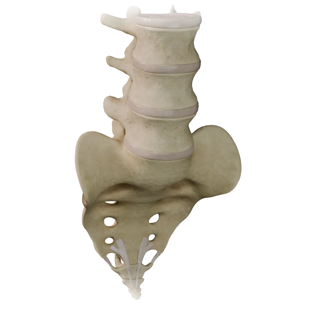
Cartilaginous joints (synchondroses) of the vertebral column
0/20

Fibrous joints of the vertebral column
0/20

Joints of thoracolumbar vertebral column
0/20
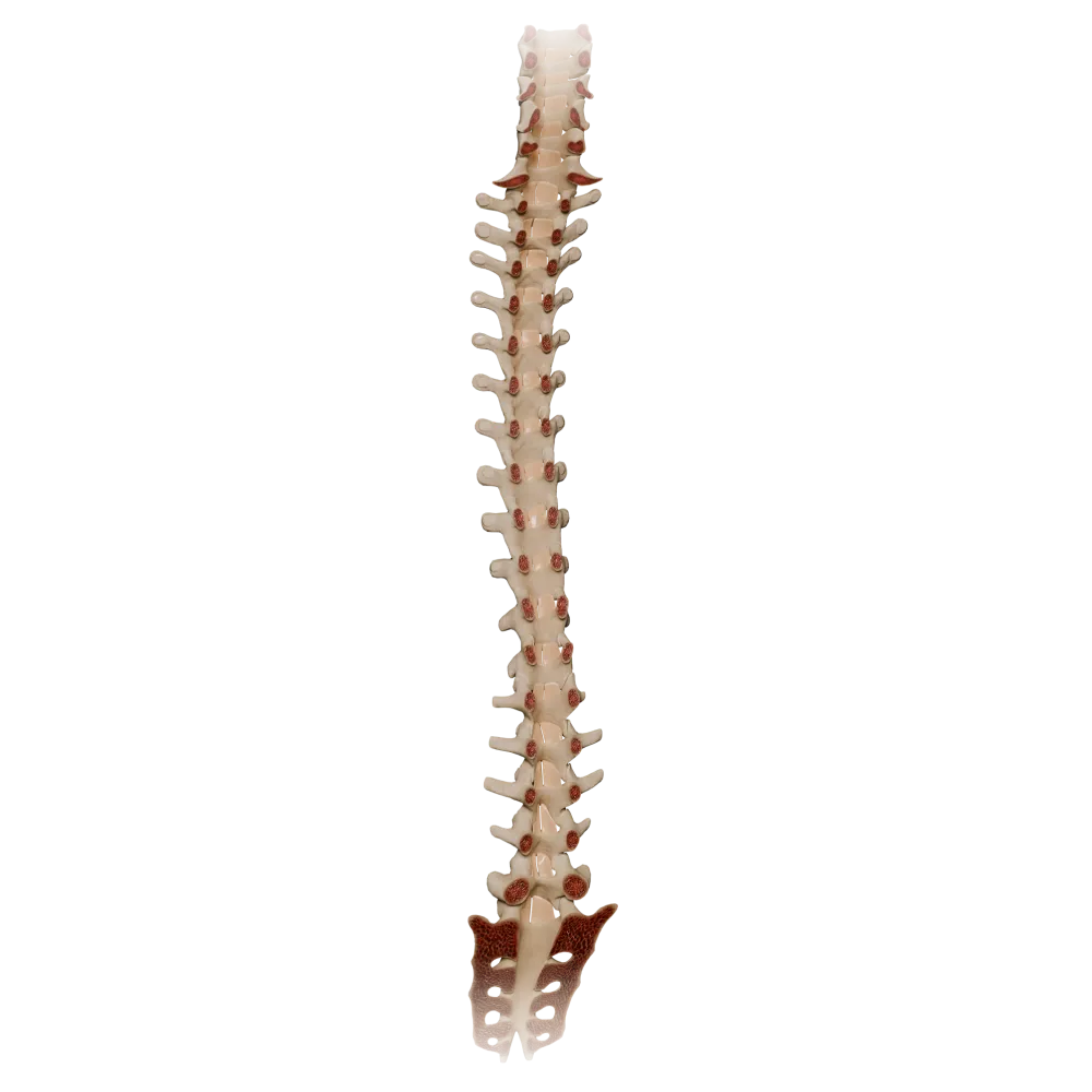
Ligamenta flava
0/20

Synovial joints of vertebral column
0/20
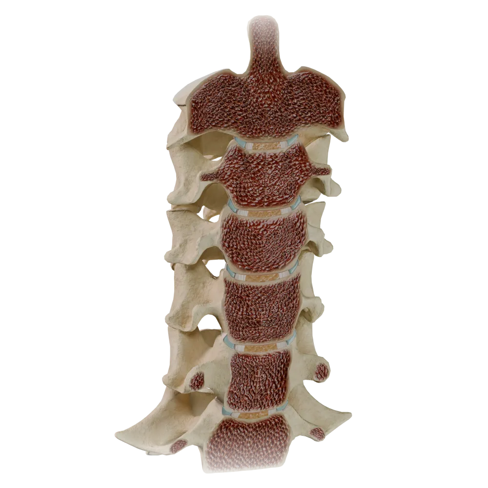
Uncovertebral joints
0/20
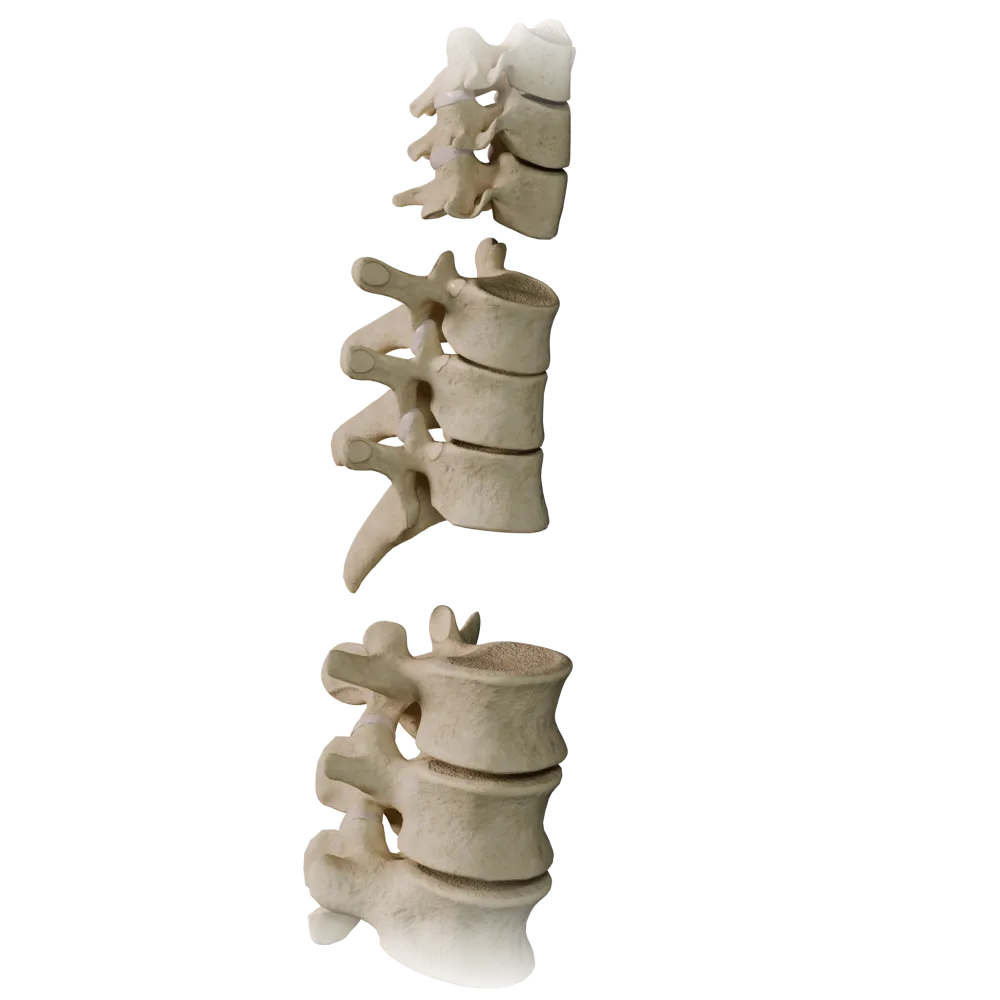
Zygapophyseal joints
0/20
Anatomy quiz on uncovertebral (uncinate) joints
Anatomy quiz on the uncovertebral (uncinate) joints of the cervical spine: structure, relationships, function.
1/20
bold
text
1. Between which anatomical structures are the uncovertebral joints formed?
-
Between the articular processes of adjacent cervical vertebrae
The uncovertebral joints (joints of Luschka) are formed between the processus uncinatus (uncinate process) on the superolateral margin of the body of the inferior vertebra and the corresponding beveled inferolateral margin of the body of the superior vertebra.
-
Between the uncinate processes of the body of the inferior vertebra and the beveled inferolateral margins of the body of the superior vertebra
The uncovertebral joints (joints of Luschka) are formed between the processus uncinatus (uncinate process) on the superolateral margin of the body of the inferior vertebra and the corresponding beveled inferolateral margin of the body of the superior vertebra.
-
Between the transverse processes of adjacent cervical vertebrae
The uncovertebral joints (joints of Luschka) are formed between the processus uncinatus (uncinate process) on the superolateral margin of the body of the inferior vertebra and the corresponding beveled inferolateral margin of the body of the superior vertebra.
-
Between the spinous processes and vertebral arches of adjacent cervical vertebrae
The uncovertebral joints (joints of Luschka) are formed between the processus uncinatus (uncinate process) on the superolateral margin of the body of the inferior vertebra and the corresponding beveled inferolateral margin of the body of the superior vertebra.
-
I find it difficult to answer
The uncovertebral joints (joints of Luschka) are formed between the processus uncinatus (uncinate process) on the superolateral margin of the body of the inferior vertebra and the corresponding beveled inferolateral margin of the body of the superior vertebra.
2. At which levels of the cervical spine are the uncovertebral joints located?
-
C1–C2
The uncovertebral joints are located at the C3–C7 levels because the vertebral bodies of C3–C7 have prominent uncinate processes (processus uncinati). They are absent at the C1–C2 level because of the distinctive anatomy of the atlas and axis.
-
C2–C4
The uncovertebral joints are located at the C3–C7 levels because the vertebral bodies of C3–C7 have prominent uncinate processes (processus uncinati). They are absent at the C1–C2 level because of the distinctive anatomy of the atlas and axis.
-
C3–C7
The uncovertebral joints are located at the C3–C7 levels because the vertebral bodies of C3–C7 have prominent uncinate processes (processus uncinati). They are absent at the C1–C2 level because of the distinctive anatomy of the atlas and axis.
-
C6–Th2
The uncovertebral joints are located at the C3–C7 levels because the vertebral bodies of C3–C7 have prominent uncinate processes (processus uncinati). They are absent at the C1–C2 level because of the distinctive anatomy of the atlas and axis.
-
I find it difficult to answer
The uncovertebral joints are located at the C3–C7 levels because the vertebral bodies of C3–C7 have prominent uncinate processes (processus uncinati). They are absent at the C1–C2 level because of the distinctive anatomy of the atlas and axis.
3. What is another name for the uncovertebral joints in the anatomical literature?
-
Schmorl's joints
The uncovertebral joints are traditionally termed the joints of Luschka (Luschka), after the German anatomist Hubert von Luschka, who described them in the nineteenth century. Schmorl's joints are herniations of the intervertebral discs into the vertebral bodies; the joints of Weitbrecht and Cruveilhier are components of the atlantoaxial articulation.
-
Joints of Luschka
The uncovertebral joints are traditionally termed the joints of Luschka (Luschka), after the German anatomist Hubert von Luschka, who described them in the nineteenth century. Schmorl's joints are herniations of the intervertebral discs into the vertebral bodies; the joints of Weitbrecht and Cruveilhier are components of the atlantoaxial articulation.
-
Joints of Weitbrecht
The uncovertebral joints are traditionally termed the joints of Luschka (Luschka), after the German anatomist Hubert von Luschka, who described them in the nineteenth century. Schmorl's joints are herniations of the intervertebral discs into the vertebral bodies; the joints of Weitbrecht and Cruveilhier are components of the atlantoaxial articulation.
-
Joints of Cruveilhier
The uncovertebral joints are traditionally termed the joints of Luschka (Luschka), after the German anatomist Hubert von Luschka, who described them in the nineteenth century. Schmorl's joints are herniations of the intervertebral discs into the vertebral bodies; the joints of Weitbrecht and Cruveilhier are components of the atlantoaxial articulation.
-
I find it difficult to answer
The uncovertebral joints are traditionally termed the joints of Luschka (Luschka), after the German anatomist Hubert von Luschka, who described them in the nineteenth century. Schmorl's joints are herniations of the intervertebral discs into the vertebral bodies; the joints of Weitbrecht and Cruveilhier are components of the atlantoaxial articulation.
4. What type of articular cartilage lines the articular surfaces of the uncovertebral joints?
-
Fibrocartilage
The articular surfaces of the uncovertebral joints are covered with hyaline cartilage, as are those of most true synovial joints. The presence of hyaline cartilage and a synovial membrane confirms their status as true joints.
-
Elastic cartilage
The articular surfaces of the uncovertebral joints are covered with hyaline cartilage, as are those of most true synovial joints. The presence of hyaline cartilage and a synovial membrane confirms their status as true joints.
-
Hyaline cartilage
The articular surfaces of the uncovertebral joints are covered with hyaline cartilage, as are those of most true synovial joints. The presence of hyaline cartilage and a synovial membrane confirms their status as true joints.
-
Mixed-type articular cartilage with a predominance of the fibrous component
The articular surfaces of the uncovertebral joints are covered with hyaline cartilage, as are those of most true synovial joints. The presence of hyaline cartilage and a synovial membrane confirms their status as true joints.
-
I find it difficult to answer
The articular surfaces of the uncovertebral joints are covered with hyaline cartilage, as are those of most true synovial joints. The presence of hyaline cartilage and a synovial membrane confirms their status as true joints.
5. What is the shape of the joint space of the uncovertebral joints?
-
Sagittally oriented and vertical
The joint space of the uncovertebral joints comprises a crescentic (semilunar) shape and is oriented in the frontal plane, corresponding to the anatomical configuration of the uncinate process and the beveled margin of the superior vertebra.
-
Crescent-shaped and oriented in the frontal plane
The joint space of the uncovertebral joints comprises a crescentic (semilunar) shape and is oriented in the frontal plane, corresponding to the anatomical configuration of the uncinate process and the beveled margin of the superior vertebra.
-
Horizontal and parallel to the endplates of the vertebral bodies
The joint space of the uncovertebral joints comprises a crescentic (semilunar) shape and is oriented in the frontal plane, corresponding to the anatomical configuration of the uncinate process and the beveled margin of the superior vertebra.
-
Oblique, at a 45° angle to the horizontal plane
The joint space of the uncovertebral joints comprises a crescentic (semilunar) shape and is oriented in the frontal plane, corresponding to the anatomical configuration of the uncinate process and the beveled margin of the superior vertebra.
-
I find it difficult to answer
The joint space of the uncovertebral joints comprises a crescentic (semilunar) shape and is oriented in the frontal plane, corresponding to the anatomical configuration of the uncinate process and the beveled margin of the superior vertebra.
6. What is located immediately laterally to the uncovertebral joint in the cervical spine?
-
Vertebral artery in the sulcus arteriae vertebralis
Immediately laterally to the uncovertebral joint, the vertebral artery and vertebral vein pass through the transverse foramen (foramen transversarium, canalis transversarius) together with the sympathetic periarterial plexus. This determines the important neurovascular relationships of this joint.
-
Vertebral artery and vertebral vein in the canalis transversarius
Immediately laterally to the uncovertebral joint, the vertebral artery and vertebral vein pass through the transverse foramen (foramen transversarium, canalis transversarius) together with the sympathetic periarterial plexus. This determines the important neurovascular relationships of this joint.
-
Anterior spinal artery
Immediately laterally to the uncovertebral joint, the vertebral artery and vertebral vein pass through the transverse foramen (foramen transversarium, canalis transversarius) together with the sympathetic periarterial plexus. This determines the important neurovascular relationships of this joint.
-
Internal carotid artery
Immediately laterally to the uncovertebral joint, the vertebral artery and vertebral vein pass through the transverse foramen (foramen transversarium, canalis transversarius) together with the sympathetic periarterial plexus. This determines the important neurovascular relationships of this joint.
-
I find it difficult to answer
Immediately laterally to the uncovertebral joint, the vertebral artery and vertebral vein pass through the transverse foramen (foramen transversarium, canalis transversarius) together with the sympathetic periarterial plexus. This determines the important neurovascular relationships of this joint.
7. Which structure is located medially to and separated from the uncovertebral joint?
-
Anterior longitudinal ligament
The intervertebral disc (discus intervertebralis), with its annulus fibrosus, is located medially to the uncovertebral joint. The uncinate processes effectively bound the disc laterally, preventing lateral protrusion of its nucleus pulposus.
-
Intervertebral disc and annulus fibrosus
The intervertebral disc (discus intervertebralis), with its annulus fibrosus, is located medially to the uncovertebral joint. The uncinate processes effectively bound the disc laterally, preventing lateral protrusion of its nucleus pulposus.
-
Posterior longitudinal ligament
The intervertebral disc (discus intervertebralis), with its annulus fibrosus, is located medially to the uncovertebral joint. The uncinate processes effectively bound the disc laterally, preventing lateral protrusion of its nucleus pulposus.
-
Interspinal ligament
The intervertebral disc (discus intervertebralis), with its annulus fibrosus, is located medially to the uncovertebral joint. The uncinate processes effectively bound the disc laterally, preventing lateral protrusion of its nucleus pulposus.
-
I find it difficult to answer
The intervertebral disc (discus intervertebralis), with its annulus fibrosus, is located medially to the uncovertebral joint. The uncinate processes effectively bound the disc laterally, preventing lateral protrusion of its nucleus pulposus.
8. Which movement in the cervical spine is primarily restricted by the uncovertebral joints?
-
Flexion and extension
The uncovertebral joints act as guides, restricting excessive lateral displacement (lateral translation) of the bodies of adjacent cervical vertebrae relative to one another. They thereby stabilize the intervertebral disc and the segment as a whole.
-
Lateral displacement and lateral translation of the vertebral bodies
The uncovertebral joints act as guides, restricting excessive lateral displacement (lateral translation) of the bodies of adjacent cervical vertebrae relative to one another. They thereby stabilize the intervertebral disc and the segment as a whole.
-
Axial rotation about the vertical axis
The uncovertebral joints act as guides, restricting excessive lateral displacement (lateral translation) of the bodies of adjacent cervical vertebrae relative to one another. They thereby stabilize the intervertebral disc and the segment as a whole.
-
Anteroposterior translation of the vertebral bodies
The uncovertebral joints act as guides, restricting excessive lateral displacement (lateral translation) of the bodies of adjacent cervical vertebrae relative to one another. They thereby stabilize the intervertebral disc and the segment as a whole.
-
I find it difficult to answer
The uncovertebral joints act as guides, restricting excessive lateral displacement (lateral translation) of the bodies of adjacent cervical vertebrae relative to one another. They thereby stabilize the intervertebral disc and the segment as a whole.
9. Based on the structure of the joint capsule and the presence of a synovial membrane, to which type of joint do the uncovertebral joints belong?
-
Synchondrosis
The uncovertebral joints are classified as synovial joints (diarthroses): they comprise a joint capsule, synovial membrane, joint cavity, and articular cartilage. This distinguishes them from symphyses and synchondroses, which do not comprise a joint cavity.
-
Symphysis (semi-joint)
The uncovertebral joints are classified as synovial joints (diarthroses): they comprise a joint capsule, synovial membrane, joint cavity, and articular cartilage. This distinguishes them from symphyses and synchondroses, which do not comprise a joint cavity.
-
Synovial joint (diarthrosis)
The uncovertebral joints are classified as synovial joints (diarthroses): they comprise a joint capsule, synovial membrane, joint cavity, and articular cartilage. This distinguishes them from symphyses and synchondroses, which do not comprise a joint cavity.
-
Synostosis
The uncovertebral joints are classified as synovial joints (diarthroses): they comprise a joint capsule, synovial membrane, joint cavity, and articular cartilage. This distinguishes them from symphyses and synchondroses, which do not comprise a joint cavity.
-
I find it difficult to answer
The uncovertebral joints are classified as synovial joints (diarthroses): they comprise a joint capsule, synovial membrane, joint cavity, and articular cartilage. This distinguishes them from symphyses and synchondroses, which do not comprise a joint cavity.
10. The entrance to the intervertebral foramen is located posterior to the uncovertebral joint. Which nerve exits through this foramen at the corresponding segmental level?
-
Sympathetic preganglionic nerve
The spinal nerve of the corresponding segment (nn. spinales C3–C8) exits through the intervertebral foramen (foramen intervertebrale), which is located posterolaterally to the uncovertebral joint. The uncovertebral joint forms the anteromedial wall of this foramen.
-
Spinal nerve of the corresponding segment
The spinal nerve of the corresponding segment (nn. spinales C3–C8) exits through the intervertebral foramen (foramen intervertebrale), which is located posterolaterally to the uncovertebral joint. The uncovertebral joint forms the anteromedial wall of this foramen.
-
Recurrent nerve of Luschka (sinuvertebral nerve)
The spinal nerve of the corresponding segment (nn. spinales C3–C8) exits through the intervertebral foramen (foramen intervertebrale), which is located posterolaterally to the uncovertebral joint. The uncovertebral joint forms the anteromedial wall of this foramen.
-
Accessory nerve (cranial nerve XI)
The spinal nerve of the corresponding segment (nn. spinales C3–C8) exits through the intervertebral foramen (foramen intervertebrale), which is located posterolaterally to the uncovertebral joint. The uncovertebral joint forms the anteromedial wall of this foramen.
-
I find it difficult to answer
The spinal nerve of the corresponding segment (nn. spinales C3–C8) exits through the intervertebral foramen (foramen intervertebrale), which is located posterolaterally to the uncovertebral joint. The uncovertebral joint forms the anteromedial wall of this foramen.
11. Which ligament of the cervical spine is immediately adjacent to the anterior surface of the cervical vertebral bodies, including the region of the uncinate processes?
-
Posterior longitudinal ligament
The anterior longitudinal ligament (ligamentum longitudinale anterius) is adjacent to the anterior surface of the cervical vertebral bodies and intervertebral discs, including the anterior surface of the bases of the uncinate processes. It is one of the anatomical landmarks used to describe the relationships of the uncovertebral region.
-
Nuchal ligament
The anterior longitudinal ligament (ligamentum longitudinale anterius) is adjacent to the anterior surface of the cervical vertebral bodies and intervertebral discs, including the anterior surface of the bases of the uncinate processes. It is one of the anatomical landmarks used to describe the relationships of the uncovertebral region.
-
Anterior longitudinal ligament
The anterior longitudinal ligament (ligamentum longitudinale anterius) is adjacent to the anterior surface of the cervical vertebral bodies and intervertebral discs, including the anterior surface of the bases of the uncinate processes. It is one of the anatomical landmarks used to describe the relationships of the uncovertebral region.
-
Ligamenta flava
The anterior longitudinal ligament (ligamentum longitudinale anterius) is adjacent to the anterior surface of the cervical vertebral bodies and intervertebral discs, including the anterior surface of the bases of the uncinate processes. It is one of the anatomical landmarks used to describe the relationships of the uncovertebral region.
-
I find it difficult to answer
The anterior longitudinal ligament (ligamentum longitudinale anterius) is adjacent to the anterior surface of the cervical vertebral bodies and intervertebral discs, including the anterior surface of the bases of the uncinate processes. It is one of the anatomical landmarks used to describe the relationships of the uncovertebral region.
12. From which embryonic tissues do the uncinate processes of the cervical vertebrae develop?
-
From the lateral plate mesoderm
Like all structures of the vertebral bodies, the uncinate processes develop from the sclerotomal portion of the somites, a medial derivative of the paravertebral mesoderm.
-
From the sclerotomal portion of the somites (paravertebral mesoderm)
Like all structures of the vertebral bodies, the uncinate processes develop from the sclerotomal portion of the somites, a medial derivative of the paravertebral mesoderm.
-
From the neural crest
Like all structures of the vertebral bodies, the uncinate processes develop from the sclerotomal portion of the somites, a medial derivative of the paravertebral mesoderm.
-
From the visceral mesoderm
Like all structures of the vertebral bodies, the uncinate processes develop from the sclerotomal portion of the somites, a medial derivative of the paravertebral mesoderm.
-
I find it difficult to answer
Like all structures of the vertebral bodies, the uncinate processes develop from the sclerotomal portion of the somites, a medial derivative of the paravertebral mesoderm.
13. At what age do the uncovertebral joints attain their definitive morphology as true synovial joints?
-
By the time of birth (intrauterine)
The uncovertebral joints develop postnatally: the uncinate processes begin to develop after birth and attain complete formation, with development of a synovial cavity, by 9–14 years of age. In neonates, these joints are absent as differentiated synovial structures.
-
By 6–8 years of age
The uncovertebral joints develop postnatally: the uncinate processes begin to develop after birth and attain complete formation, with development of a synovial cavity, by 9–14 years of age. In neonates, these joints are absent as differentiated synovial structures.
-
By 9–14 years of age
The uncovertebral joints develop postnatally: the uncinate processes begin to develop after birth and attain complete formation, with development of a synovial cavity, by 9–14 years of age. In neonates, these joints are absent as differentiated synovial structures.
-
By 18–25 years of age
The uncovertebral joints develop postnatally: the uncinate processes begin to develop after birth and attain complete formation, with development of a synovial cavity, by 9–14 years of age. In neonates, these joints are absent as differentiated synovial structures.
-
I find it difficult to answer
The uncovertebral joints develop postnatally: the uncinate processes begin to develop after birth and attain complete formation, with development of a synovial cavity, by 9–14 years of age. In neonates, these joints are absent as differentiated synovial structures.
14. Which nerve provides innervation to the capsule of the uncovertebral joint?
-
Ventral branch of the spinal nerve
The capsule of the uncovertebral joint is innervated by the recurrent meningeal branch of the spinal nerve, which enters the intervertebral foramen.
-
Recurrent meningeal nerve, nerve of Luschka
The capsule of the uncovertebral joint is innervated by the recurrent meningeal branch of the spinal nerve, which enters the intervertebral foramen.
-
Dorsal branch of the spinal nerve
The capsule of the uncovertebral joint is innervated by the recurrent meningeal branch of the spinal nerve, which enters the intervertebral foramen.
-
Phrenic nerve
The capsule of the uncovertebral joint is innervated by the recurrent meningeal branch of the spinal nerve, which enters the intervertebral foramen.
-
I find it difficult to answer
The capsule of the uncovertebral joint is innervated by the recurrent meningeal branch of the spinal nerve, which enters the intervertebral foramen.
15. What is the normal shape of the uncinate process (processus uncinatus) when the body of a cervical vertebra is viewed anteriorly?
-
Rounded, in the form of a tubercle
The processus uncinatus comprises a hook-shaped (semilunar) configuration: it is directed medially and superiorly from the superolateral margin of the vertebral body, embracing the inferolateral margin of the body of the superior vertebra. This configuration determines its function as a guiding structure.
-
Pyramidal, with a broad base
The processus uncinatus comprises a hook-shaped (semilunar) configuration: it is directed medially and superiorly from the superolateral margin of the vertebral body, embracing the inferolateral margin of the body of the superior vertebra. This configuration determines its function as a guiding structure.
-
Hook-shaped (semilunar), directed medially and superiorly
The processus uncinatus comprises a hook-shaped (semilunar) configuration: it is directed medially and superiorly from the superolateral margin of the vertebral body, embracing the inferolateral margin of the body of the superior vertebra. This configuration determines its function as a guiding structure.
-
Flat, with a broad horizontal superior surface
The processus uncinatus comprises a hook-shaped (semilunar) configuration: it is directed medially and superiorly from the superolateral margin of the vertebral body, embracing the inferolateral margin of the body of the superior vertebra. This configuration determines its function as a guiding structure.
-
I find it difficult to answer
The processus uncinatus comprises a hook-shaped (semilunar) configuration: it is directed medially and superiorly from the superolateral margin of the vertebral body, embracing the inferolateral margin of the body of the superior vertebra. This configuration determines its function as a guiding structure.
16. At which cervical vertebral levels are the uncinate processes best developed and most anatomically prominent?
-
C3
The uncinate processes are most prominent at the C5–C6 levels, the segments with the greatest range of motion in the cervical spine. The increased load at these levels results in more robust development of the guiding structures of the uncovertebral region.
-
C4
The uncinate processes are most prominent at the C5–C6 levels, the segments with the greatest range of motion in the cervical spine. The increased load at these levels results in more robust development of the guiding structures of the uncovertebral region.
-
C5–C6
The uncinate processes are most prominent at the C5–C6 levels, the segments with the greatest range of motion in the cervical spine. The increased load at these levels results in more robust development of the guiding structures of the uncovertebral region.
-
C7
The uncinate processes are most prominent at the C5–C6 levels, the segments with the greatest range of motion in the cervical spine. The increased load at these levels results in more robust development of the guiding structures of the uncovertebral region.
-
I find it difficult to answer
The uncinate processes are most prominent at the C5–C6 levels, the segments with the greatest range of motion in the cervical spine. The increased load at these levels results in more robust development of the guiding structures of the uncovertebral region.
17. What is the structural nature of the joint capsule of the uncovertebral joint?
-
A thick fibrous capsule reinforced by several ligaments
The joint capsule of the uncovertebral joints is thin and lax, with a well-developed synovial membrane that produces synovial fluid. The absence of robust ligamentous reinforcement accounts for the relative mobility of these joints.
-
A thin, lax capsule with a well-developed synovial membrane
The joint capsule of the uncovertebral joints is thin and lax, with a well-developed synovial membrane that produces synovial fluid. The absence of robust ligamentous reinforcement accounts for the relative mobility of these joints.
-
A fibrocartilaginous rim without a synovial lining
The joint capsule of the uncovertebral joints is thin and lax, with a well-developed synovial membrane that produces synovial fluid. The absence of robust ligamentous reinforcement accounts for the relative mobility of these joints.
-
A bilayered capsule with an inner layer of fibrocartilage
The joint capsule of the uncovertebral joints is thin and lax, with a well-developed synovial membrane that produces synovial fluid. The absence of robust ligamentous reinforcement accounts for the relative mobility of these joints.
-
I find it difficult to answer
The joint capsule of the uncovertebral joints is thin and lax, with a well-developed synovial membrane that produces synovial fluid. The absence of robust ligamentous reinforcement accounts for the relative mobility of these joints.
18. What functional role do the uncinate processes perform in relation to the intervertebral disc in the cervical spine?
-
They increase disc compression under axial loading
The uncinate processes are located lateral to the intervertebral disc and form lateral barriers that mechanically prevent lateral displacement and protrusion of the annulus fibrosus. They thereby protect the adjacent neurovascular structures from compression by the disc.
-
They form lateral “barriers” that restrict lateral disc protrusion
The uncinate processes are located lateral to the intervertebral disc and form lateral barriers that mechanically prevent lateral displacement and protrusion of the annulus fibrosus. They thereby protect the adjacent neurovascular structures from compression by the disc.
-
They provide disc nutrition through diffusion of synovial fluid
The uncinate processes are located lateral to the intervertebral disc and form lateral barriers that mechanically prevent lateral displacement and protrusion of the annulus fibrosus. They thereby protect the adjacent neurovascular structures from compression by the disc.
-
They transmit the load directly to the vertebral bodies, bypassing the disc
The uncinate processes are located lateral to the intervertebral disc and form lateral barriers that mechanically prevent lateral displacement and protrusion of the annulus fibrosus. They thereby protect the adjacent neurovascular structures from compression by the disc.
-
I find it difficult to answer
The uncinate processes are located lateral to the intervertebral disc and form lateral barriers that mechanically prevent lateral displacement and protrusion of the annulus fibrosus. They thereby protect the adjacent neurovascular structures from compression by the disc.
19. Which term is used in Terminologia Anatomica to designate the uncinate process of a cervical vertebra?
-
Cervical spinous process
According to Terminologia Anatomica, the uncinate process of a cervical vertebra is designated by the term processus uncinatus (from Latin uncus, meaning hook). Processus mamillaris pertains to the lumbar vertebrae; processus articularis is the articular process; processus spinosus is the spinous process.
-
Lateral articular process
According to Terminologia Anatomica, the uncinate process of a cervical vertebra is designated by the term processus uncinatus (from Latin uncus, meaning hook). Processus mamillaris pertains to the lumbar vertebrae; processus articularis is the articular process; processus spinosus is the spinous process.
-
Uncinate process
According to Terminologia Anatomica, the uncinate process of a cervical vertebra is designated by the term processus uncinatus (from Latin uncus, meaning hook). Processus mamillaris pertains to the lumbar vertebrae; processus articularis is the articular process; processus spinosus is the spinous process.
-
Mammillary process
According to Terminologia Anatomica, the uncinate process of a cervical vertebra is designated by the term processus uncinatus (from Latin uncus, meaning hook). Processus mamillaris pertains to the lumbar vertebrae; processus articularis is the articular process; processus spinosus is the spinous process.
-
I find it difficult to answer
According to Terminologia Anatomica, the uncinate process of a cervical vertebra is designated by the term processus uncinatus (from Latin uncus, meaning hook). Processus mamillaris pertains to the lumbar vertebrae; processus articularis is the articular process; processus spinosus is the spinous process.
20. Which type of cells produces the synovial fluid of the uncovertebral joint?
-
Chondrocytes of the articular cartilage
Synovial fluid is produced by two types of synoviocytes: type A (macrophage-like, phagocytic) and type B (fibroblast-like, secretory), which are cells of the synovial membrane of the joint capsule. This applies to all synovial joints, including the uncovertebral joints.
-
Type A and type B synoviocytes (phagocytic and secretory cells of the synovial membrane)
Synovial fluid is produced by two types of synoviocytes: type A (macrophage-like, phagocytic) and type B (fibroblast-like, secretory), which are cells of the synovial membrane of the joint capsule. This applies to all synovial joints, including the uncovertebral joints.
-
Fibrocytes of the joint capsule
Synovial fluid is produced by two types of synoviocytes: type A (macrophage-like, phagocytic) and type B (fibroblast-like, secretory), which are cells of the synovial membrane of the joint capsule. This applies to all synovial joints, including the uncovertebral joints.
-
Osteoblasts of the subchondral bone
Synovial fluid is produced by two types of synoviocytes: type A (macrophage-like, phagocytic) and type B (fibroblast-like, secretory), which are cells of the synovial membrane of the joint capsule. This applies to all synovial joints, including the uncovertebral joints.
-
I find it difficult to answer
Synovial fluid is produced by two types of synoviocytes: type A (macrophage-like, phagocytic) and type B (fibroblast-like, secretory), which are cells of the synovial membrane of the joint capsule. This applies to all synovial joints, including the uncovertebral joints.
Retake this quiz?
Your current progress will be reset.
