Odontogenic Sinusitis: Classification, Etiology, Clinical Presentation, Diagnosis, and Treatment
A detailed overview of odontogenic maxillary sinusitis: causes, symptoms of acute and chronic inflammation, diagnosis, and modern treatment approaches.


Anesthesia
Pain management and sedation techniques

Angiology
Arterial and venous pathologies

Cardiology
Acquired and congenital heart diseases

Dentistry
Diseases of teeth, gums, and the oral cavity

Dermatology
Disorders of the skin and subcutaneous tissue

Embryology
Stages of embryogenesis and congenital anomalies

Endocrinology
Disorders of the glands and hormonal imbalance

Gastroenterology
Stomach, intestinal, and digestive diseases

Gynecology
Diseases of female reproductive organs

Hematology
Hematopoiesis and blood-related disorders

Hepatology
Liver, gallbladder, and biliary tract diseases

Histology
Microscopic tissue and cell structures

Infectious diseases
Bacterial, viral, and parasitic infections

Neurology
Brain, spinal cord, and peripheral nerve disorders

Obstetrics
Pregnancy complications and abnormal fetal positions

Oncology
Cancer types, benign and malignant tumors

Ophthalmology
Conditions affecting the eyes and vision

Orthopedics
Bone, joint, and soft tissue disorders

Otorhinolaryngology
Ear, nose, and throat diseases

Pediatrics
Child health, development, and clinical conditions

Physiology
Biological processes within organs and systems

Pulmonology
Lung and respiratory tract diseases

Traumatology
Acute injuries and musculoskeletal trauma

Urology
Urinary tract and male reproductive disorders
Anesthesia
Pain management and sedation techniques
Angiology
Arterial and venous pathologies
Cardiology
Acquired and congenital heart diseases
Dentistry
Diseases of teeth, gums, and the oral cavity
Dermatology
Disorders of the skin and subcutaneous tissue
Embryology
Stages of embryogenesis and congenital anomalies
Endocrinology
Disorders of the glands and hormonal imbalance
Gastroenterology
Stomach, intestinal, and digestive diseases
Gynecology
Diseases of female reproductive organs
Hematology
Hematopoiesis and blood-related disorders
Hepatology
Liver, gallbladder, and biliary tract diseases
Histology
Microscopic tissue and cell structures
Infectious diseases
Bacterial, viral, and parasitic infections
Neurology
Brain, spinal cord, and peripheral nerve disorders
Obstetrics
Pregnancy complications and abnormal fetal positions
Oncology
Cancer types, benign and malignant tumors
Ophthalmology
Conditions affecting the eyes and vision
Orthopedics
Bone, joint, and soft tissue disorders
Otorhinolaryngology
Ear, nose, and throat diseases
Pediatrics
Child health, development, and clinical conditions
Physiology
Biological processes within organs and systems
Pulmonology
Lung and respiratory tract diseases
Traumatology
Acute injuries and musculoskeletal trauma
Urology
Urinary tract and male reproductive disorders
Specialties
AnesthesiaAngiologyCardiologyDentistryDermatologyEmbryologyEndocrinologyGastroenterologyGynecologyHematologyHepatologyHistologyInfectious diseasesNeurologyObstetricsOncologyOphthalmologyOrthopedicsOtorhinolaryngologyPediatricsPhysiologyPulmonologyTraumatologyUrology
This article is for informational purposes only
The content on this website, including text, graphics, and other materials, is provided for informational purposes only. It is not intended as advice or guidance. Regarding your specific medical condition or treatment, please consult your healthcare provider.
External ear injuries include mechanical damage to the auricle and external auditory canal. These conditions vary by depth of lesion, etiology, and mechanism of injury.
Superficial external ear injuries:
3D models of superficial external ear injuries:
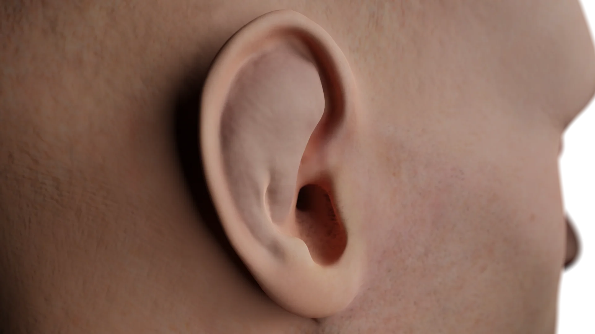 Othematoma of the external ear
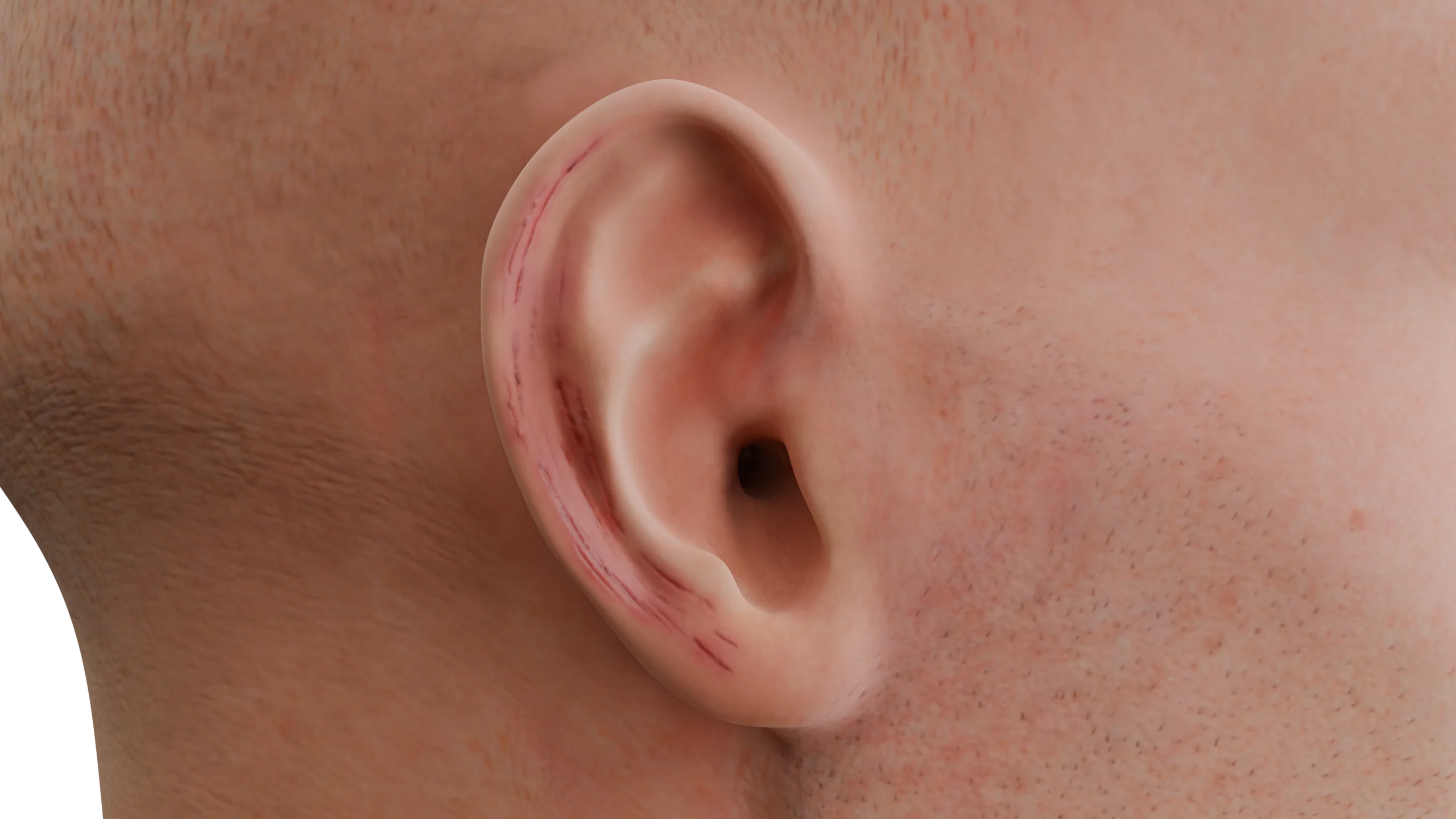 Auricular abrasions
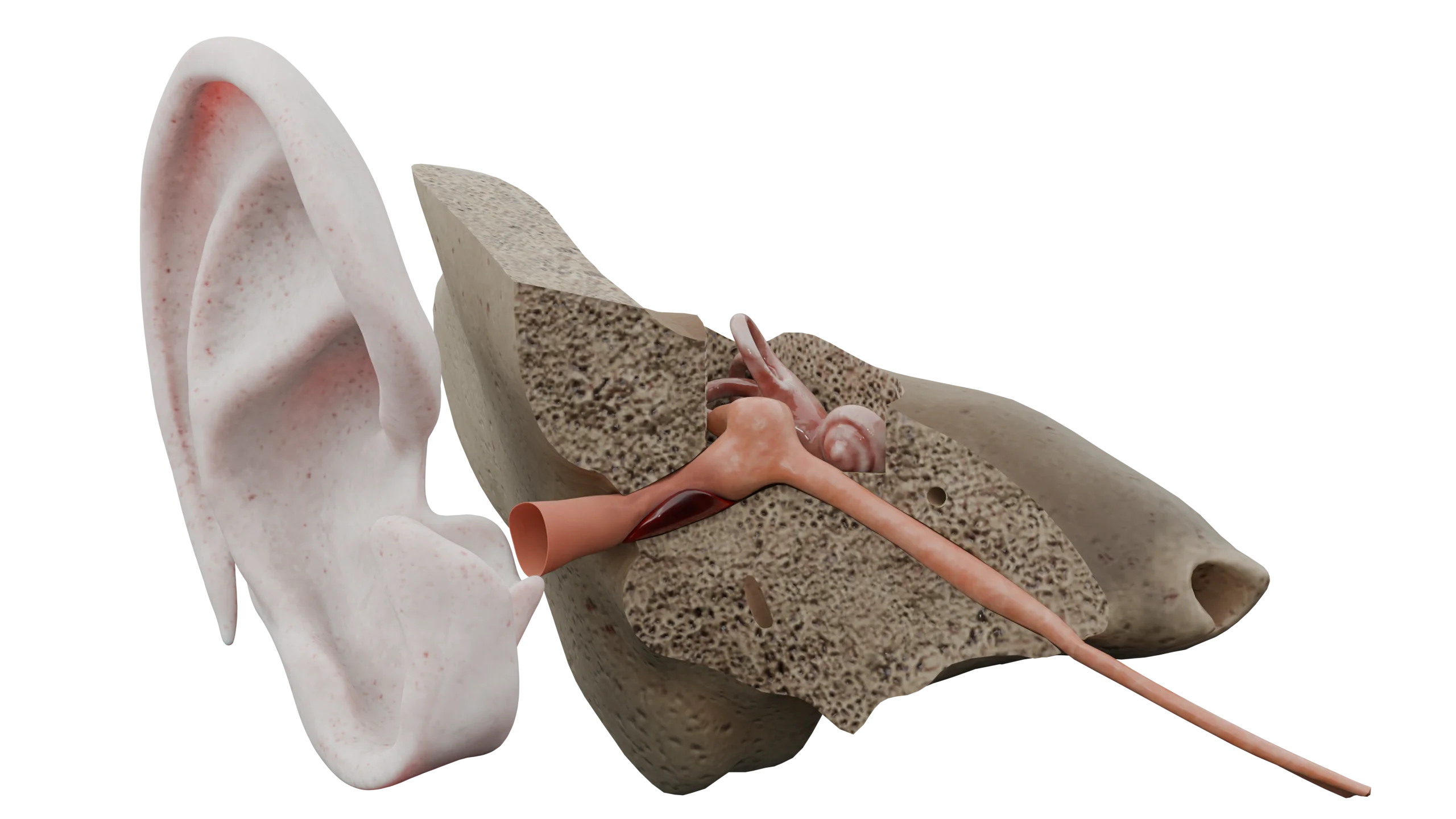 Hematoma of the external auditory canal
Deep external ear injuries:
3D models of deep external ear injuries:
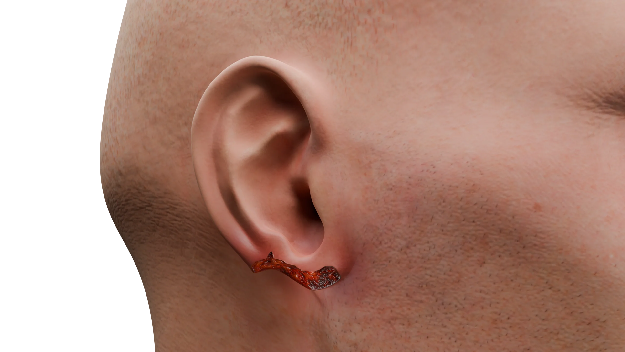 Avulsion of the ear lobule
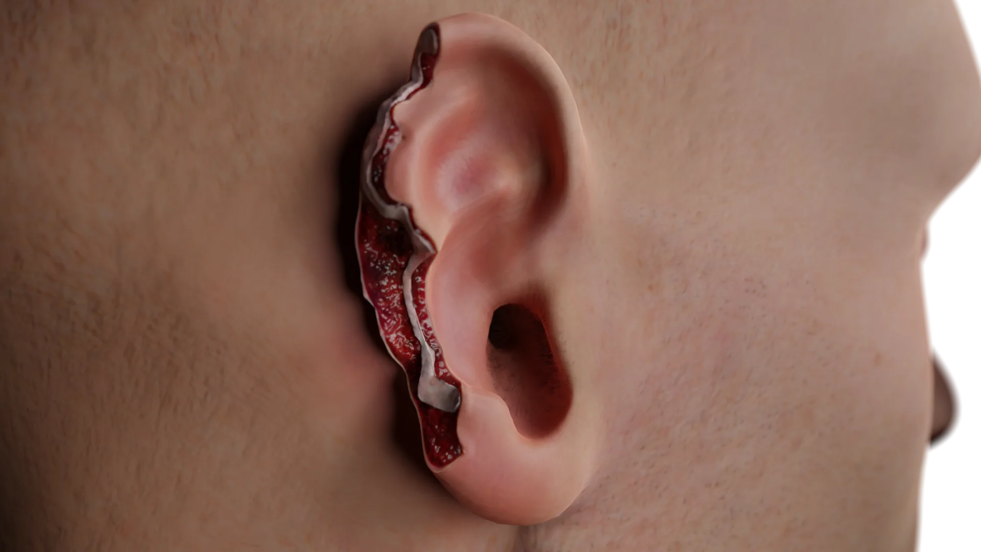 Segmental auricular avulsion
Burns of the auricle:
Auricular frostbites:
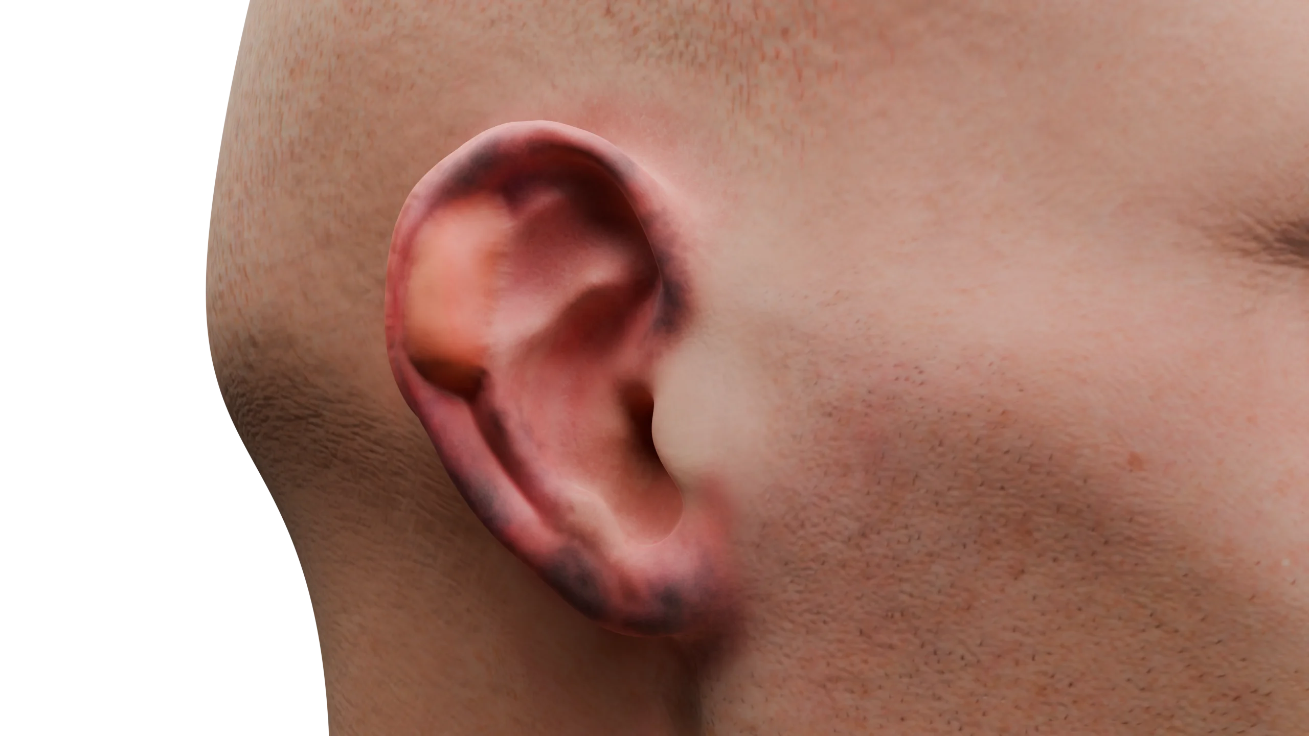
Superficial and deep injuries of the auricle occur due to mechanical impact (trauma, blows, falls, bites, car accidents).
Injuries to the auditory canal often occur during attempts at self-cleaning of earwax or the entry of foreign objects.
The extent of damage depends on the force of impact and the nature of the injury. Burns and frostbites occur due to thermal damaging factors (fire, cold).
Mechanical and thermal factors impact the auricle, causing tissue damage of varying depths.
Othematoma occurs after blunt trauma with significant force. In this situation, the walls of the vessels located between the cartilage and the perichondrium rupture, forming a limited blood accumulation that deforms the outer appearance of the auricle. The auricle itself gains a bluish tint and swells.
A contusion occurs with less severe blunt trauma. It is characterized by concussion of the soft tissues. Smaller vessels are more severely damaged, skin integrity is not compromised, and hyperemia and infiltration of the auricle is noted.
Auricular and external acoustic meatus abrasions are characterized by partial damage to the epidermis. They quickly become covered with hemorrhagic crusts, and slight bleeding may occur.
A hematoma of the auditory canal is characterized by damage to the subcutaneous vessels. This results in localized blood accumulation that protrudes and narrows the canal’s lumen, with varying degrees of compromise. The skin at the hematoma site becomes cyanotic.
Deep injuries of the auricle occur from various causes: sports and household injuries, workplace incidents (improper handling of tools), animal or human bites, falls, as well as careless wearing of piercings.
Auricular avulsions in daily life are rare; they typically occur due to traffic accidents, combat sports (e.g., boxing), or street fights. This type of injury is characterized by compromised skin integrity, damage to the underlying tissues, subcutaneous fat, vessels, sometimes cartilage, as well as pronounced bleeding and swelling.
Burns occur under extreme high temperatures, open flames, hot objects, and substances.
Auricular burns may also damage the external auditory canal.
Auricular frostbites frequently happen in extremely low temperatures, as the auricles are among the body’s most protruding parts.
With superficial ear injuries, acute pain occurs at the moment of trauma, with subsequent discomfort and dull pain. The auricle swells, changes color, and then gradually returns to its normal appearance during recovery. When abrasions compromise the skin integrity, scant bleeding occurs; later, they become covered with hemorrhagic crusts and heal under a scab.
Othematoma is characterized by pronounced pain and alteration in the appearance of the auricle. It protrudes, fluctuates at the site of effusion, more often on the outer surface, in the area of the scaphoid and triangular fossae. The skin acquires a cyanotic color. Without appropriate treatment and drainage of the hematoma, the auricle often becomes misshapen due to cartilage transformation resembling ‘cauliflower ear’ or ‘boxer’s ear’. If a large hematoma occurs in the ear canal, obstructing the lumen, conductive hearing loss may occur.
Deep auricular injuries are characterized by severe tenderness and profuse bleeding. The wound edges gape, revealing underlying tissues, and in some cases, cartilage or bone. The configuration of the wound edges varies depending on the injuring factor, with foreign objects, dirt, and earring remnants possible inside.
Burns of the auricle manifest differently according to the stage, as described above. It’s noteworthy that isolated ear burns are extremely rare, often accompanying injuries to the scalp, face, neck, and other body parts. Severe burn areas can lead to burn disease development. Common symptoms include severe tenderness, although bleeding is absent. In third and fourth-degree burns, the bottom of the wound is painless, and a necrotic scab forms.
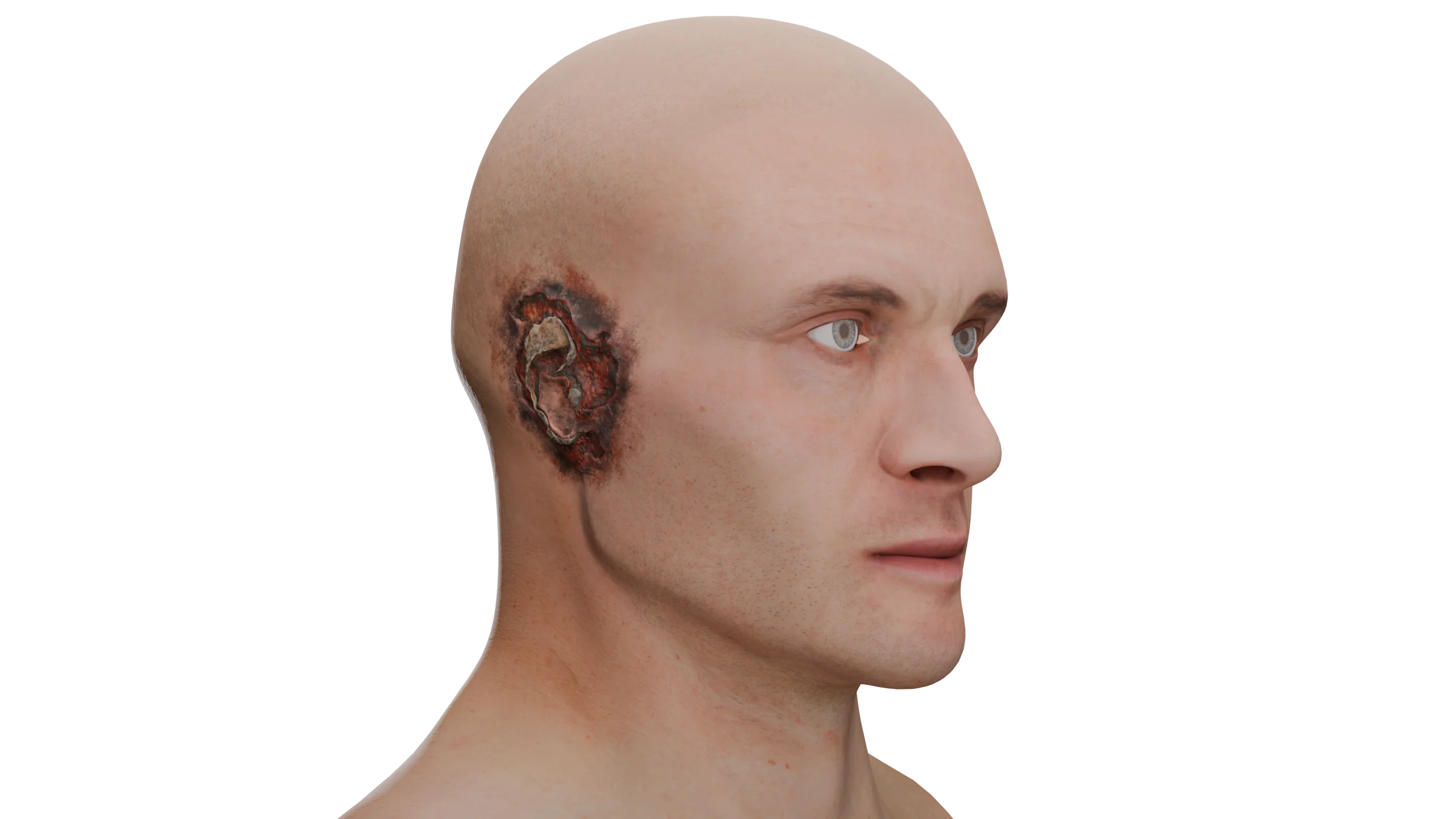
Clinical changes in frostbite have also been described earlier. A characteristic feature is the absence of pain during frostbite. Discomfort arises upon warming the affected tissues: itching, burning, significant pain, numbness, or paresthesia may occur.
For the diagnosis of traumatic injuries to the auricle and external auditory canal, in most cases, collecting the medical history and examination are sufficient. Sometimes an X-ray or CT of the temporal bone and temporomandibular joint is conducted to assess the extent of injury.
Find more scientifically accurate content on our social media



Auricular and acoustic meatus abrasions are treated with antiseptic solutions. If infection occurs, antibiotic ointments may be used. Under the crusts, wounds rapidly epithelize and heal.
Otohematoma needs to be punctured under sterile conditions, drained, and a compression dressing applied, which is sutured to the auricle for large hematomas. Systemic antibacterial drugs are prescribed to prevent secondary infection and development of perichondritis.
Ear canal hematomas are also drained, and the canal is tightly packed with ear tampons that are soaked with antiseptic solutions daily.
For deep ear injuries, primary surgical treatment is performed, and the wound edges are sutured. In case of partial avulsion, non-viable tissues are excised as sparingly as possible, and the cartilage, subcutaneous fat, and skin are sutured separately.
For complete auricle avulsions, the viability of the amputated section is assessed. It is recommended that no more than 6 hours pass since the trauma and that the avulsed auricle segment is kept cold. If all necessary conditions are met, replantation is performed, with prescribed antibiotic therapy, thromboembolic prophylaxis, and daily dressings.
Burns are washed, remnants of burnt clothing and hair are removed, non-viable tissues are excised, then the wounds are treated with antiseptic solutions, cooled, and covered with sterile dressings. Before dressing changes, it is mandatory to adequately anesthetize the patient.
For 2nd and 3rd-degree burns, large blisters are opened and excised; for 3rd and 4th-degree burns, the scab is excised to expose viable tissues. During daily dressings, antimicrobial and wound-healing ointments are used.
To prevent cicatricial stenosis of the external auditory canal, it must be tightly tamponaded with sterile dressings and daily soaked with antiseptic solutions. If bacterial infection occurs, systemic broad-spectrum antibacterial therapy is prescribed based on sensitivity. Patients with 3B or 4th-degree burns or extensive burns are admitted to surgical or burn departments for suitable treatment. Skin grafting is performed later if necessary.
The treatment scheme for frostbite corresponds to that for burns, but initially, the frostbitten body part needs to be warmed. The victim is placed in a warm room and given plenty of warm drinks. The auricle should be gradually rewarmed without harsh methods or rubbing. Sterile warmed solutions of furacilin, saline, warm compresses, and gentle massage with warm clean hands can be used for this purpose.
In case of severe pain, the patient needs adequate analgesia. Damaged tissues are rinsed and treated with antiseptic solutions. If blisters and scabs are present, necrotic tissues are excised and dressings with antimicrobial and wound-healing ointments are applied.
For 3rd and 4th-degree frostbite, systemic antibacterial therapy is prescribed. If indicated, these patients are also admitted to surgical or burn departments. Over time, neuropathy may develop, accompanied by numbness and cold sensitivity.
All patients with wounds, burns, or frostbite receive urgent tetanus immunization; rabies prophylaxis is administered in cases of bites.
1. What complications may arise from ear injuries?
2. How can complications after an ear injury be prevented?
3. When is surgical intervention necessary?
4. Is it possible to restore the auricle after injury?
References
1.
VOKA 3D Anatomy & Pathology – Complete Anatomy and Pathology 3D Atlas. VOKA 3D Anatomy & Pathology.
Available from: https://catalog.voka.io/
2.
Sclafani AP, Dyleski RA, Pitman MJ, Schantz SP. Total otolaryngology—head and neck surgery. New York: Thieme Medical Publishers; 2015. ISBN: 978-1-60406-646-3.
3.
Behrbohm H, Kaschke O, Nawka T, Swift A. Bolezni ukha, gorla i nosa [Ear, nose, and throat diseases]. 2nd ed. Moscow: MEDpress-inform; 2016. 776 p. [In Russian.] ISBN 978-5-00030-322-1.
4.
Hohman MH, Jamal Z, Krogmann RJ, et al. Auricular Hematoma. [Updated 2024 May 1]. In: StatPearls [Internet]. Treasure Island (FL): StatPearls Publishing; 2025 Jan-.
Available from: https://www.ncbi.nlm.nih.gov/books/NBK531499/
5.
Trauma Management of the Auricle. Facial Plast Surg. 2015 Aug;31(4):382-5. doi: 10.1055/s-0035-1562882. Epub 2015 Sep 15. PMID: 26372713.
6.
Kraenzlin FS, Mushin OP, Ayazi S, Loree J, Bell DE. Epidemiology and Outcomes of Auricular Burn Injuries. J Burn Care Res. 2018 Apr 20;39(3):326-331. doi: 10.1097/BCR.0000000000000586. PMID: 28557871.
Make VOKA your preferred source
See more VOKA articles in Google Search
Table of Contents









Summarize article with AI
Choose your preferable AI assistant:
Link successfully copied to clipboard
Thank you!
Your message is sent!
Our experts will contact you shortly. If you have any additional questions, please contact us at info@voka.io