Chronic Laryngitis: Etiology, Classification, Diagnosis and Treatment
Danata A.Otorhinolaryngologist, MD
13 min read·July 23, 2025
This article is for informational purposes only
The content on this website, including text, graphics, and other materials, is provided for informational purposes only. It is not intended as advice or guidance. Regarding your specific medical condition or treatment, please consult your healthcare provider.
Chronic laryngitis belongs to a group of persistent inflammatory laryngeal conditions lasting longer than 3 weeks (primarily develops due to prolonged exposure to various irritants). Forms of the disease vary by etiology (cause) and the nature of pathomorphological changes in the mucosa — ranging from catarrhal inflammation to hyperplasia or atrophy. The precise identification of the form defines the treatment strategy and prognosis.
Classification
The primary clinical, morphological, and etiological forms of chronic laryngitis include:
chronic catarrhal laryngitis is the most common form of chronic laryngeal inflammation, characterized by persistent, moderate inflammation of the mucous membrane without atrophy or hypertrophy;
reflux laryngitis (posterior laryngitis) is an inflammation of the laryngeal mucosa caused by the reflux of stomach contents into the laryngopharynx;
atrophic laryngitis is a chronic inflammatory condition of the larynx characterized by thinning and atrophy of the mucous membrane;
laryngomycosis is a fungal infection of the larynx;
chronic hyperplastic laryngitis is a type of chronic laryngitis characterized by excessive growth of laryngeal tissue.
Chronic Catarrhal Laryngitis
Etiology of Chronic Catarrhal Laryngitis
The most common causes of chronic catarrhal laryngitis are smoking, excessive vocal strain, and allergens. The inflammatory process may spread from the nasopharynx or oropharynx, or from the lower respiratory tract. Recurrent sinusitis, rhinitis, tracheitis, and bronchitis may contribute to chronic laryngitis.
In patients with chronic laryngitis, the following bacteria are present: Staphylococcus aureus, Haemophilus influenzae, Candida albicans, Moraxella nonliquefaciens, Neisseria meningitidis, Streptococcus pneumoniae, Lactobacillus spp, Helicobacter pylori.
Anatomy of Chronic Catarrhal Laryngitis
Chronic catarrhal laryngitis arises as a result of exposure to irritants. The laryngeal epithelium undergoes hyperplasia, with submucosal edema and catarrh observed. The mucosa becomes smooth and hyperemic. The vocal folds are also hyperemic, thickened, and fail to completely close during phonation; mucus is present in the piriform sinuses.
Clinical Manifestations of Chronic Catarrhal Laryngitis
Leading symptoms of chronic catarrhal laryngitis include:
Hoarseness and changes in vocal timbre;
Rapid vocal fatigue;
Dry cough;
Throat tickling.
Diagnosis of Chronic Catarrhal Laryngitis
Diagnosis is based on anamnesis and clinical examination. Depending on the clinic’s equipment, indirect laryngoscopy or flexible video laryngoscopy is performed.
Treatment of Chronic Catarrhal Laryngitis
The first and most important step in treatment is identifying and removing the causal factor of the inflammation. Smoking cessation and reducing vocal strain are recommended. If the infection extends from the upper or lower airway, it is imperative to manage the primary source.
To control inflammation, inhalations with saline solutions, antibacterial agents, or glucocorticosteroids as needed are advised.
To alleviate inflammation, inhalations with saline solutions, antibiotics, and glucocorticosteroids, if necessary, are advised.
Reflux Laryngitis
Etiology of Reflux Laryngitis
In reflux laryngitis, the etiological factor is cardia insufficiency, causing hydrochloric acid reflux into the upper airway.
Anatomy of Reflux Laryngitis
Reflux laryngitis is characterized by hyperemia and edema in the interariespace, with mucus accumulation in the piriform sinuses.
Possible heartburn (although not a definitive symptom).
Symptoms worsen at night and during sleep.
Diagnosis of Reflux Laryngitis
Diagnosis is based on anamnesis and clinical examination. Indirect laryngoscopy or video laryngoscopy is performed depending on the equipment. In reflux laryngitis, consulting a gastroenterologist, with esophagogastroduodenoscopy and esophageal pH monitoring, is essential.
Treatment of Reflux Laryngitis
In reflux laryngitis, prescribed therapy by a gastroenterologist aims to reduce gastric acid secretion. To reduce laryngeal inflammation, saline inhalations, antibiotics, and glucocorticosteroids, if necessary, are indicated.
Atrophic Laryngitis
Etiology of Atrophic Laryngitis
Atrophic laryngitis is more commonly seen in elderly patients, during hormonal changes (such as menopause), or after long-term exposure to harmful working conditions (high dustiness, harmful chemicals).
Anatomy of Atrophic Laryngitis
In atrophic laryngitis, the number of mucous glands in the larynx diminishes. The mucosa appears parchment-like, thin, dull, and covered with thick, viscous mucus. The vocal folds are also thin and fail to close completely when phonating, leaving an oval glottal gap.
Clinical Manifestations of Atrophic Laryngitis
Atrophic laryngitis is characterized by the following symptoms:
Dry, hacking cough and throat tickling;
Hoarseness of varying intensity, potentially leading to aphonia;
Globus sensation.
Diagnosis of Atrophic Laryngitis
Diagnosis is based on anamnesis and clinical examination. Depending on the clinic’s equipment, indirect laryngoscopy or flexible video laryngoscopy is performed.
Treatment of Atrophic Laryngitis
For atrophic laryngitis, consistent hydration of the mucosa by saline inhalations or oil-based sprays is advised. Mucosal irrigation with iodine solutions incites a mild irritant response, enhancing mucus production.
Fungal Laryngitis
Etiology of Fungal Laryngitis
Fungal laryngitis typically results from chronicization of acute fungal laryngitis. It develops due to poor or incorrect therapy for fungi such as Candida, Aspergillus, Penicillium. More common in immunocompromised patients or those chronically using inhaled corticosteroids.
Anatomy of Fungal Laryngitis
In cases of fungal laryngitis caused by Candida, white, curd-like deposits are present on the hyperemic mucosa, most commonly on the epiglottis. In aspergillosis, black colonies may be seen on the irritated mucosal surface.
Clinical Manifestations of Fungal Laryngitis
Fungal laryngitis presents with the following symptoms:
Dysphagia;
Hoarseness;
Pharyngeal itching;
Halitosis (unpleasant mouth odor).
Diagnosis of Fungal Laryngitis
Diagnosis is based on medical history and physical examination. Depending on available equipment and protocols, indirect laryngoscopy or flexible video laryngoscopy may be performed. To identify the causative pathogen and assess its antimicrobial susceptibility, a bacteriological examination of laryngeal mucosal samples is performed.
Treatment of Fungal Laryngitis
Fungal laryngitis is typically managed with local or systemic antifungal agents.
To hydrate the laryngeal mucosa, saline inhalations are recommended.
Chronic Hyperplastic Laryngitis
This group of conditions encompasses widespread hyperplastic laryngitis and chronic edematous-polypoid laryngitis (Reinke’s edema). Notably, some authors classify Reinke’s edema as a benign laryngeal tumor.
Etiology of Chronic Hyperplastic Laryngitis
This group of laryngeal disorders occurs predominantly in individuals with voice-dependent professions who misuse or overstrain their vocal cords. It is also commonly observed among smokers and individuals with chronic alcohol use. Less frequently, cases occur in individuals with harmful profession-related factors (dust, paint solvents, textiles, and leather).
Anatomic Pathology of Chronic Hyperplastic Laryngitis
Diffuse hyperplastic laryngitis is marked by extensive hyperplasia of the laryngeal mucosa. Various regions of the larynx become symmetrically thickened. Various regions of the larynx become symmetrically thickened. The most commonly affected areas include the free edges of the vocal folds, the arytenoid cartilages, and the interarytenoid region. Hyperplastic growths may lead to significant narrowing of the glottic airway. The mucosa appears hyperemic and uneven.
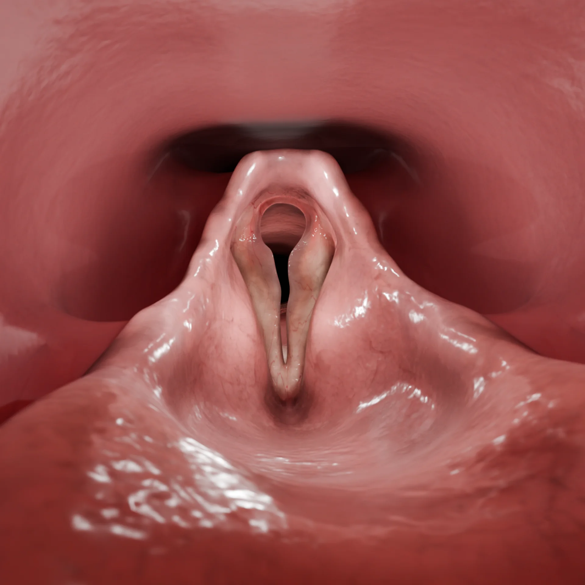
In edematous–polypoid laryngitis, there is bilateral, gelatinous swelling occurs along the entire free edge of the vocal folds. These folds fail to close completely during phonation, and the glottic airway may become narrowed due to mucosal overgrowth.
Clinical Manifestations of Chronic Hyperplastic Laryngitis
The following common symptoms are characteristic of all diseases in this group:
Hoarseness and changes in vocal timbre;
Persistent dry cough;
Globus sensation.
In cases of pronounced mucosal hyperplasia, symptoms may progress to aphonia or laryngeal stenosis secondary to significant airway narrowing.
Anatomic Pathology of Chronic Hyperplastic Laryngitis
Diagnosis is based on medical history and physical examination. Depending on available equipment and protocols, indirect laryngoscopy or flexible video laryngoscopy may be performed.
3D Animation: Chronic Edematous–Polypoid Laryngitis
A biopsy combined with microscopic examination — most commonly carried out intraoperatively — is required.
Find more scientifically accurate content on our social media
Subscribe and don’t miss out the latest resources
Treatment of Chronic Hyperplastic Laryngitis
Initial management for all patients includes eliminating contributing factors such as tobacco and alcohol use, excessive vocal strain, and occupational exposure to harmful irritants. Individuals in voice-dependent professions should consult a phoniatrist for targeted vocal therapy.
The treatment approach is typically multimodal and includes both medical therapy and surgical excision of hyperplastic lesions.
Conservative therapy:
Inhalations with isotonic solutions, corticosteroids, and when necessary, antiseptic or antibacterial agents;
Intralaryngeal instillation of medications.
Surgical treatment consists of the mechanical removal of altered tissues. Several methods are distinguished:
the “cold” method involves excising tissue using micro-instruments such as forceps, bite-out tools, and microdebriders. On the one hand, this approach provides tissue samples for histopathological analysis. On the other, it poses certain risks, including potentially excessive tissue removal and damage to the vocal folds, which may result in permanent voice dysfunction;
CO2 laser is the gold standard for treatment, leaving minimal scarring and promoting rapid healing. Prior to the procedure, a preliminary histopathological analysis of the tissues (biopsy) should be performed.
Postoperative voice rest is essential to support optimal healing and reduce the risk of recurrence.
FAQ
1. What are the characteristic symptoms of chronic laryngitis?
Chronic laryngitis manifests as hoarseness, rapid vocal fatigue, changes in vocal timbre, and also dry cough and throat irritation. A long-standing disease may lead to aphonia, along with a globus sensation.
2. What can be the cause of chronic laryngitis?
The primary causes of chronic laryngitis include smoking, excessive vocal strain, exposure to allergens, and chronic inflammatory diseases of the upper respiratory tract. The condition may also be associated with gastroesophageal reflux.
3. What complications can arise with chronic laryngitis?
Potential complications of chronic laryngitis include aphonia (loss of voice), laryngeal stenosis, and the development of precancerous lesions in the absence of prolonged therapy.
4. What clinical recommendations exist for treating chronic laryngitis?
Smoking cessation and reduction of vocal strain are strongly recommended. Regular ENT follow-up is advised, along with saline or corticosteroid inhalations. In cases of bacterial infection, antibiotic therapy may be administered as indicated. For individuals in voice-related professions, vocal therapy with a phoniatrist may be beneficial.
References
1.
VOKA 3D Anatomy & Pathology – Complete Anatomy and Pathology 3D Atlas. VOKA 3D Anatomy & Pathology.
Available from: https://catalog.voka.io/
2.
Sclafani AP, Dyleski RA, Pitman MJ, Schantz SP. Total otolaryngology—head and neck surgery. New York: Thieme; 2015. ISBN: 978-1-60406-646-3.
3.
Behrbohm H, Kaschke O, Nawka T, Swift A. Ear, Nose, and Throat Diseases. 2nd ed. Moscow: MEDpress-inform; 2016. 776 p. [In Russian.] ISBN 978-5-00030-322-1.
4.
Toward an Understanding of the Pathophysiology of Chronic Laryngitis. Perspect ASHA Spec Interest Groups. 2016 Mar;1(3):14-25. doi: 10.1044/persp1.sig3.14. Epub 2016 Mar 1. PMID: 32864454; PMCID: PMC8951218.
5.
Stein DJ, Noordzij JP. Incidence of chronic laryngitis. Ann Otol Rhinol Laryngol. 2013 Dec;122(12):771-4. doi: 10.1177/000348941312201207. PMID: 24592580.
6.
Zhukhovitskaya A, Verma SP. Identification and Management of Chronic Laryngitis. Otolaryngol Clin North Am. 2019 Aug;52(4):607-616. doi: 10.1016/j.otc.2019.03.004. Epub 2019 May 14. PMID: 31101358.
7.
Endoscopic approach to hyperplastic laryngeal lesions: a literature review and personal experience. The Egyptian Journal of Otolaryngology [Internet]. 2023 Aug 21;39(1).
Available from: https://doi.org/10.1186/s43163-023-00490-4
St. Petersburg FL 33702, 7901 4th St N STE 300, USA
Thank you!
Your message is sent! Our experts will contact you shortly. If you have any additional questions, please contact us at info@voka.io
Cookie Consent
We use cookies to enhance your browsing experience, analyze site traffic, and deliver content. Please choose whether you accept all cookies or wish to reject non-essential tracking.
Cookie Preferences
Manage your cookie preferences below:
Essential cookies enable basic functions and are necessary for the proper function of the website.
Name
Description
Duration
Geolocation Config
This cookie is used to store the consent settings based on the visitor's location.
30 days
Cookie Preferences
This cookie is used to store the user's cookie consent preferences.
30 days
Google reCAPTCHA helps protect websites from spam and abuse by verifying user interactions through challenges.
Name
Description
Duration
_GRECAPTCHA
Google reCAPTCHA sets a necessary cookie (_GRECAPTCHA) when executed for the purpose of providing its risk analysis.
179 days
Statistics cookies collect information anonymously. This information helps us understand how visitors use our website.
Google Analytics is a powerful tool that tracks and analyzes website traffic for informed marketing decisions.
ID used to identify users for 24 hours after last activity
24 hours
_gat
Used to monitor number of Google Analytics server requests when using Google Tag Manager
1 minute
_gac_
Contains information related to marketing campaigns of the user. These are shared with Google AdWords / Google Ads when the Google Ads and Google Analytics accounts are linked together.
90 days
__utma
ID used to identify users and sessions
2 years after last activity
__utmt
Used to monitor number of Google Analytics server requests
10 minutes
__utmb
Used to distinguish new sessions and visits. This cookie is set when the GA.js javascript library is loaded and there is no existing __utmb cookie. The cookie is updated every time data is sent to the Google Analytics server.
30 minutes after last activity
__utmc
Used only with old Urchin versions of Google Analytics and not with GA.js. Was used to distinguish between new sessions and visits at the end of a session.
End of session (browser)
__utmz
Contains information about the traffic source or campaign that directed user to the website. The cookie is set when the GA.js javascript is loaded and updated when data is sent to the Google Anaytics server
6 months after last activity
__utmv
Contains custom information set by the web developer via the _setCustomVar method in Google Analytics. This cookie is updated every time new data is sent to the Google Analytics server.
2 years after last activity
__utmx
Used to determine whether a user is included in an A / B or Multivariate test.
18 months
_ga
ID used to identify users
2 years
_gali
Used by Google Analytics to determine which links on a page are being clicked
30 seconds
Clarity is a web analytics service that tracks and reports website traffic.