Diseases of the Nasopharynx: Classification, Causes, Diagnosis, and Treatment
Danata A.Otorhinolaryngologist, MD
13 min read·April 04, 2025
This article is for informational purposes only
The content on this website, including text, graphics, and other materials, is provided for informational purposes only. It is not intended as advice or guidance. Regarding your specific medical condition or treatment, please consult your healthcare provider.
Nasopharyngeal diseases are a common group of pathologies, especially in children. They are associated with impaired function of lymphoid tissue located in this area, and can lead to a significant deterioration in the quality of life, nasal breathing difficulties, sleep disorders, hearing loss and other complications. The most common are hypertrophy of adenoids and adenoiditis, and less commonly hypertrophy of the tubal tonsils. These conditions require timely diagnosis and comprehensive treatment, including both conservative and surgical methods.
Definition of nasopharyngeal diseases
Adenoid hypertrophy is a pathologic overgrowth of the nasopharyngeal tonsils that occurs more often in children.
Hypertrophy of tubal ridges is a pathologic overgrowth of the lymphoid tissue of tubal tonsils located in the area of the opening of the auditory tubes (tubal ridges).
Adenoiditis is an inflammation of the nasopharyngeal tonsil.
Classification of diseases of the nasopharynx
Adenoid Hypertrophy:
1nd degree hypertrophy;
2nd degree hypertrophy;
3rd degree hypertrophy;
4nd degree hypertrophy.
Hypertrophy of the tubal shafts.
Inflammation of the adenoids:
acute adenoiditis;
chronic adenoiditis.
Etiology
The nasopharyngeal tonsil (adenoids) typically enlarges around the age of 3 years. Hypertrophy occurs in children until puberty, after which regression occurs, and by the age of 20, the tonsil is defined as a small formation. This condition is quite common—about 35 cases per 1000, accounting for over 50% of all visits to the otorhinolaryngologist. Sometimes adenoids are found in younger children and may persist into adulthood. The tubal tonsil can also enlarge in preschool children, but this is quite rare.
The causes of adenoid and tubal tonsil hypertrophy are identical, as both are composed of lymphoid tissue and located in the nasopharynx. The most common factor is infectious: bacteria, viruses, and fungi cause antigenic stimulation, but immature lymphoid tissue produces insufficient antibodies, leading to compensatory hypertrophy. Subsequently, after immune maturation, tissue size decreases.
Additionally, household and food allergens, as well as gastroesophageal reflux disease (GERD), in which hydrochloric acid vapors irritate nasopharyngeal tissues, contribute to the enlargement of adenoids and tubal tonsils. According to research, the size of the nasopharyngeal tonsils is also affected by parental smoking and genetic predisposition.
Viruses and bacteria play a leading role in the development of adenoiditis. Most common pathogens:
RS-viruses;
adenoviruses;
influenza and parainfluenza viruses;
herpes viruses (including Epstein-Barr virus);
streptococci (St. pneumoniae, St. pyogenes);
staphylococci (S. aureus, S. epidermidis);
Pseudomonas aeruginosa;
Moraxella catarrhalis;
activation of the conditional-pathogenic microflora of the nasopharynx.
Accompanying pathologies, such as recurrent or chronic diseases of the upper respiratory tract (rhinitis, sinusitis, bronchitis), GERD, and allergies can contribute to the development of disease and worsen its course. The presence of hormonal or immune disorders (diabetes mellitus, thyroid pathology, HIV infection) also adversely affects the development of the disease. Lack of a history of breastfeeding and vitamin D deficiency also exacerbate and prolong the course of adenoiditis.
Do not forget about environmental conditions: dry air, inadequate temperature control, cluttered premises, and working in harmful enterprises negatively affect the state of the upper respiratory tract and worsen the disease manifestation.
Anatomy
Adenoids are located in the upper vault of the nasopharynx. They are classified according to their size and projection into the nasopharynx lumen:
Grade 1 adenoids — located in the upper part of the nasopharynx and cover ⅓ of the vomer;
Grade 2 adenoids — occupy ½ of the nasopharynx and cover ½ of the vomer;
Grade 3 adenoids — cover ⅔ of the nasopharynx, leaving small space for breathing;
Grade 4 adenoids — completely block the nasopharyngeal lumen, descend into the oropharynx, and cover the vomer.
Nasopharyngeal tonsil is a heterogeneous elastic lumpy pink-colored substance, 5–7 mm thick and 20–25 mm in diameter, with longitudinal grooves of varying sizes.
Tubal tonsils are small areas of lymphoid tissue (up to 7 mm) located cranially near the openings of the auditory tubes. If they are hypertrophied, the openings are blocked, and the exit from the eustachian tube is obstructed.
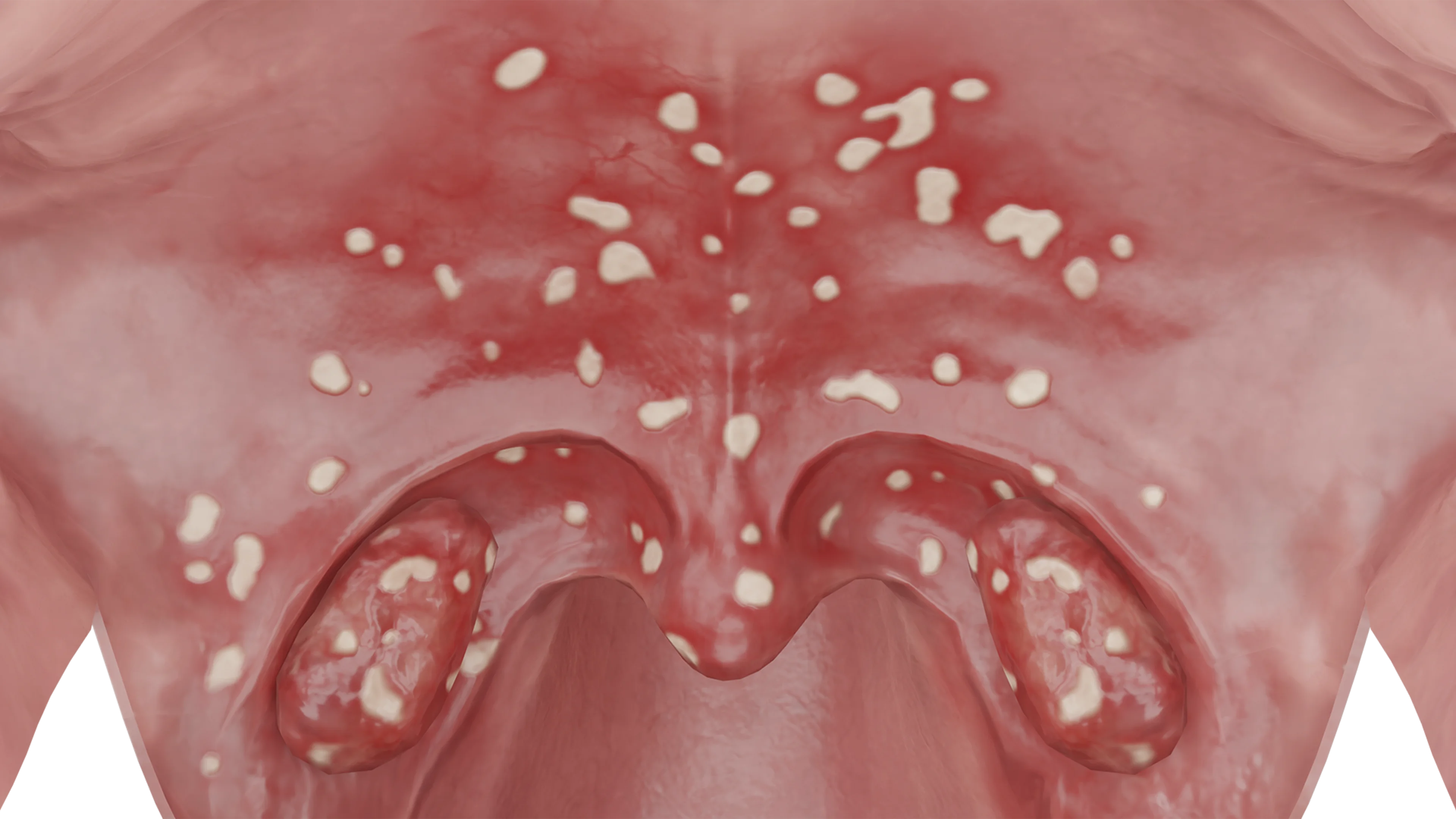
In inflammation of the nasopharyngeal tonsil, its barrier function is compromised, the epithelial cilia are destroyed, adenoid vegetations become hyperemic and infiltrated, covered with fibrinous plaque. A large amount of serous or mucopurulent content is found in the grooves. Mucus drainage is also noted along the posterior pharyngeal wall. Lymphoid follicles enlarge, and hyperemia of the posterior palatine arches and lateral pharyngeal wall is observed. Adenoiditis is divided into catarrhal, exudative-serous, and mucous-purulent based on the nature of the discharge.
Hypertrophied adenoids are characterized by a diverse clinical picture. The most common complaints from parents are constant nasal breathing difficulties, snoring, and noisy breathing. In young children, eating becomes difficult due to impaired nasal breathing. With a pronounced degree of hypertrophy, nasality develops. Due to breathing disorders, children sleep worse at night, wake up frequently, and some experience obstructive sleep apnea syndrome (OSAS), with breathing pauses up to 1 minute. This leads to increased fatigue and reduced ability to perform tasks.
There is also the notion of habitus adenoideus, or adenoid-type face. Such children have a flattened nasal bridge, slightly open mouth, malocclusion (mandibular prognathism), protruding upper incisors, gothic palate, small exophthalmos, and elongated face shape.
Hypertrophied tubal tonsils do not cause clinical manifestations by themselves. Their influence on the airways is mediated by blocking the auditory tube.
Hypertrophied adenoids, like tubal tonsils, obstruct the auditory tube openings, leading to tubotympanitis, recurrent acute otitis, or slow-moving exudative otitis. In some cases, conductive hearing loss develops, which may be the only complaint.
The location of adenoids in relation to the auditory tube opening – 3D model
Clinically, adenoiditis is classified as acute (up to 7–10 days), subacute (10 days to 1 month), and chronic (over 1 month). However, in practice, this classification is conditional. Since edema of lymphoid tissue occurs in adenoiditis, the clinical picture resembles that of adenoid hypertrophy. In addition to the abovementioned complaints, mucous or purulent nasal discharge, mucus dripping down the posterior pharyngeal wall, accompanied by coughing, is typical. Deterioration is noted at night.
Acute adenoiditis is characterized by febrile body temperature, general intoxication, pain in the depth of the nose and head with radiation to the eyes and ears. Regional lymph nodes are enlarged, their soreness is noted.
Chronic adenoiditis features subfebrile body temperature, suffocating night cough, and concomitant middle ear pathology, accompanied by conductive hearing loss.
Diagnosis
Initially, complaints are gathered, medical history is clarified, and otorhinolaryngoscopy is performed. To determine the degree of nasopharyngeal obstruction and examine the auditory tube openings, posterior rhinoscopy and nasopharyngeal endoscopy are performed. Manual examination is conducted rarely. To determine the degree of hypertrophy of adenoid vegetations, lateral view nasopharyngeal radiography is performed. In inflammation of the nasopharyngeal tonsil, a microbiological study for microflora and antibiotic sensitivity is conducted.
Find more scientifically accurate content on our social media
Subscribe and don’t miss out the latest resources
Treatment
Conservative therapy is initially given to children with hypertrophied nasopharyngeal or tubal tonsils. Intranasal glucocorticosteroids (mometasone furoate) are prescribed for at least 1 month with follow-up evaluation. If the results are positive, further use of the drug is recommended according to the scheme.
In the absence of effect, and in the presence of complications (OSA, hearing loss, chronic otitis), surgical treatment is recommended. Under local or general anesthesia, an adenotomy is performed, during which the hypertrophied lymphoid tissue is excised with a Beckman adenotome.
Hypertrophied lymphoid tissue of the nasopharyngeal tonsil in adults is subject to mandatory surgical removal with subsequent pathohistologic examination.
The tubal tonsils are subject only to conservative treatment with the use of topical glucocorticosteroids. Their surgical excision is not performed, as in most cases it leads to scarring of the aperture of the auditory tubes. In cases of significant eustachian tube obstruction and presence of exudate in the middle ear, accompanied by hearing loss, balloon dilation is performed.
To treat inflamed nasopharyngeal tonsils, antibacterial drugs are used as topical sprays or systemically, considering the pathogen’s sensitivity. In addition to etiotropic therapy, regular sanitation of the nasal cavity and nasopharynx from pathological contents is recommended with saline or seawater solutions. Vasoconstrictive sprays are used to reduce swelling. It is necessary to carry out general strengthening measures. With recurrent adenoiditis or middle ear complications, surgical treatment should be considered.
FAQ
1. What are adenoids and where are they located?
Adenoids (nasopharyngeal tonsil) are a collection of lymphoid tissue in the nasopharynx. They are part of the immune system and help protect the body from infections, but pathological overgrowth can cause breathing and hearing problems.
2. At what age is adenoid hypertrophy more common?
Adenoids typically enlarge in children aged 3–7 years, and begin to regress after 10–12 years (involution). However, in some individuals, they persist into adulthood.
3. What symptoms indicate enlarged adenoids?
Main signs include: • constant nasal congestion, mouth breathing; • snoring and restless sleep; • nasal voice; • frequent otitis media and hearing loss; • adenoid facies (with prolonged course).
4. What is the difference between adenoid hypertrophy and adenoiditis?
Hypertrophy refers to tissue enlargement without inflammation. Adenoiditis is inflammation of the adenoids, often accompanied by purulent discharge, fever, and intoxication.
5. Is it always necessary to remove adenoids?
No, not always. Initially, conservative treatment (drops, washing, physiotherapy) is applied. Surgery (adenotomy) is necessary for 3rd or 4th degree hypertrophy with breathing difficulties, frequent otitis media, and hearing loss, or sleep apnea (breathing cessation during sleep).
6. Can adenoids regrow after removal?
Yes, in 5–10% of cases, recurrence is possible, especially in children under 3–4 years old.
7. What chronic nasopharyngeal diseases can develop due to adenoids?
With prolonged presence of hypertrophied adenoids or frequent adenoiditis, the following may develop: • chronic adenoiditis (persistent inflammation of the nasopharyngeal tonsil); • chronic rhinosinusitis (inflammation of the nasal sinuses); • chronic tubo-otitis (inflammation of the auditory tube); • chronic pharyngitis (inflammation of the pharynx).
References
1.
VOKA 3D Anatomy & Pathology – Complete Anatomy and Pathology 3D Atlas. VOKA 3D Anatomy & Pathology.
Available from: https://catalog.voka.io/
2.
Sclafani AP, Dyleski RA, Pitman MJ, Schantz SP. Total otolaryngology—head and neck surgery. New York: Thieme Medical Publishers; 2015. ISBN: 978-1-60406-646-3.
3.
Behrbohm H, Kaschke O, Nawka T, Swift A. Ear, Nose, and Throat Diseases. 2nd ed. Moscow: MEDpress-inform; 2016. 776 p. [In Russian.] ISBN 978-5-00030-322-1.
4.
Swidsinski A, Göktas O, Bessler C, Loening-Baucke V, Hale LP, Andree H, et al. Spatial organisation of microbiota in quiescent adenoiditis and tonsillitis. J Clin Pathol. 2007 Mar;60(3):253-260. doi: 10.1136/jcp.2006.037309. Epub 2006 May 12. PMID: 16698947; PMCID: PMC1860565.
5.
Evcimik MF, Dogru M, Cirik AA, Nepesov MI. Adenoid hypertrophy in children with allergic disease and influential factors. Int J Pediatr Otorhinolaryngol. 2015 May;79(5):694-7. doi: 10.1016/j.ijporl.2015.02.017. Epub 2015 Feb 25. PMID: 25758194.
St. Petersburg FL 33702, 7901 4th St N STE 300, USA
Thank you!
Your message is sent! Our experts will contact you shortly. If you have any additional questions, please contact us at info@voka.io
Cookie Consent
We use cookies to enhance your browsing experience, analyze site traffic, and deliver content. Please choose whether you accept all cookies or wish to reject non-essential tracking.
Cookie Preferences
Manage your cookie preferences below:
Essential cookies enable basic functions and are necessary for the proper function of the website.
Name
Description
Duration
Geolocation Config
This cookie is used to store the consent settings based on the visitor's location.
30 days
Cookie Preferences
This cookie is used to store the user's cookie consent preferences.
30 days
Google reCAPTCHA helps protect websites from spam and abuse by verifying user interactions through challenges.
Name
Description
Duration
_GRECAPTCHA
Google reCAPTCHA sets a necessary cookie (_GRECAPTCHA) when executed for the purpose of providing its risk analysis.
179 days
Statistics cookies collect information anonymously. This information helps us understand how visitors use our website.
Google Analytics is a powerful tool that tracks and analyzes website traffic for informed marketing decisions.
ID used to identify users for 24 hours after last activity
24 hours
_gat
Used to monitor number of Google Analytics server requests when using Google Tag Manager
1 minute
_gac_
Contains information related to marketing campaigns of the user. These are shared with Google AdWords / Google Ads when the Google Ads and Google Analytics accounts are linked together.
90 days
__utma
ID used to identify users and sessions
2 years after last activity
__utmt
Used to monitor number of Google Analytics server requests
10 minutes
__utmb
Used to distinguish new sessions and visits. This cookie is set when the GA.js javascript library is loaded and there is no existing __utmb cookie. The cookie is updated every time data is sent to the Google Analytics server.
30 minutes after last activity
__utmc
Used only with old Urchin versions of Google Analytics and not with GA.js. Was used to distinguish between new sessions and visits at the end of a session.
End of session (browser)
__utmz
Contains information about the traffic source or campaign that directed user to the website. The cookie is set when the GA.js javascript is loaded and updated when data is sent to the Google Anaytics server
6 months after last activity
__utmv
Contains custom information set by the web developer via the _setCustomVar method in Google Analytics. This cookie is updated every time new data is sent to the Google Analytics server.
2 years after last activity
__utmx
Used to determine whether a user is included in an A / B or Multivariate test.
18 months
_ga
ID used to identify users
2 years
_gali
Used by Google Analytics to determine which links on a page are being clicked
30 seconds
Clarity is a web analytics service that tracks and reports website traffic.




.webp)