Vulvitis: Predisposing Factors, Clinical Manifestations, Diagnosis, and Treatment
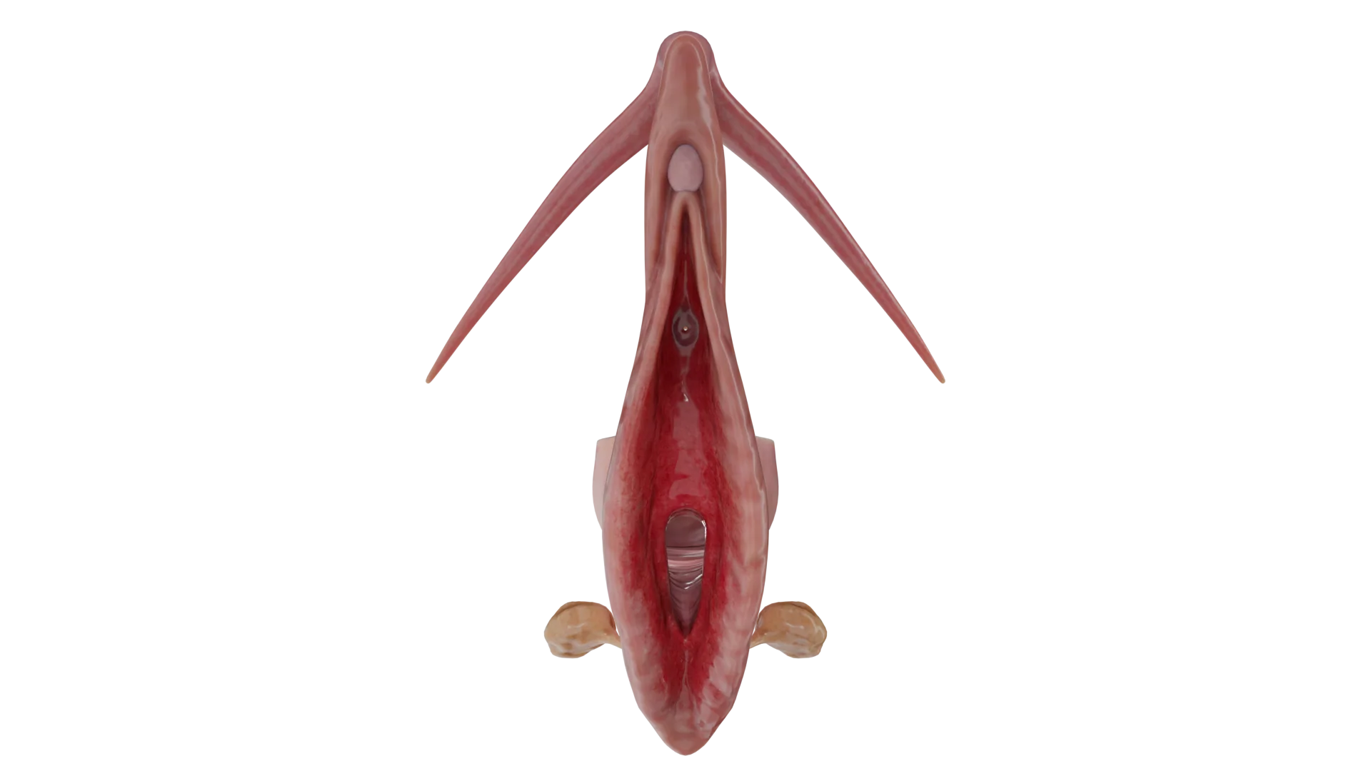
Vulvitis refers to vulvar inflammation affecting the labia, clitoris, mons pubis, and vestibule of the vagina. Clinical manifestations, diagnosis, and treatment.


Anesthesia
Pain management and sedation techniques

Angiology
Arterial and venous pathologies

Cardiology
Acquired and congenital heart diseases

Dentistry
Diseases of teeth, gums, and the oral cavity

Dermatology
Disorders of the skin and subcutaneous tissue

Embryology
Stages of embryogenesis and congenital anomalies

Endocrinology
Disorders of the glands and hormonal imbalance

Gastroenterology
Stomach, intestinal, and digestive diseases

Gynecology
Diseases of female reproductive organs

Hematology
Hematopoiesis and blood-related disorders

Hepatology
Liver, gallbladder, and biliary tract diseases

Histology
Microscopic tissue and cell structures

Infectious diseases
Bacterial, viral, and parasitic infections

Neurology
Brain, spinal cord, and peripheral nerve disorders

Obstetrics
Pregnancy complications and abnormal fetal positions

Oncology
Cancer types, benign and malignant tumors

Ophthalmology
Conditions affecting the eyes and vision

Orthopedics
Bone, joint, and soft tissue disorders

Otorhinolaryngology
Ear, nose, and throat diseases

Pediatrics
Child health, development, and clinical conditions

Physiology
Biological processes within organs and systems

Pulmonology
Lung and respiratory tract diseases

Traumatology
Acute injuries and musculoskeletal trauma

Urology
Urinary tract and male reproductive disorders
Anesthesia
Pain management and sedation techniques
Angiology
Arterial and venous pathologies
Cardiology
Acquired and congenital heart diseases
Dentistry
Diseases of teeth, gums, and the oral cavity
Dermatology
Disorders of the skin and subcutaneous tissue
Embryology
Stages of embryogenesis and congenital anomalies
Endocrinology
Disorders of the glands and hormonal imbalance
Gastroenterology
Stomach, intestinal, and digestive diseases
Gynecology
Diseases of female reproductive organs
Hematology
Hematopoiesis and blood-related disorders
Hepatology
Liver, gallbladder, and biliary tract diseases
Histology
Microscopic tissue and cell structures
Infectious diseases
Bacterial, viral, and parasitic infections
Neurology
Brain, spinal cord, and peripheral nerve disorders
Obstetrics
Pregnancy complications and abnormal fetal positions
Oncology
Cancer types, benign and malignant tumors
Ophthalmology
Conditions affecting the eyes and vision
Orthopedics
Bone, joint, and soft tissue disorders
Otorhinolaryngology
Ear, nose, and throat diseases
Pediatrics
Child health, development, and clinical conditions
Physiology
Biological processes within organs and systems
Pulmonology
Lung and respiratory tract diseases
Traumatology
Acute injuries and musculoskeletal trauma
Urology
Urinary tract and male reproductive disorders
Specialties
AnesthesiaAngiologyCardiologyDentistryDermatologyEmbryologyEndocrinologyGastroenterologyGynecologyHematologyHepatologyHistologyInfectious diseasesNeurologyObstetricsOncologyOphthalmologyOrthopedicsOtorhinolaryngologyPediatricsPhysiologyPulmonologyTraumatologyUrology
This article is for informational purposes only
The content on this website, including text, graphics, and other materials, is provided for informational purposes only. It is not intended as advice or guidance. Regarding your specific medical condition or treatment, please consult your healthcare provider.
Adenomyosis is a benign condition of the uterus that has been historically diagnosed via histological examination after hysterectomy (removal of the uterus). During the examination, ectopic endometrial glands and stroma are studied at a minimum depth of 2.5 mm below the endomyometrial junction (EMJ), accompanied by hypertrophic and hyperplastic myometrium. This condition is typically visualized using modalities such as ultrasound or MRI.
The reported prevalence of adenomyosis ranges from 5 % to 70 %. Before age 40, the condition affects 2 out of 10 women, whereas between ages 40 and 50 the prevalence increases to 8 out of 10 women. However, the true prevalence of adenomyosis is difficult to determine due to the absence of a unified definition and noninvasive diagnostic criteria. There are still no pathognomonic clinical features of adenomyosis, nor are there laparoscopic criteria that can be used to diagnose this condition.
Adenomyosis may coexist with other estrogen‑dependent benign disorders, such as endometriosis (70 % of cases), uterine fibroids (50 %), and endometrial hyperplasia (35 %).
The pathogenesis of adenomyosis remains unclear, although several theories have been proposed:
A new area of research focuses on expression of messenger RNA (mRNA) and long non‑coding RNA (lncRNA) in adenomyotic lesions.
Attempts have been made to classify adenomyosis into subtypes based on histologic findings and imaging results, but none of the proposed systems have been adopted in clinical practice. According to the simplest classification, adenomyosis is divided into diffuse and focal types depending on its distribution within the myometrium.
Diffuse adenomyosis is defined by multiple foci within the myometrium (with less than 25 % of the affected area surrounded by normal myometrium), whereas focal adenomyosis appears as isolated nodules of hypertrophic myometrium and ectopic endometrium.
However, the pathogenesis of adenomyosis remains uncertain, and the relationship between disease severity and clinical manifestations has not been established, , making it difficult to determine standardized treatment approaches.
In Russian‑language literature, a classification of adenomyosis based on depth of invasion is commonly used:
3D Models of Adenomyosis:
 Adenomyosis, Grade 1
 Adenomyosis, Grade 2
 Adenomyosis, Grade 3
 Adenomyosis, Grade 4
According to Bird’s classification, adenomyotic lesions are subdivided based on the depth of penetration, determined by the affected uterine layer, and the degree of involvement, measured by the number of endometrial glands per low‑power field.
The authors also demonstrated a direct correlation between the severity of dysmenorrhea and the depth of penetration. Thus, 4.3 % of women with Grade 1 adenomyosis reported dysmenorrhea, compared with 42.4 % of Grade 2 patients and 83.3% of Grade 3 patients.
Another histopathologic feature described in patients with deep adenomyosis is hemosiderin deposition around adenomyotic lesions. This results from bleeding within ectopic endometrial foci and suggests that hemosiderin deposition may reflect the extent and severity of adenomyosis; however, the clinical significance of this finding remains unclear.
Levghur et al. described the depth of adenomyosis as the percentage of myometrial thickness involved and distinguished:
The authors also noted dysmenorrhea in 77.8 % of patients with deep adenomyosis compared with 12.5 % in the intermediate group. Superficial myometrial foci were not associated with dysmenorrhea or menorrhagia.
Hulka et al. introduced a new category of focal adenomyosis and added the term “adenomyoma” to previous classifications.
Rasmussen et al. proposed a histologic classification based on endomyometrial biopsies obtained via transcervical resection of endometrium (TCRE). The procedure requires biopsy samples at least 5 mm deep.
Classification of Adenomyosis
| Classification Criterion | Types/Grades of Adenomyosis | Characteristics |
|---|---|---|
| By distribution within myometrium | Diffuse | Multiple foci in the myometrium (< 25 % of the affected surface surrounded by healthy myometrium) |
| Focal (adenomyoma) | Isolated nodules composed of hypertrophic myometrium and ectopic endometrium | |
| By depth of invasion (Russian-language sources) | Grade 1 | Involvement of submucosal layer |
| Grade 2 | < 50 % of myometrial thickness involved | |
| Grade 3 | Full‑thickness involvement of myometrium | |
| Grade 4 | Extension beyond uterus | |
| Bird’s classification | Class I (subbasal adenomyosis) | Lesions close to basal endometrium without deep penetration |
| Class II (mid‑myometrium) | Penetration into the mid‑myometrial layer | |
| Class III (deep adenomyosis) | > 50 % of myometrial thickness involved | |
| Depth of invasion (Levghur et al.) | Superficial (< 40 % of myometrial thickness) | Not associated with dysmenorrhea |
| Intermediate (40–80 %) | Moderate symptoms | |
| Deep (> 80 %) | Severe dysmenorrhea (77.8 % of cases) | |
| Histologic classification (Rasmussen) | Internal adenomyosis | Invasion ≥ 2 mm without contact with basal endometrium |
| Serrated JZ | Invasion > 3 mm with contact against basal endometrium | |
| Linear JZ | Involvement ≤ 3 mm or absence of invasion | |
| Additional criteria | Presence of hemosiderin | Marker of severity; clinical significance uncertain |
For clinical diagnosis, the criteria proposed by the MUSA consensus (Morphological Uterus Sonographic Assessment) based on transvaginal ultrasound findings are used. Although the MUSA group provided unified guidance for recognizing and identifying sonographic features of adenomyotic lesions, it did not lead to the development of a classification system for adenomyosis. Given that ultrasound is a subjective assessment method, standardization and classification remain challenging.
According to the MUSA classification, the sonographic features of adenomyosis can be divided into direct and indirect signs.
According to MUSA, myometrial cysts are defined as round structures within the myometrium. Their contents may be anechoic, low‑echogenic, have a “ground‑glass” appearance, or show mixed echogenicity. Cysts may be surrounded by a hyperechoic rim. There is no size requirement for myometrial cysts, and the hyperechoic rim is not mandatory. Experts recommend using color Doppler imaging to identify blood vessels, which assists in differentiating adenomyotic cysts from other myometrial cystic lesions.
Hyperechoic islands are defined as hyperechoic areas within the myometrium. They may be regular, irregular, or poorly defined. However, hyperechoic islands must not be connected to the endometrium. The minimum distance from the endometrium is not precisely defined, as it may vary individually. No minimum diameter or number of hyperechoic islands has been established.
Experts note that evaluating these features is difficult due to the lack of 3D ultrasound imaging, challenges in identifying the endometrial–myometrial interface, and the limited JZ visibility. The MUSA consensus defined this feature as follows: “Hyperechoic subendometrial lines or buds may be observed disrupting the JZ. Hyperechoic subendometrial lines are (almost) perpendicular to the endometrial cavity and are connected to the endometrium. However, experts note that any form of endometrial tissue invasion into the myometrium may be a sign of adenomyosis, even if it does not appear as lines or buds.”
A globular uterus is diagnosed when the perimetrium contour diverges from the cervix in at least two directions (anterior, posterior, or lateral), rather than following a trajectory parallel to the endometrium. The uterine diameters (length, width, depth) are approximately equal, producing a characteristic spherical shape. Consensus was reached that this sign may be falsely positive in cases of uterine fibroids or intracavitary anomalies.
This parameter is assessed by comparing the thickness of the anterior and posterior uterine walls. A ratio close to 1 indicates symmetry, whereas a ratio above or below 1 indicates asymmetry, although this assessment is subjective. A difference of more than 5 mm between the walls is considered an indirect sign. Note that uterine asymmetry may also be associated with transient uterine contractions or uterine fibroids.
This type of shadowing is characterized by hyperechoic linear bands, sometimes alternating with hypoechoic ones. Fan‑shaped shadowing is best evaluated in grayscale mode. Diagnostic difficulties may arise due to other lesions that produce shadowing, such as uterine fibroids or cesarean‑section scar fibrosis.
Translesional vascularity is defined as blood vessels running perpendicular to the uterine cavity/perimetrium and traversing the lesion. This pattern is likely present in diffuse adenomyosis. Circular vascularity, typically seen around uterine fibroids, may also occur in adenomyosis. Although intralesional vessels may be seen in uterine fibroids, translesional vascularity (vessels crossing the lesion) is an atypical feature. This sign helps differentiate adenomyosis from uterine fibroids.
Several challenges exist in defining this criterion. First, the JZ is difficult to evaluate without 3D imaging. According to the MUSA group, the JZ may appear irregular due to cystic areas, hyperechoic spots, or hyperechoic lines. The degree of irregularity is expressed as the difference between the maximal and minimal JZ thickness.
Second, the extent of irregularity is subjectively assessed as the percentage of the JZ that appears irregular (less than 50 % or more than 50 %). The JZ should be evaluated using 3D ultrasound in sagittal, transverse, and coronal planes. Measuring the JZ thickness is not a mandatory diagnostic criterion.
An interrupted JZ is diagnosed when a JZ portion cannot be visualized on either 2D or 3D ultrasound in any scanning plane. A continuous JZ means that it is clearly visible in all planes on 2D or 3D ultrasound.
A pooled analysis of studies showed that MRI has a sensitivity of approximately 78 % and a specificity of 93 % for diagnosing adenomyosis. Although transvaginal ultrasound is reported to have similar sensitivity and specificity, ultrasound results are too heterogeneous to combine. Thus, MRI‑based systems provide greater objectivity and consistency in the classification of adenomyosis. MRI allows visualization of the zonal anatomy of the uterus and clear JZ depiction, enabling diagnosis of lesions in any part of the endometrium and myometrium. The most comprehensive recent classification is the system proposed by Kobayashi, which includes five components and evaluates them accordingly.
MRI‑Based Classification of Adenomyosis (Kobayashi System, 2020)
| Criterion | Grade | Description |
|---|---|---|
| Area Affected | А | Internal adenomyosis, JZ thickness > 12 mm |
| В | External adenomyosis, JZ thickness < 8 mm | |
| Lesion Size | A1 or B1 | Less than one‑third of the uterine wall thickness; predominantly focal |
| A2 or B2 | Less than two‑thirds of the uterine wall thickness; may be focal or diffuse | |
| A3 or B3 | More than two‑thirds of the uterine wall thickness; predominantly diffuse | |
| Coexisting Pathologies | C0–C5 | C0 — none, C1 — peritoneal endometriosis, C2 — ovarian endometrioma, C3 — deep infiltrating endometriosis, C4 — uterine fibroids, C5 — other |
| Location | D1–D5 | D1 — anterior uterine wall, D2 — posterior uterine wall, D3 — left lateral wall, D4 — right lateral wall, D5 — uterine fundus |
The final score is then reported as a four‑letter code with corresponding numbers based on MRI findings.
In patients with abnormal uterine bleeding, hysteroscopy can be a valuable diagnostic method that provides direct visualization of the uterine cavity and allows tissue sampling for histological examination. Although the diagnosis cannot be based on visual inspection alone, several features have been identified that may suggest adenomyosis: marked hypervascularization on the endometrial surface, irregular endometrium with small openings (the so‑called “strawberry pattern”), and fibrotic and/or hemorrhagic cystic lesions. More detailed information can be obtained during histological evaluation of biopsy samples taken using a resectoscope with a diathermic loop.
Adenomyosis is asymptomatic in approximately 30 % of cases. The most common clinical symptoms include menorrhagia (up to 50 % of patients), dysmenorrhea, metrorrhagia, abnormal uterine bleeding, chronic pelvic pain, dyspareunia, and infertility. The exact mechanism linking adenomyosis and infertility remains unclear. Several contributing factors have been proposed and can be grouped into four potential pathways:
Note that endometriosis occurs in 54–90 % of patients with adenomyosis. Therefore, infertility cannot be attributed solely to adenomyosis, as coexisting endometriosis — well known to impair fertility — may be the primary cause.
Find more scientifically accurate content on our social media



1. What is adenomyosis?
2. What causes adenomyosis?
3. What are the symptoms of adenomyosis?
4. How is adenomyosis diagnosed?
5. What is the grading system for adenomyosis?
6. Can you get pregnant with adenomyosis?
7. How is adenomyosis different from endometriosis?
8. What are the risks of adenomyosis?
9. At what stage of adenomyosis is hysterectomy performed?
References
1.
VOKA 3D Anatomy & Pathology – Complete Anatomy and Pathology 3D Atlas [Internet]. VOKA 3D Anatomy & Pathology.
Available from: https://catalog.voka.io/
2.
Harmsen MJ, Van den Bosch T, de Leeuw RA, Dueholm M, Exacoustos C, Valentin L, et al. Consensus on revised definitions of Morphological Uterus Sonographic Assessment (MUSA) features of adenomyosis: results of modified Delphi procedure. Ultrasound Obstet Gynecol. 2022 Jul;60(1):118-131. doi: 10.1002/uog.24786. PMID: 34587658; PMCID: PMC9328356.
3.
Gordts S, Grimbizis G, Campo R. Symptoms and classification of uterine adenomyosis, including the place of hysteroscopy in diagnosis. Fertil Steril. 2018;109:380-388.e1.
4.
Nirgianakis K, Kalaitzopoulos DR, Schwartz ASK. Fertility, pregnancy and neonatal outcomes of patients with adenomyosis: a systematic review and meta-analysis. Reprod Biomed Online. 2021;42:185-206.
5.
Munro, M.G. Classification and reporting systems for adenomyosis J Minim Invasive Gynecol. 2020; 27:296-308.
6.
Van den Bosch T, de Bruijn AM, de Leeuw RA. Sonographic classification and reporting system for diagnosing adenomyosis. Ultrasound Obstet Gynecol. 2019;53:576-582.
7.
Maxim M, Dason ES, Chan C. Current diagnosis and management of adenomyosis in Canada: a survey of Canadian gynaecologists. J Endometr Pelvic Pain Disord. 2022;14:98-105.
8.
Loring M, Chen TY, Isaacson KB. A systematic review of adenomyosis: it is time to reassess what we thought we knew about the disease. J Minim Invasive Gynecol. 2021;28:644-655.
9.
Song SY, Lee SY, Kim HY. Long-term efficacy and feasibility of levonorgestrel-releasing intrauterine device use in patients with adenomyosis. Med (Baltim). 2020;99:e20421.
10.
Neriishi K, Hirata T, Fukuda S. Long-term dienogest administration in patients with symptomatic adenomyosis. J Obstet Gynaecol Res. 2018;44:1439-1444.
11.
Vannuccini S, Luisi S, Tosti C. Role of medical therapy in the management of uterine adenomyosis. Fertil Steril. 2018;109:398-405.
12.
Matsushima T, Akira S, Fukami T. Efficacy of hormonal therapies for decreasing uterine volume in patients with adenomyosis. Gynecol Minim Invasive Ther. 2018;7:119-123.
13.
Andreeva E, Absatarova Y. Triptorelin for the treatment of adenomyosis: a multicenter observational study of 465 women in Russia. Int J Gynaecol Obstet. 2020;151:347-354.
14.
Matsushima T, Akira S, Yoneyama K. Recurrence of uterine adenomyosis after administration of gonadotropin-releasing hormone agonist and the efficacy of dienogest. Gynecol Endocrinol. 2020;36:521-524.
15.
de Bruijn AM, Smink M, Lohle PNM. Uterine artery embolization for the treatment of adenomyosis: a systematic review and meta-analysis. J Vasc Interv Radiol. 2017;28:1629-1642.e1.
16.
Osada H. Uterine adenomyosis and adenomyoma: the surgical approach. Fertil Steril. 2018;109:406-417.
Make VOKA your preferred source
See more VOKA articles in Google Search
Table of Contents









Summarize article with AI
Choose your preferable AI assistant:
Link successfully copied to clipboard
Thank you!
Your message is sent!
Our experts will contact you shortly. If you have any additional questions, please contact us at info@voka.io