Systemic Enamel Hypoplasia: Etiology, Classification, Clinical Presentation, and Treatment
Developmental Enamel Defects: Systemic Hypoplasia. Overview of the Etiology, Clinical Forms, Diagnosis, and Aesthetic Restoration Techniques for Teeth.


Anesthesia
Pain management and sedation techniques

Angiology
Arterial and venous pathologies

Cardiology
Acquired and congenital heart diseases

Dentistry
Diseases of teeth, gums, and the oral cavity

Dermatology
Disorders of the skin and subcutaneous tissue

Embryology
Stages of embryogenesis and congenital anomalies

Endocrinology
Disorders of the glands and hormonal imbalance

Gastroenterology
Stomach, intestinal, and digestive diseases

Gynecology
Diseases of female reproductive organs

Hematology
Hematopoiesis and blood-related disorders

Hepatology
Liver, gallbladder, and biliary tract diseases

Histology
Microscopic tissue and cell structures

Infectious diseases
Bacterial, viral, and parasitic infections

Neurology
Brain, spinal cord, and peripheral nerve disorders

Obstetrics
Pregnancy complications and abnormal fetal positions

Oncology
Cancer types, benign and malignant tumors

Ophthalmology
Conditions affecting the eyes and vision

Orthopedics
Bone, joint, and soft tissue disorders

Otorhinolaryngology
Ear, nose, and throat diseases

Pediatrics
Child health, development, and clinical conditions

Physiology
Biological processes within organs and systems

Plastic surgery
Reconstructive and aesthetic procedures

Pulmonology
Lung and respiratory tract diseases

Traumatology
Acute injuries and musculoskeletal trauma

Urology
Urinary tract and male reproductive disorders
Anesthesia
Pain management and sedation techniques
Angiology
Arterial and venous pathologies
Cardiology
Acquired and congenital heart diseases
Dentistry
Diseases of teeth, gums, and the oral cavity
Dermatology
Disorders of the skin and subcutaneous tissue
Embryology
Stages of embryogenesis and congenital anomalies
Endocrinology
Disorders of the glands and hormonal imbalance
Gastroenterology
Stomach, intestinal, and digestive diseases
Gynecology
Diseases of female reproductive organs
Hematology
Hematopoiesis and blood-related disorders
Hepatology
Liver, gallbladder, and biliary tract diseases
Histology
Microscopic tissue and cell structures
Infectious diseases
Bacterial, viral, and parasitic infections
Neurology
Brain, spinal cord, and peripheral nerve disorders
Obstetrics
Pregnancy complications and abnormal fetal positions
Oncology
Cancer types, benign and malignant tumors
Ophthalmology
Conditions affecting the eyes and vision
Orthopedics
Bone, joint, and soft tissue disorders
Otorhinolaryngology
Ear, nose, and throat diseases
Pediatrics
Child health, development, and clinical conditions
Physiology
Biological processes within organs and systems
Plastic surgery
Reconstructive and aesthetic procedures
Pulmonology
Lung and respiratory tract diseases
Traumatology
Acute injuries and musculoskeletal trauma
Urology
Urinary tract and male reproductive disorders
Specialties
AnesthesiaAngiologyCardiologyDentistryDermatologyEmbryologyEndocrinologyGastroenterologyGynecologyHematologyHepatologyHistologyInfectious diseasesNeurologyObstetricsOncologyOphthalmologyOrthopedicsOtorhinolaryngologyPediatricsPhysiologyPlastic surgeryPulmonologyTraumatologyUrology
This article is for informational purposes only
The content on this website, including text, graphics, and other materials, is provided for informational purposes only. It is not intended as advice or guidance. Regarding your specific medical condition or treatment, please consult your healthcare provider.
Dental caries, also known as tooth decay, is an infection that leads to the mineral breakdown and destruction of the hard tissues of the teeth.
The condition tends to develop when a combination of factors is in play. These may include a pathological bacterial plaque, high consumption of readily fermentable carbohydrates, susceptible teeth, prolonged exposure to unfavorable conditions, etc.
Microorganisms colonize tooth surfaces not subject to friction from the tongue, cheeks, and food fibers, forming a biofilm known as dental plaque. Typical sites of decay are found where the plaque accumulates.
Frequent and prolonged consumption of easily fermentable carbohydrates increases the proportion of cariesogenic microflora in the biofilm, which can rapidly metabolize food carbohydrates to form organic acids and can also survive in low pH conditions. The most commonly founf strains are Streptococcus mutans and Lactobacilli, as well as Streptococcus sobrinus, Streptococcus oralis, Actinomyces israelii, Actinomyces gerencseriae, Bifidobacteria, and Prevotella. Streptococcus mutans also synthesizes intra- and extracellular adhesive polysaccharides (glucans and fructans) that promote prolonged retention of bacteria on tooth surfaces.
Teeth are constantly bathed in saliva containing mineral substances. Saliva, due to its buffering capacity, helps maintain a neutral pH in the oral cavity. At neutral pH levels (≈7), the processes of demineralization (dissolution of mineral component crystals) and remineralization (partial or complete restoration of mineral component crystals) of enamel and dentin are in balance.
As bacteria process food carbohydrates (glucose, fructose, sucrose, maltose, etc.), acids build up, thus reducing pH.
Minerals in the hard tissues of the teeth start to dissolve, as soon as the pH drops to 5.2–5.5. This gradual process of mineral loss caused by dissolution under acidic conditions is called demineralization, the driver of tooth decay.
The amount of acid bacteria produce increases when carbohydrates are consumed more often and food debris containing them remains longer on the tooth surface. Consequently, the pH decreases and the enamel loses more minerals. Upon reaching the dentin, in addition to continued acid production, bacteria also begin proteolytic activity. The process causes the organic component of the dentin, collagen, to degrade.
Caries progression can be slowed or stopped by reducing the frequency and duration of acid attacks, for example, by improving hygiene or limiting sugar consumption. To this end, fluorine compounds (also known as fluoride) are especially useful. They promote remineralization, slow down the reverse process of demineralization, decrease enamel solubility, and reinforce its resistance to acids.
3D Models of Tooth Decay Stages according to Radiographic Classification:
 Stage E1
 Stage E2
 Stage D1
 Stage D2
 Stage D3
3D Models of Tooth Decay Localiztion according to Black Classification:
 Class I Cavity
  Class II Cavity
  Class III Cavity
  Class IV Cavity
  Class V Cavity
 Carious lesion always begins on the tooth surface that directly contacts the oral cavity: on the enamel surface, exposed root cementum, or exposed dentin. The most typical locations for tooth decay are pits and fissures, lateral tooth surfaces, and neck areas. Intensive decay process, in combination with poor hygiene, may impact “immune areas”, including smooth surfaces, tooth contour, and cusps.
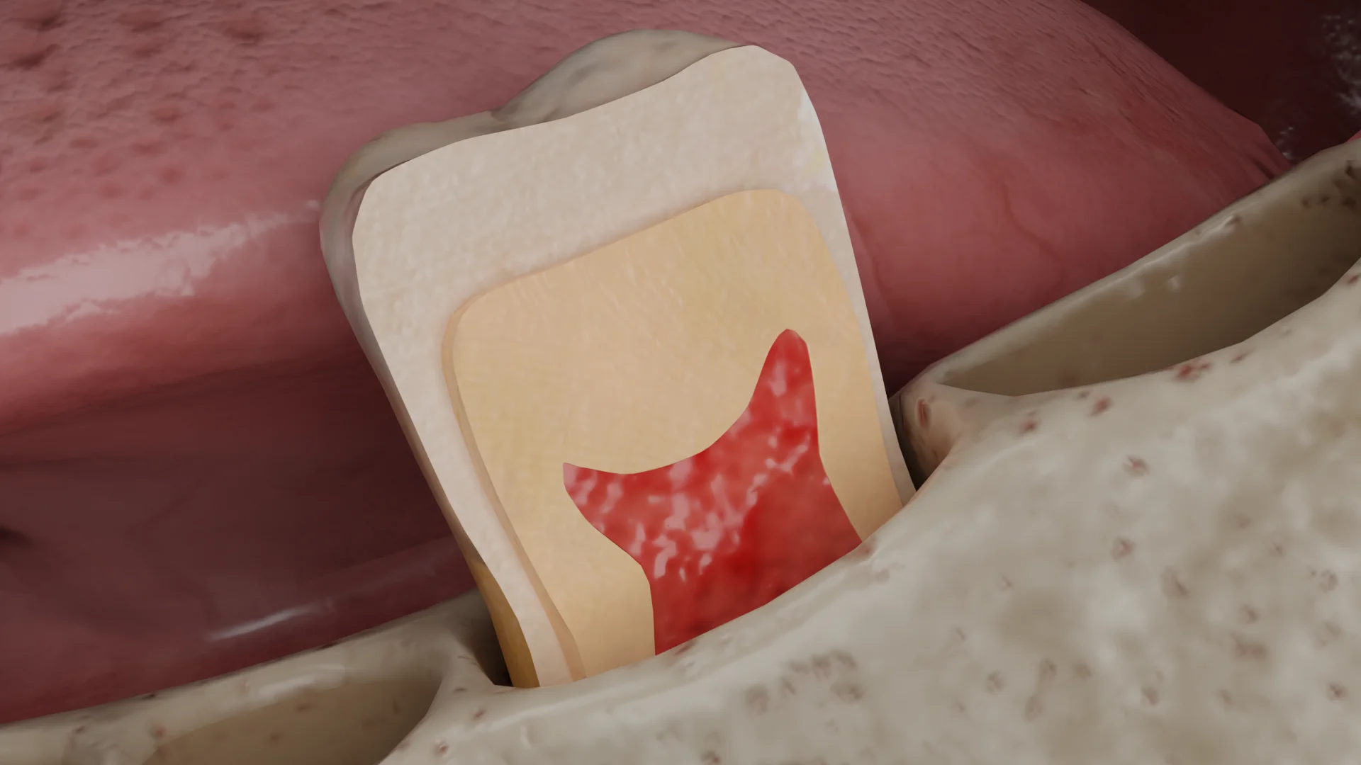
This is an initial stage of decay (E1). After plaque has been removed and the tooth is dry, the enamel is visually integral with a dull white spot of demineralization. At a later stage (E2), the white spot is noticeable even on the moist enamel surface. The spot can be stained in various shades of brown by food dyes. In the enamel, the lesion has the shape of a cone with its apex directed towards the enamel-dentin junction (EDJ). When the process reaches the EDJ and penetrates the dentin, the further direction of the lesion spread corresponds to the course of dentinal tubules.
In this case, the demineralized enamel in the lesion area may show no evidence of decay (a so-called pseudo-intact enamel), thus preventing bacteria from reaching the demineralization focus. Macroscopically, this defect looks like a pigmented fissure or spot on the enamel surface that covers darker edges of the affected dentin.
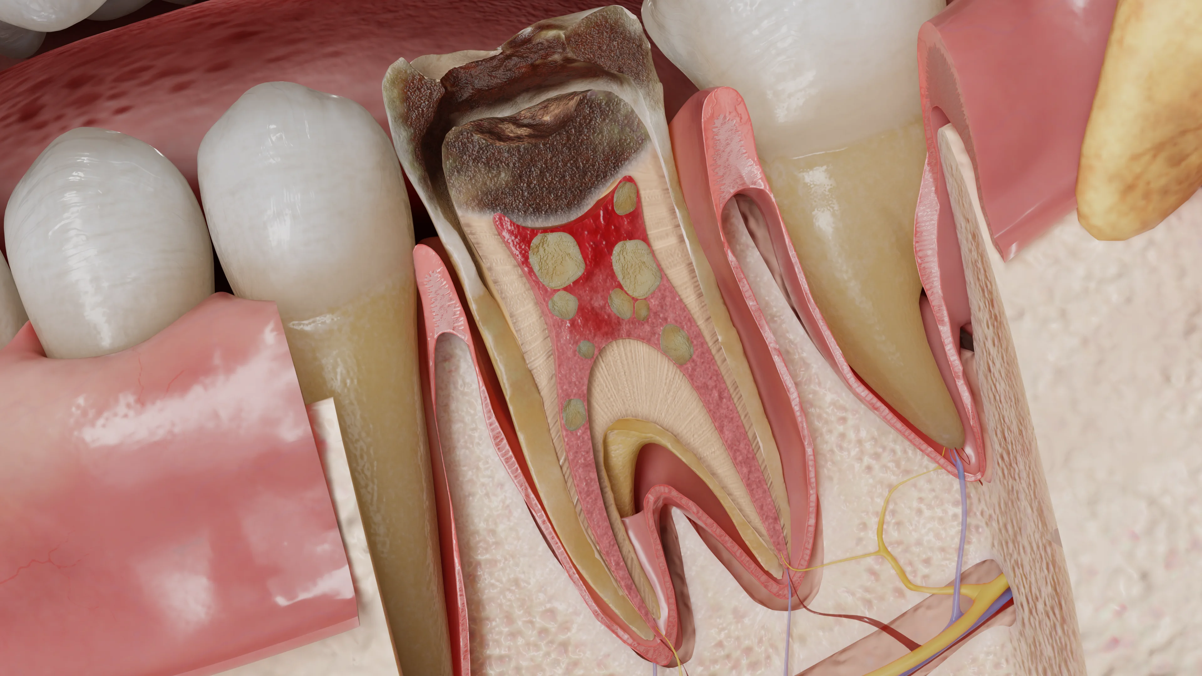
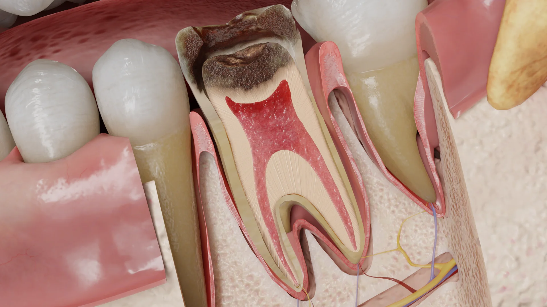
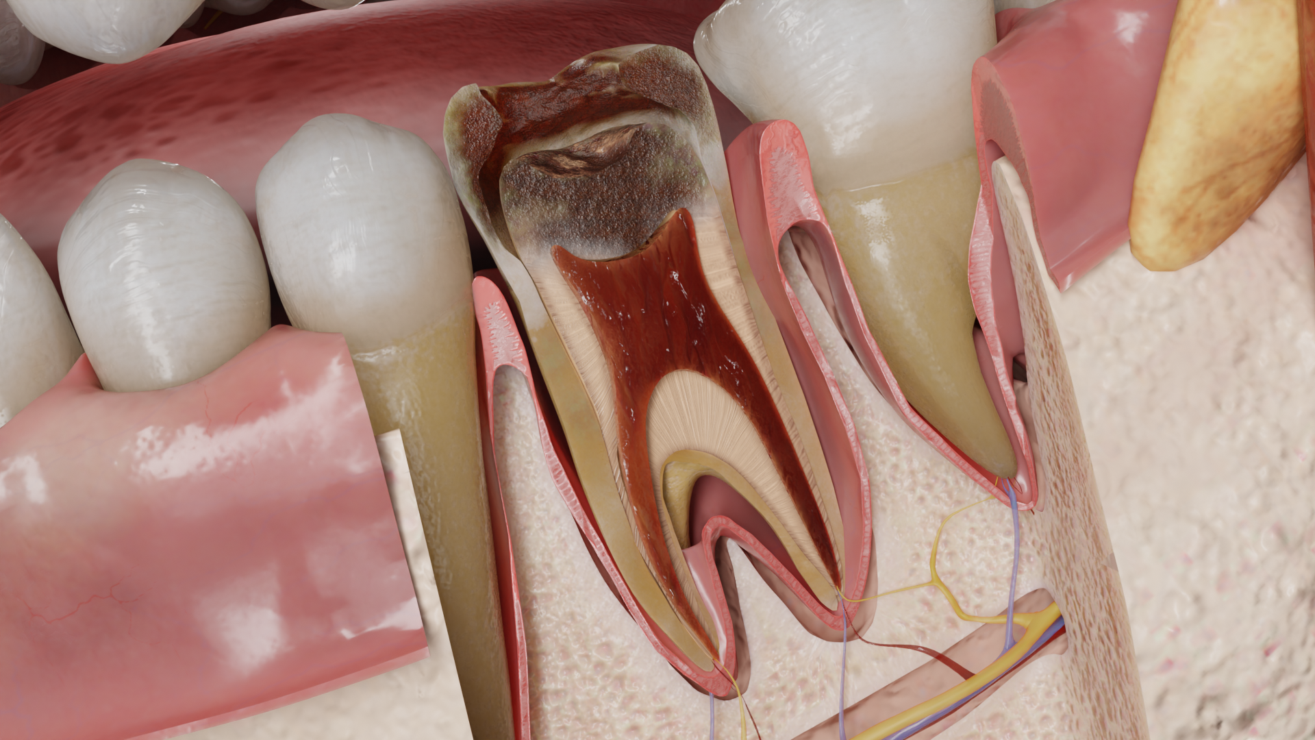
When the damaged enamel loses around 30–40 % of its minerals, it becomes highly porous and fragile. At this point, it can break easily and create a cavity. Bacteria rush deep into the defect, under the influence of their proteases, the demineralized dentin becomes necrotic (bacterial penetration zone), and the demineralization zone spreads deeper towards the pulp. Around the demineralization focus, reactive dentin sclerosis occurs. The outer (D1), middle (D2), and inner (D3) thirds of dentin are sequentially affected. Macroscopically, an active lesion looks like a cavity filled with soft, sticky, yellowish-brown necrotic dentin that is easily removed by scraping. An arrested lesion, on the other hand, presents with a denser and darker-colored dentin.
Cementum caries in the form of a cavity filled with softened dentin develops on the tooth root surface in the presence of gingival recession and exposed root cementum. Its development follows the same mechanisms as crown caries.

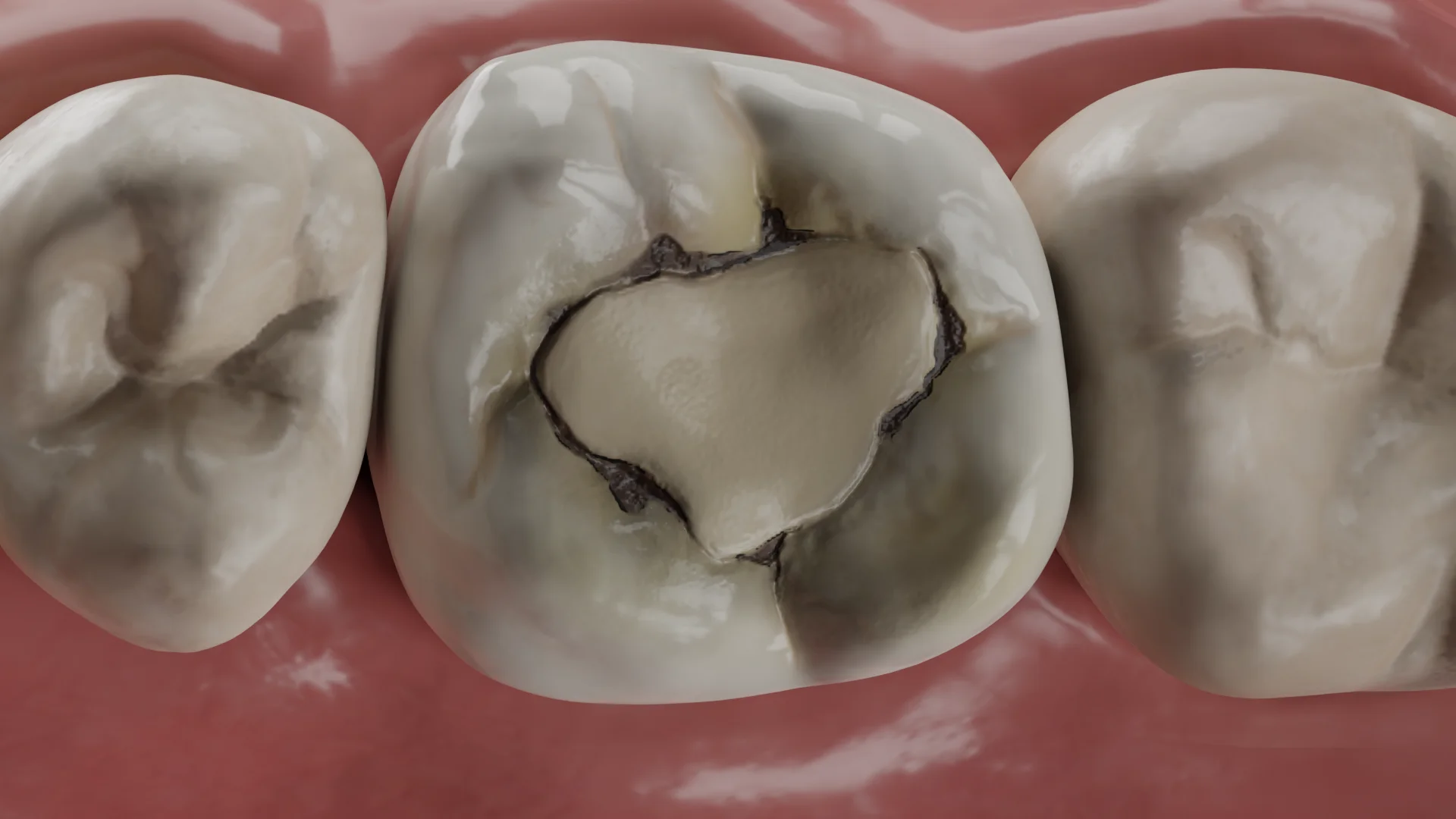
Secondary caries develops as linear staining, a spot, or cavity at the border between tooth tissues and restoration with compromised marginal integrity. This type of lesion is characterized by a superficial demineralized area at the interface between the enamel and the restored tooth, in combination with wall decay. The surface focus spreads along the enamel prisms towards the EDJ and then along the dentinal tubules. The ‘wall’ lesion is a narrow slit-like cavity, later spreading along the dentinal tubules and in the lateral direction.
Enamel caries (E1, E2) do not cause complaints; the patient may be concerned about the aesthetic defect. Clinically, a white or pigmented spot on the enamel is detected.
Dentin caries (D1–D3), cementum caries, and secondary caries: there may be no specific complaints, but a patient may be concerned about a cavity in their tooth, a cosmetic imperfection, short-term localized tenderness after chemical, thermal, mechanical irritation, food impaction, or difficulties when trying to use dental floss. Upon clinical examination, a range of signs may be identified. These may include a pigmented pit or fissure, edges of previous restorations, a probe stuck in them (class I according to Black classification, secondary caries), a gray shadow under the marginal ridge, inflammation of interdental papilla adjacent to the affected tooth, food impaction in the interdental space (class II), dark spots upon transillumination (class II, III), and visible decay cavities made of softened dentin (class I–V, cementum caries).
Find more scientifically accurate content on our social media



In cases of enamel caries without cavities, the defects are treated using remineralization or infiltration methods. The treatment is only effective when combined with proper personal hygiene, nutrition, and follow-up.
Dentin caries, cementum caries, or secondary caries require more invasive techniques, including the preparation of tooth tissues and dental filling using various dental restorative materials.
1. Which microorganisms cause dental caries?
2. How does enamel caries differ from dentin caries?
What are the main methods for treating dental caries?
4. Caries vs. Pulpitis: What are the differences?
Key Differences:
1. Localization:
• Caries affects the enamel and dentin;
• Pulpitis involves the dental pulp.
2. Symptoms:
• Caries: Short-term pain occurs only when exposed to stimuli (cold, heat, sweets);
• Pulpitis: Prolonged pain (lasting more than 5 seconds) may be triggered by temperature stimuli or occur spontaneously and is often characterized as radiating.
3. Treatment:
• Caries: Cavity preparation and filling, remineralization;
• Pulpitis: Root canal treatment (endodontic therapy).
References
1.
VOKA 3D Anatomy & Pathology – Complete Anatomy and Pathology 3D Atlas. VOKA 3D Anatomy & Pathology.
Available from: https://catalog.voka.io/
2.
Toumba KJ, Twetman S, Splieth C, Parnell C, Van Loveren C, Lygidakis NΑ. Guidelines on the use of fluoride for caries prevention in children: an updated EAPD policy document. European Archives of Paediatric Dentistry [Internet]. 2019 Oct 20;20(6):507–516.
Available from: https://doi.org/10.1007/s40368-019-00464-2
3.
Meyer-Lueckel H. Caries management: Science and clinical practice. Stuttgart: Thieme Medical Publishers; 2013.
4.
Li M. Contemporary approach to dental caries. Rijeka: IntechOpen; 2012.
5.
Xuedong Z. Dental caries: Principles and management. Berlin: Springer; 2016.
6.
Kidd EAM, Fejerskov O. Essentials of dental caries. Oxford: Oxford University Press; 2016.
7.
Eden E. Evidence-based caries prevention. Cham: Springer; 2018.
8.
De Oliveira Carrilho MR. Root caries: From prevalence to therapy. Basel: Karger Medical and Scientific Publishers; 2017.
9.
Askar H, Krois J, Göstemeyer G, Bottenberg P, Zero D, Banerjee A, Schwendicke F. Secondary caries: what is it, and how it can be controlled, detected, and managed? Clinical Oral Investigations [Internet]. 2020 Apr 17;24(5):1869–1876.
Available from: https://doi.org/10.1007/s00784-020-03268-7
10.
Askar H, Tu YK, Paris S, Yeh YC, Schwendicke F. Risk of caries adjacent to different restoration materials: Systematic review of in situ studies. Journal of Dentistry [Internet]. 2016 Oct 5;56:1–10.
Available from: https://doi.org/10.1016/j.jdent.2016.09.011
Make VOKA your preferred source
See more VOKA articles in Google Search
Table of Contents









Summarize article with AI
Choose your preferable AI assistant:
Link successfully copied to clipboard
Thank you!
Your message is sent!
Our experts will contact you shortly. If you have any additional questions, please contact us at info@voka.io