Mitral Valve Stenosis: Etiology, Pathogenesis, Symptoms, Diagnosis, and Treatment
Oleg K.Cardiovascular surgeon, MD
13 min read·April 01, 2026
This article is for informational purposes only
The content on this website, including text, graphics, and other materials, is provided for informational purposes only. It is not intended as advice or guidance. Regarding your specific medical condition or treatment, please consult your healthcare provider.
Mitral stenosis is a pathologic narrowing of the mitral valve orifice that impairs diastolic filling of the left ventricle and increases pressure in the left atrium and pulmonary circulation.
In countries with advanced healthcare systems, mitral stenosis is rare and is seen predominantly in patients with a history of rheumatic fever. In developing regions, however, it remains an important cause of valvular heart disease. Women are affected more often than men. Symptoms usually become apparent many years later, often over a period of 10–30 years.
Normal mitral valve. 1 — posterior leaflet; 2 — anterior leaflet; 3 — anterolateral papillary muscle; 4 — posteromedial papillary muscle — 3D Model
Etiology
Rheumatic heart disease is the leading cause of mitral stenosis, accounting for about 90% of cases. In countries with advanced healthcare systems, the incidence of rheumatic mitral stenosis has declined.
Degenerative mitral stenosis is associated with calcification of the mitral annulus. It is the second most common cause of mitral stenosis. The condition is seen predominantly in older patients.
Radiation-induced valvular heart disease develops 10–20 years after mediastinal irradiation and is caused by fibrosis and thickening of valvular structures.
Congenital forms of mitral stenosis are rare and usually present in childhood.
Infiltrative and systemic disorders, including Fabry disease, mucopolysaccharidoses, carcinoid heart disease, endomyocardial fibrosis, and systemic lupus erythematosus, are extremely rare causes.
True mitral stenosis should be distinguished from nonvalvular obstruction of mitral inflow, which may occur in left atrial myxoma, thrombosis, large infective vegetations, or degeneration of a mitral bioprosthesis.
Pathogenesis
Valvular and Anatomic Changes
In rheumatic heart disease, chronic immune-mediated inflammation leads to leaflet fibrosis, commissural fusion, and thickening and shortening of the chordae. This produces central narrowing of the mitral orifice, with doming of the leaflets. Over time, turbulent flow promotes progressive calcification and valvular rigidity, thereby reinforcing the stenosis.
In degenerative calcification of the mitral annulus, fibrocalcific change extends from the annulus to the base of the leaflets, restricting leaflet mobility without the typical commissural fusion.
Narrowing of the mitral orifice increases the diastolic pressure gradient between the left atrium and the left ventricle.
Chronic pressure overload raises left atrial pressure. As the valve area decreases, left ventricular filling becomes progressively limited and cardiac output falls.
Prolonged pressure overload causes left atrial dilatation, hypertrophy, and fibrosis. This promotes atrial fibrillation and creates conditions favorable for left atrial thrombus formation.
Elevated left atrial pressure is transmitted to the pulmonary veins and capillaries, causing post-capillary pulmonary hypertension and pulmonary vascular remodeling.
As the disease progresses, right ventricular overload develops, followed by right-sided heart failure and functional tricuspid regurgitation.
Clinical manifestations
A prolonged asymptomatic phase;
Dyspnea on exertion and, later, at rest. As the disease progresses, orthopnea and paroxysmal nocturnal dyspnea may occur;
Reduced exercise tolerance and fatigue, related to limited cardiac output and impaired diastolic filling of the left ventricle;
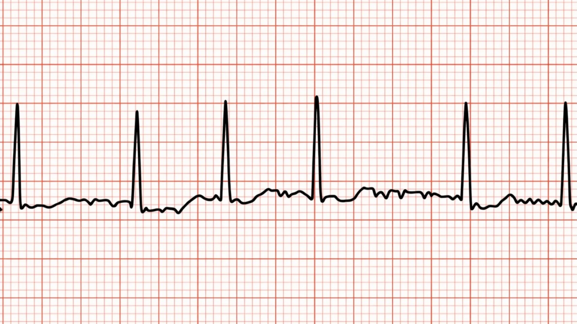
Atrial fibrillation, which may be the first clinical sign of the disease. It often causes abrupt hemodynamic deterioration because of tachycardia and loss of the atrial contribution to ventricular filling;
Example of atrial fibrillation on ECG
Hemoptysis, resulting from pulmonary hypertension and rupture of bronchial veins;
Systemic embolic events, related to the high risk of thrombus formation in the left atrium;
Signs of right-sided heart failure, including lower-extremity edema and ascites;
Hoarseness (Ortner’s syndrome), caused by compression of the recurrent laryngeal nerve by an enlarged left atrium.
Diagnosis
There are no specific laboratory markers for mitral stenosis. BNP or NT-proBNP may be used to assess the severity of heart failure.
Transthoracic echocardiography (TTE) is the key diagnostic method for mitral stenosis.
Morphologic assessment: leaflet thickening and partial fusion; shortening and fibrosis of the chordal apparatus; restricted leaflet mobility; doming of the anterior leaflet in diastole; left atrial size; presence of thrombus in the left atrium; and the condition of the right-sided chambers.
Classification by effective orifice area: ≤ 1.0 cm2 indicates very severe stenosis with particularly high risk; ≤ 1.5 cm2 indicates severe stenosis; > 1.5 cm2 suggests a hemodynamically less significant lesion.
Mean transmitral gradient: severe stenosis is generally associated with a mean gradient greater than 10 mm Hg. p.
Doppler assessment: used to measure transmitral flow velocity and calculate peak and mean gradients. It also helps estimate systolic pulmonary artery pressure from the tricuspid regurgitation jet and identify associated mitral regurgitation.
Stress echocardiography (exercise or dobutamine) is performed when there is a discrepancy between symptom severity and resting TTE findings. The study assesses the increase in transmitral gradient and pulmonary hypertension.
Transesophageal echocardiography (TEE)
TEE provides detailed assessment of leaflet and commissural morphology.
It is performed before balloon mitral commissurotomy to exclude thrombus in the left atrium and left atrial appendage and to further assess the degree of mitral regurgitation.
Spontaneous echo contrast in the left atrium is a marker of marked blood stasis and high thromboembolic risk. It is often associated with a small mitral valve area and a high gradient.
CT angiography is useful for assessing thoracic anatomy, the aorta, and the vessels of the lower extremities, and for choosing the optimal surgical access when mitral valve surgery is planned.
Cardiac catheterization is rarely required and is reserved for cases in which noninvasive findings are inconclusive.
Find more scientifically accurate content on our social media
Subscribe and don’t miss out the latest resources
Treatment
Medical therapy
Medical therapy does not eliminate the obstruction. It is used to control symptoms, stabilize hemodynamics, manage complications, and prepare the patient for intervention when needed.
Diuretics
Loop diuretics such as furosemide reduce pulmonary congestion and relieve dyspnea.
They are effective in pulmonary hypertension and right ventricular failure.
Excessive diuresis may reduce cardiac output and worsen fatigue.
Heart rate control
Beta-blockers are first-line agents. They prolong diastole and reduce the transmitral gradient.
Nondihydropyridine calcium channel blockers (verapamil, diltiazem) are alternatives when beta-blockers are contraindicated.
Ivabradine may be considered in patients with sinus rhythm who cannot tolerate beta-blockers. It lowers heart rate without a negative inotropic effect.
Digoxin may be considered in patients with atrial fibrillation, uncontrolled ventricular rate, or concomitant systolic dysfunction.
Atrial fibrillation
Loss of atrial contraction and tachycardia in mitral stenosis may rapidly lead to pulmonary edema.
Electrical cardioversion is required in hemodynamically unstable patients.
Anticoagulation
Anticoagulation is indicated in patients with mitral stenosis and atrial fibrillation, as well as in those with thrombus in the left atrium and/or left atrial appendage or a history of embolism, regardless of rhythm.
Because the evidence base for direct oral anticoagulants in this setting remains limited, the treatment of choice remains a vitamin K antagonist (warfarin) with a target INR of 2.0–3.0.
Percutaneous balloon mitral commissurotomy (PBMC) is the treatment of choice in patients with rheumatic mitral stenosis and favorable valve anatomy.
Schematic representation of percutaneous balloon mitral commissurotomy
Indications
Symptomatic severe mitral stenosis, most often of rheumatic origin;
Asymptomatic severe stenosis in the presence of pulmonary hypertension (> 50 mm Hg) or atrial fibrillation;
No indication for open surgery;
Suitable anatomy, including mobile leaflets, minimal calcification, limited subvalvular involvement, and an overall favorable morphologic profile.
Contraindications
Thrombus in the left atrium or left atrial appendage;
Moderate or severe mitral regurgitation;
Marked calcification;
Previous unsuccessful PBMC.
Technique
A balloon catheter is advanced through the interatrial septum into the left atrium and inflated across the mitral orifice, splitting the fused commissures.
Surgical Therapy
Surgical commissurotomy and valve-preserving repair
Indicated in patients with anatomy unfavorable for PBMC, left atrial thrombus, or combined valvular disease;
The procedure includes commissurotomy, repair of the subvalvular apparatus, and annuloplasty when needed.
Mitral valve replacement
Indicated in severe valvular deformity, marked calcification, mixed valvular disease, or recurrent stenosis after repeat interventions;
In addition to median sternotomy, mitral valve surgery may be performed via a right mini-thoracotomy with peripheral cardiopulmonary bypass in selected patients with suitable anatomy and adequate peripheral vascular access.
Mechanical prosthetic valves are recommended for younger and middle-aged patients (up to 60 years of age) with expected long-term survival. They require lifelong vitamin K antagonist therapy and are less desirable in women planning pregnancy.
Bioprosthetic valves are recommended in older patients (65 years of age and older) and in women of reproductive age. Their durability is limited by structural degeneration and the potential need for repeat intervention.
Mechanical (left) and bioprosthetic (right) valves
FAQ
1. What is the main cause of mitral stenosis?
In most cases, mitral stenosis is caused by rheumatic heart disease, which leads to progressive narrowing of the mitral valve orifice.
2. How is severe mitral stenosis defined?
Mitral stenosis is generally considered severe when the mitral valve area is 1.5 cm² or less, especially when this is accompanied by symptoms or signs of pulmonary hypertension.
3. Why do symptoms worsen with tachycardia in mitral stenosis?
Tachycardia shortens diastole, increases the transmitral gradient, and raises pressure in the left atrium and pulmonary veins. As a result, symptoms may worsen rapidly.
4. What is the main diagnostic method for mitral stenosis?
Transthoracic echocardiography (TTE) is the principal method for assessing mitral valve area, transmitral gradients, and the severity of pulmonary hypertension.
5. When is transesophageal echocardiography required?
Transesophageal echocardiography (TEE) is performed to exclude thrombus in the left atrium or left atrial appendage and to further evaluate valve anatomy before balloon commissurotomy or surgery.
6. How is symptomatic severe mitral stenosis treated?
The treatment of choice is percutaneous balloon mitral commissurotomy in patients with favorable valve anatomy. If this procedure is contraindicated or not suitable, surgical treatment is recommended.
7. When is surgery preferred in mitral stenosis?
Surgery is indicated when valve anatomy is unfavorable for percutaneous treatment, when significant mitral regurgitation is present, or when thrombus is found in the left atrium.
8. How are patients with mitral stenosis and atrial fibrillation managed?
Management is based on ventricular rate control and anticoagulation. In rheumatic mitral stenosis, anticoagulation with a vitamin K antagonist remains the standard approach.
9. Why are direct oral anticoagulants not recommended in rheumatic mitral stenosis?
Because the available evidence remains insufficient, current guidance continues to favor vitamin K antagonists over direct oral anticoagulants in rheumatic mitral stenosis.
10. Which type of valve prosthesis is preferred when the mitral valve is replaced?
Bioprosthetic valves are generally preferred in patients older than 65 years. In younger patients, mechanical prostheses are often favored, taking into account the need for lifelong anticoagulation.
References
1.
VOKA 3D Anatomy & Pathology – Complete Anatomy and Pathology 3D Atlas. VOKA 3D Anatomy & Pathology.
Available from: https://catalog.voka.io/
2.
Praz, F., et al. (2025). 2025 ESC/EACTS guidelines for the management of valvular heart disease. European Heart Journal, 46(44), 4635–4736. https://doi.org/10.1093/eurheartj/ehaf194
3.
Otto, C. M., et al. (2021). 2020 ACC/AHA guideline for the management of patients with valvular heart disease: A report of the American College of Cardiology/American Heart Association Joint Committee on Clinical Practice Guidelines. Journal of the American College of Cardiology 77(4), e25–e197. https://doi.org/10.1016/j.jacc.2020.11.018
4.
Harb, S. C., & Griffin, B. P. (2017). Mitral valve disease: A comprehensive review. Current Cardiology Reports, 2017 Aug;19(8):73. https://doi.org/10.1007/s11886-017-0883-5
5.
Kumar, K., & Simpson, T. (2024). Transcatheter therapy for mitral valve stenosis. Interventional Cardiology Clinics, 13(2), 271–278. https://doi.org/10.1016/j.iccl.2024.01.003
6.
Wunderlich, N. C., et al. (2019). Rheumatic mitral valve stenosis: Diagnosis and treatment options. Current Cardiology Reports, 21(3), 14. https://doi.org/10.1007/s11886-019-1099-7
7.
Al-Taweel, A., et al. (2019). Degenerative mitral valve stenosis: Diagnosis and management. Echocardiography, 36(10), 1901–1909. https://doi.org/10.1111/echo.14495
8.
Robinson, S., et al. (2021). The assessment of mitral valve disease: A guideline from the British Society of Echocardiography. Echo Research & Practice, 8(1), G87–G136. https://doi.org/10.1530/ERP-20-0034
St. Petersburg FL 33702, 7901 4th St N STE 300, USA
Thank you!
Your message is sent! Our experts will contact you shortly. If you have any additional questions, please contact us at info@voka.io
Cookie Consent
We use cookies to enhance your browsing experience, analyze site traffic, and deliver content. Please choose whether you accept all cookies or wish to reject non-essential tracking.
Cookie Preferences
Manage your cookie preferences below:
Essential cookies enable basic functions and are necessary for the proper function of the website.
Name
Description
Duration
Geolocation Config
This cookie is used to store the consent settings based on the visitor's location.
30 days
Cookie Preferences
This cookie is used to store the user's cookie consent preferences.
30 days
Google reCAPTCHA helps protect websites from spam and abuse by verifying user interactions through challenges.
Name
Description
Duration
_GRECAPTCHA
Google reCAPTCHA sets a necessary cookie (_GRECAPTCHA) when executed for the purpose of providing its risk analysis.
179 days
Statistics cookies collect information anonymously. This information helps us understand how visitors use our website.
Google Analytics is a powerful tool that tracks and analyzes website traffic for informed marketing decisions.
ID used to identify users for 24 hours after last activity
24 hours
_gat
Used to monitor number of Google Analytics server requests when using Google Tag Manager
1 minute
_gac_
Contains information related to marketing campaigns of the user. These are shared with Google AdWords / Google Ads when the Google Ads and Google Analytics accounts are linked together.
90 days
__utma
ID used to identify users and sessions
2 years after last activity
__utmt
Used to monitor number of Google Analytics server requests
10 minutes
__utmb
Used to distinguish new sessions and visits. This cookie is set when the GA.js javascript library is loaded and there is no existing __utmb cookie. The cookie is updated every time data is sent to the Google Analytics server.
30 minutes after last activity
__utmc
Used only with old Urchin versions of Google Analytics and not with GA.js. Was used to distinguish between new sessions and visits at the end of a session.
End of session (browser)
__utmz
Contains information about the traffic source or campaign that directed user to the website. The cookie is set when the GA.js javascript is loaded and updated when data is sent to the Google Anaytics server
6 months after last activity
__utmv
Contains custom information set by the web developer via the _setCustomVar method in Google Analytics. This cookie is updated every time new data is sent to the Google Analytics server.
2 years after last activity
__utmx
Used to determine whether a user is included in an A / B or Multivariate test.
18 months
_ga
ID used to identify users
2 years
_gali
Used by Google Analytics to determine which links on a page are being clicked
30 seconds
Clarity is a web analytics service that tracks and reports website traffic.