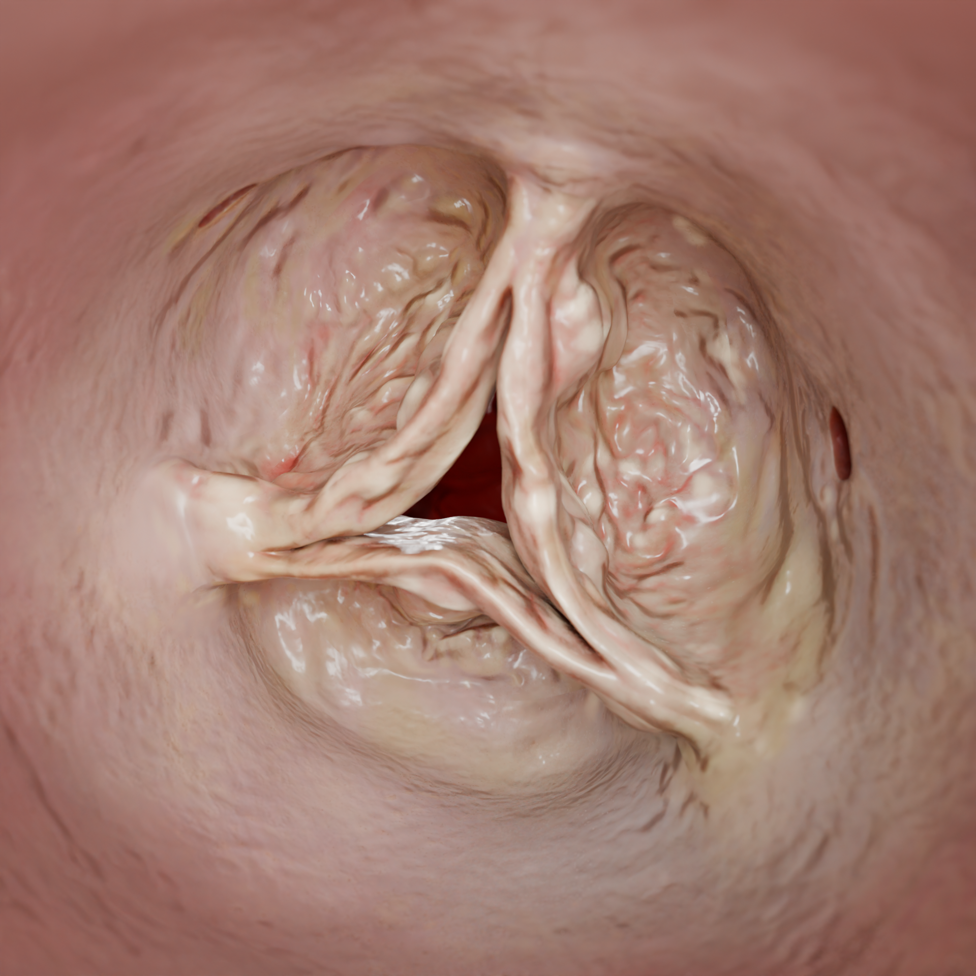
Aortic Stenosis: Etiology, Pathophysiology, Symptoms, Severity, Diagnosis, and Treatment
Oleg K.Cardiovascular surgeon, MD
15 min read·April 14, 2025
This article is for informational purposes only
The content on this website, including text, graphics, and other materials, is provided for informational purposes only. It is not intended as advice or guidance. Regarding your specific medical condition or treatment, please consult your healthcare provider.
Aortic valve stenosis, or aortic stenosis (AS), is a heart valve disease in which the aortic valve is narrowed, obstructing normal blood flow from the left ventricle.
3D Animation: Rheumatic Aortic Valve Stenosis
3D Animation: Degenerative Aortic Valve Stenosis
Epidemiology
Aortic stenosis is the most common form of valvular heart disease. Its prevalence increases significantly with age. Among people older than 75 years, the prevalence of AS may reach 12–13%, while critical aortic stenosis is observed in 3–4% of older patients.
Men are affected by AS more often than women.
In high-income countries with well-developed healthcare systems, AS is diagnosed more frequently. This is associated with better access to medical care and wider availability of diagnostic testing. In developing countries, data on the prevalence of AS are limited, which may reflect underdiagnosis and shorter life expectancy.
In the past, the main cause of aortic stenosis was rheumatic fever, which led to deformation and fusion of the valve cusps. With advances in medicine and a decline in the incidence of rheumatic disease in developed countries, the pattern of etiologic factors has changed. Today, the leading cause of AS is age-related degenerative valve disease, including calcification and fibrosis of the cusps. Atherosclerosis has also become increasingly important in the development of the disease.
The growing number of patients with AS worldwide is primarily related to population aging, reduced mortality from other diseases, and improved availability and quality of diagnostic methods.
Rheumatic Aortic Valve Stenosis — 3D ModelDegenerative Aortic Valve Stenosis — 3D Model
Etiology
Degenerative changes: age-related calcification of the aortic valve cusps, leading to thickening and reduced mobility.
Congenital anomalies: a bicuspid aortic valve, which predisposes patients to earlier calcification and, consequently, to AS.
Rheumatic fever: causes fusion and deformation of the valve cusps.
Less commonly, AS may be associated with conditions such as chronic kidney disease, carcinoid syndrome, Paget disease, and systemic lupus erythematosus.
3D Animation: Development of Aortic Valve Stenosis
Pathophysiology
Narrowing of the valve orifice increases resistance to blood ejection. As a result, systolic pressure in the left ventricle (LV) rises, leading to concentric hypertrophy, meaning thickening of the ventricular walls without significant enlargement of the chamber.
Hypertrophy reduces LV compliance, or distensibility, which impairs diastolic relaxation. Increased diastolic pressure in the LV is transmitted to the left atrium (LA), causing LA hypertrophy and dilatation. Increased diastolic pressure in the left ventricle is transmitted to the left atrium, causing left atrial hypertrophy and dilatation. However, these compensatory mechanisms cannot fully offset the progressive increase in venous pressure within the pulmonary vessels. This leads to congestion in the pulmonary circulation and may trigger signs of heart failure.
In the early stages of AS, cardiac output is maintained by the Frank–Starling mechanism, according to which the force of myocardial contraction increases as myocardial stretch increases. Over time, however, prolonged pressure overload and progressive hypertrophy deplete myocardial reserve. Contractile function then declines, and systolic dysfunction develops.
LV wall hypertrophy increases myocardial oxygen demand. At the same time, elevated diastolic pressure limits coronary blood flow and reduces the efficiency of coronary perfusion. This may lead to myocardial ischemia even in the absence of coronary atherosclerosis.
Decompensated valve disease leads to dilatation of the LV cavity and mitral annulus, resulting in mitral regurgitation.
Clinical Manifestations
The disease may remain asymptomatic for a long time. Over time, the combination of the changes described above leads to the characteristic symptoms of AS: dyspnea, angina, and syncope.
Dyspnea, for instance, may occur due to blood congestion in the pulmonary veins.
Angina develops as a result of ischemia of the hypertrophied myocardium.
Syncope occurs because of reduced cardiac output and insufficient cerebral blood flow.
The onset of these symptoms indicates an unfavorable prognosis and the need to consider aortic valve replacement. Cardiac asthma, meaning episodes of severe breathlessness due to pulmonary congestion, lower-extremity edema, and hepatomegaly (liver enlargement) indicate progression to the decompensated stage and require immediate medical attention.
Diagnosis
Echocardiography is the main diagnostic method for AS. It allows assessment of aortic valve anatomy, the degree of valve calcification, valve area, pressure gradient, and LV size and function. Transthoracic echocardiography (TTE) is recommended for the initial assessment. If TTE does not provide sufficient information, transesophageal echocardiography (TEE) is indicated.
Classification of Aortic Stenosis Severity by Echocardiographic Criteria
Parameter
Mild AS
Moderate AS
Severe AS
Critical AS
Peak velocity (m/s)
< 3.0
3.0–3.9
≥ 4.0
≥ 5.0
Mean gradient (mmHg)
< 20
20–39
≥ 40
≥ 60
Aortic valve area (AVA) (cm²)
> 1.5
1.0–1.5
≤ 1.0
≤ 0.6
Indexed AVA (cm²/m²)
> 0,85
0.6–0.85
≤ 0.6
≤ 0.4
The following parameters are used to evaluate AS (the figures given below indicate cutoffs for severe AS):
Peak aortic jet velocity across the aortic valve (Vmax): a value ≥ 4.0 m/s indicates severe AS.
Mean transvalvular pressure gradient (MPG; ΔPm): a value ≥ 40 mmHg indicates severe AS. p. indicates severe AS.
Aortic valve area (AVA): a value ≤ 1.0 cm², or indexed AVA ≤ 0.6 cm²/m², indicates severe AS.
In patients with low cardiac output and reduced LV ejection fraction (LVEF < 50%), dobutamine stress echocardiography is recommended to distinguish true severe AS from pseudo-severe AS.
MRI and CT may be used as additional imaging methods, including for planning the operative approach.
CT angiography (CTA) is the gold-standard imaging modality before transcatheter aortic valve implantation (TAVI; in U.S. practice, transcatheter aortic valve replacement, or TAVR). It allows assessment of the anatomy of the aortic root and ascending aorta, the degree and extent of valve and vascular calcification, the risk of coronary artery obstruction, and the feasibility of vascular access.
MRI can detect and quantify myocardial fibrosis, which is a marker of decompensated aortic stenosis.
Measurement of B-type natriuretic peptide (BNP), historically called brain natriuretic peptide, or N-terminal pro–B-type natriuretic peptide (NT-proBNP) may be useful for risk stratification in patients with AS. If elevated, these markers are associated with a worse prognosis and may serve as an additional rationale for intervention in asymptomatic patients.
Treatment of Aortic Stenosis
Risk factor modification may help slow the progression of AS and reduce the risk of complications. This includes blood pressure control, maintenance of normal blood lipid levels through diet and lipid-lowering therapy, complete smoking cessation, strict blood glucose control, and maintenance of a healthy weight through balanced nutrition and regular physical activity.
Medication Therapy
At present, no medication has been shown to slow the progression of AS. However, medical therapy may be useful for controlling symptoms and managing concomitant conditions. Diuretics may be used to reduce pulmonary congestion in patients with heart failure. Beta-blockers may help control heart rate and blood pressure, especially in patients with concomitant coronary artery disease. Angiotensin-converting enzyme inhibitors or angiotensin II receptor blockers may be used in patients with heart failure or hypertension. It is important to note that these medications should be used with caution. Excessive reduction in blood pressure may worsen organ perfusion in patients with severe AS.
Aortic valve replacement is indicated in the following cases:
Symptomatic severe AS.
Asymptomatic severe AS with LV systolic dysfunction, defined as LVEF < 50%, or a positive exercise test.
Very severe AS, defined as a mean gradient ≥ 60 mmHg p. or peak velocity > 5.0 m/s.
Rapid clinical deterioration or a sharp increase in the blood BNP level.
Transcatheter aortic valve implantation (TAVI) is a minimally invasive method of aortic valve replacement. The transfemoral arterial approach is used most often. However, when the femoral arteries are too small, tortuous, or affected by atherosclerosis, alternative catheter access routes may be used: transapical access through the LV apex, transaortic access through the ascending aorta, or transsubclavian access through the subclavian artery. Both balloon-expandable and self-expanding transcatheter valves are used.
TAVI should be preferred in patients aged ≥ 75 years and/or those at high surgical risk, defined as EuroSCORE II > 8%, and/or when open surgery is not possible because of comorbidities.
Contraindications to TAVI
Inability to access the aortic valve through the vessels, for example because of severe atherosclerosis or small arterial diameter.
Unfavorable valve or aortic anatomy that makes the procedure technically unfeasible.
Life expectancy is estimated to be less than 1 year or no improvement in the quality of life is expected following the procedure.
3D Animation: Transcatheter Aortic Valve Implantation
Find more scientifically accurate content on our social media
Subscribe and don’t miss out the latest resources
Surgical Aortic Valve Replacement
Open surgical aortic valve replacement (SAVR) remains the standard treatment for many patients. Mechanical or bioprosthetic valves may be implanted. The procedure is performed using cardiopulmonary bypass. Depending on anatomical features, SAVR may be performed through full median sternotomy, ministernotomy, or right anterior minithoracotomy.
Contraindications to SAVR include high surgical risk or comorbidities that make the operation unsafe.
3D Animation: Aortic Valve Replacement
FAQ
1. Why does aortic valve stenosis occur?
The most common scenarios include: • Age-related degenerative changes. • Congenital bicuspid aortic valve. • Sequelae of rheumatic fever. Less commonly, AS may be associated with conditions such as chronic kidney disease, carcinoid syndrome, Paget disease, and systemic lupus erythematosus.
2. What auscultatory findings are typical of aortic valve stenosis?
On auscultation, the following findings may be detected: • A harsh systolic ejection murmur, loudest at the right upper sternal border, in the second right intercostal space, and radiating to the carotid arteries. • A diminished aortic component of the second heart sound due to reduced mobility of the valve cusps. • Paradoxical splitting of the second heart sound in severe AS.
3. How does hemodynamics change in aortic valve stenosis?
• Aortic valve narrowing → Increased pressure in the left ventricle → Concentric LV hypertrophy → Reduced LV compliance → Increased pressure in the left atrium → Pulmonary venous congestion. • Increased myocardial oxygen demand + impaired coronary perfusion → Myocardial ischemia. • With progression → Systolic dysfunction → LV dilatation and mitral regurgitation → Heart failure.
4. What symptoms are typical in patients with aortic valve stenosis?
A characteristic “symptom triad” is seen: 1. Dyspnea, due to pulmonary congestion. 2. Angina, due to ischemia of the hypertrophied myocardium. 3. Syncope, usually during physical exertion. Fatigue, weakness, and dizziness may also occur as a result of reduced cardiac output.
5. How does TAVI differ from surgery?
Transcatheter aortic valve implantation (TAVI) is performed using a catheter and, in most cases, does not require opening the chest. However, it has specific indications. Open surgical aortic valve replacement remains the standard treatment for patients at low surgical risk.
References
1.
VOKA 3D Anatomy & Pathology – Complete Anatomy and Pathology 3D Atlas. VOKA 3D Anatomy & Pathology.
Available from: https://catalog.voka.io/
2.
Vahanian, A., Beyersdorf, F., Praz, F., Milojevic, M., Baldus, S., Bauersachs, J., Capodanno, D., Conradi, L., De Bonis, M., De Paulis, R., Delgado, V., Freemantle, N., Gilard, M., Haugaa, K. H., Jeppsson, A., Jüni, P., Pierard, L., Prendergast, B. D., Sádaba, J. R., … Wojakowski, W. (2022). 2021 ESC/EACTS guidelines for the management of valvular heart disease. European Heart Journal, 43(7), 561–632. https://doi.org/10.1093/eurheartj/ehab395
3.
Chen, J., Li, W., & Xiang, M. (2020). Burden of valvular heart disease, 1990–2017: Results from the Global Burden of Disease Study 2017. Journal of Global Health, 2020 Sep 8;10(2):020404. doi:10.7189/jogh.10.020404.
4.
Joseph, J., Naqvi, S. Y., Giri, J., & Goldberg, S. (2017). Aortic stenosis: Pathophysiology, diagnosis, and therapy. The American Journal of Medicine, 130(3), 253–263. doi:10.1016/j.amjmed.2016.10.005
5.
Zheng, K. H., Tzolos, E., & Dweck, M. R. (2020). Pathophysiology of aortic stenosis and future perspectives for medical therapy. Cardiology Clinics, 38(1), 1–12. doi:10.1016/j.ccl.2019.09.010
6.
Kanwar, A., Thaden, J. J., & Nkomo, V. T. (2018). Management of patients with aortic valve stenosis. Mayo Clin Proc. 2018 Apr;93(4):488–508 doi:10.1016/j.mayocp.2018.01.020
7.
Howard, C., Jullian, L., Joshi, M., Noshirwani, A., Bashir, M., & Harky, A. (2019). TAVI and the future of aortic valve replacement. Journal of Cardiac Surgery, 34(12), 1577–1590. https://doi.org/10.1111/jocs.14226
50 Franklin Street, STE 203 Boston, MA 02110 United States
Thank you!
Your message is sent! Our experts will contact you shortly. If you have any additional questions, please contact us at info@voka.io
Cookie Consent
We use cookies to enhance your browsing experience, analyze site traffic, and deliver content. Please choose whether you accept all cookies or wish to reject non-essential tracking.
Cookie Preferences
Manage your cookie preferences below:
Essential cookies enable basic functions and are necessary for the proper function of the website.
Name
Description
Duration
Geolocation Config
This cookie is used to store the consent settings based on the visitor's location.
30 days
Cookie Preferences
This cookie is used to store the user's cookie consent preferences.
30 days
Google reCAPTCHA helps protect websites from spam and abuse by verifying user interactions through challenges.
Name
Description
Duration
_GRECAPTCHA
Google reCAPTCHA sets a necessary cookie (_GRECAPTCHA) when executed for the purpose of providing its risk analysis.
179 days
Statistics cookies collect information anonymously. This information helps us understand how visitors use our website.
Google Analytics is a powerful tool that tracks and analyzes website traffic for informed marketing decisions.
Used to monitor number of Google Analytics server requests
10 minutes
__utmb
Used to distinguish new sessions and visits. This cookie is set when the GA.js javascript library is loaded and there is no existing __utmb cookie. The cookie is updated every time data is sent to the Google Analytics server.
30 minutes after last activity
__utmc
Used only with old Urchin versions of Google Analytics and not with GA.js. Was used to distinguish between new sessions and visits at the end of a session.
End of session (browser)
__utmz
Contains information about the traffic source or campaign that directed user to the website. The cookie is set when the GA.js javascript is loaded and updated when data is sent to the Google Anaytics server
6 months after last activity
__utmv
Contains custom information set by the web developer via the _setCustomVar method in Google Analytics. This cookie is updated every time new data is sent to the Google Analytics server.
2 years after last activity
__utmx
Used to determine whether a user is included in an A / B or Multivariate test.
18 months
_ga
ID used to identify users
2 years
_gali
Used by Google Analytics to determine which links on a page are being clicked
30 seconds
_ga_
ID used to identify users
2 years
_gid
ID used to identify users for 24 hours after last activity
24 hours
_gat
Used to monitor number of Google Analytics server requests when using Google Tag Manager
1 minute
_gac_
Contains information related to marketing campaigns of the user. These are shared with Google AdWords / Google Ads when the Google Ads and Google Analytics accounts are linked together.
90 days
__utma
ID used to identify users and sessions
2 years after last activity
Clarity is a web analytics service that tracks and reports website traffic.