Chemodectoma (Paraganglioma): Etiology, Pathogenesis, Classification, Diagnosis, and Treatment Methods
Oleg K.Cardiovascular surgeon, MD
10 min read·May 07, 2025
This article is for informational purposes only
The content on this website, including text, graphics, and other materials, is provided for informational purposes only. It is not intended as advice or guidance. Regarding your specific medical condition or treatment, please consult your healthcare provider.
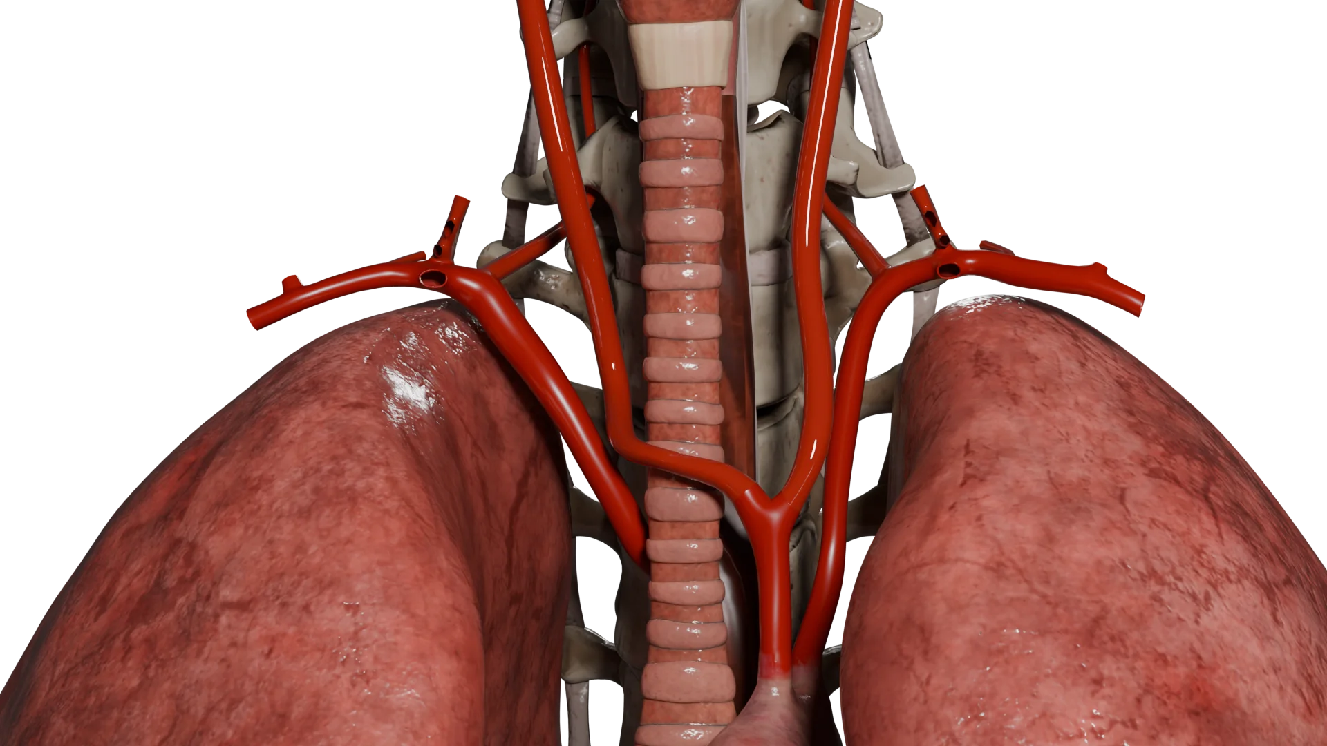
Carotid body paraganglioma (chemodectoma) is a rare, mostly benign tumor arising from the chemoreceptor cells of the carotid body located at the bifurcation of the common carotid artery.
The average age of diagnosis is about 45 years old.
The prevalence is high in Latin American countries, especially in Mexico, where women account for up to 90% of cases.
Residents of high altitude areas have an increased risk of developing chemodectomas due to chronic hypoxia.
Etiology
Genetic mutations — mutations in the genes encoding the subunits of the succinate dehydrogenase complex (SDH), involved in the mitochondrial respiratory chain and cellular metabolism, play a major role. Key genes:
SDHD — most commonly associated with multiple head and neck paragangliomas; inherited paternally;
SDHB — associated with more aggressive forms and an increased risk of malignancy;
SDHC, SDHA, SDHAF2 — less frequently involved, but can also contribute to tumor development.
Syndromes, associated with chemodectomas:
von Hippel-Lindau syndrome (VHL);
multiple endocrine neoplasia type 2 (MEN2);
neurofibromatosis type 1 (NF1).
Chronic hypoxia — living at an altitude of more than 2000 m above sea level, as well as conditions such as chronic obstructive pulmonary disease and congenital heart defects, can stimulate carotid body hyperplasia.
Family history — about 10% of cases are familial.
3D animation — chemodectoma type I
3D animation — chemodectoma type III
Pathogenesis
Hereditary mutations (SDHx)
Mutations SDH (a key enzyme of complex II of the mitochondrial respiratory chain and Krebs cycle) → accumulation of succinate (acts as oncometabolite) → inactivation of prolyl hydroxylases (PHDs), enzymes controlling degradation of HIF-1α (hypoxia-inducible factor) → accumulation of HIF-1α → pseudohypoxia → activation of VEGF (angiogenesis) / GLUT1 (glycolysis) / PDGF (cell proliferation) → neoplastic transformation of chemoreceptor cells and tumor formation.
Chronic hypoxia
Hypoxia (altitude, COPD) → decreased pO₂ → activation of carotid receptors → cell hyperplasia → accumulation of HIF-1α → angiogenesis and neoplasia.
As a result, the tumor gradually increases in size. As it grows, it may demonstrate locally invasive characteristics, to the extent of surrounding or compressing nearby anatomical structures such as the internal and external carotid arteries, vagus, hypoglossal, and glossopharyngeal nerves. This can cause accompanying neurological and vascular symptoms.
Classification of paraganglioma
William Shamblin proposed dividing carotid body paragangliomas into three types (Shamblin classification):
Type I: limited tumors up to 3.5 cm, loosely connected to arterial walls;
Type II: 3.5–5 cm, partially encompass carotid arteries, have a denser fusion with arterial walls;
Type III: more than 5 cm, enclosing carotid arteries and/or adjacent vessels and nerves with a markedly dense connection to these structures.
Additionally, chemodectomas can be classified by etiology: sporadic (up to 85%), familial (10–15%), hyperplastic (1–5%).
Paraganglioma Type I
Paraganglioma Type III
Clinical manifestations
For long periods of time, symptoms may be absent. As the tumor grows, complaints may appear:
Palpable tumor — slow-growing, painless, pulsating mass on the lateral surface of the neck (in the region of the sternocleidomastoid muscle);
Neurological symptoms — hoarseness, dysphagia, tongue numbness due to compression of cranial nerves (IX–XII);
Complaints related to catecholamine production — palpitations, episodes of hypertension, sweating, headache, tremor;
Rare symptoms — dizziness, fainting due to carotid sinus compression.
Diagnosis of paraganglioma
Physical examination: firm, pulsating tumor on the neck, moves horizontally but not vertically.
Ultrasound with Doppler: hypervascular tumor at the carotid bifurcation.
MRI with contrast: characteristic “salt-and-pepper” appearance on T1-weighted images.
CT with contrast: determination of the degree of vessel invasion, evaluation of Shamblin classification. Construction of a 3D model for planning surgery.
Angiography: identification of the “harp sign”—divergence of the internal and external carotid arteries. Allows for assessing the need/possibility of embolization in this area as a pre-surgery stage.
PET-CT with 68Ga-DOTATATE: if multiple or metastatic foci are suspected.
Laboratory tests: determine plasma and urine levels of metanephrines and normetanephrines if a secreting tumor is suspected.
Genetic testing for mutations in SDH genes in the presence of family history or multiple tumors.
Find more scientifically accurate content on our social media
Subscribe and don’t miss out the latest resources
Treatment
Treatment of carotid chemodectoma depends on tumor size, clinical symptomatology, risk of complications, functional activity, genetic profile, and Shamblin grade. The main treatment modalities are surgical excision, preoperative embolization, and in some cases radiotherapy or chemotherapy.
Preoperative embolization
Preoperative embolization is performed to reduce tumor vascularization, decrease intraoperative blood loss, and facilitate tumor dissection during surgery.
Indications:
shamblin grade II-III tumors;
tumor diameter > 3 cm;
presence of significant arterial blood flow (according to CT/angiography);
planned resection with vascular reconstruction.
Contraindications:
shamblin I tumors;
absence of an arterial component (low vascularization);
anastomoses with cerebral circulation – risk of cerebral embolization.
Selective angiography of the external carotid artery is performed. Embolizing agents are introduced through a microcatheter: polyvinyl alcohol particles (PVA), microspheres, less often glue or coils. Control angiography confirms blood flow reduction. Surgery is performed within 24-48 hours after embolization as long as the effect persists.
Extracapsular resection of the tumor. Standard for Shamblin I-II, vascular preservation, minimal risk.
Carotid resection with reconstruction. More often at Shamblin III. After resection of the carotid artery site, a prosthesis (Dacron, PTFE) is performed.
Neuromonitoring and microsurgery. It is used when close to cranial nerves (IX-XII).
Possible complications:
bleeding (especially with inadequate embolization);
damage to cranial nerves (IX-XII) – up to 30% in large tumors;
stroke (when collateral blood flow is impaired);
recurrence (rare, with incomplete resection).
Chemotherapy
It is not the standard treatment for carotid chemodectoma, as most tumors are slow growing and benign in nature. However, in rare cases of malignant course (rapid progression, metastatic lesions) or in inoperable metastatic forms, systemic therapy may be used.
Radionuclide therapy ¹⁷⁷Lu-DOTATATE (PRRT)
Prescribed in the presence of somatostatin receptor expression by PET with ⁶⁸⁸Ga-DOTATATE.
FAQ
1. Is carotid chemodectoma a malignant tumor?
In most cases, it is not. The carotid chemodectoma is considered a benign, slowly growing tumor. However, with the presence of an SDHB gene mutation, there is a potential for malignancy with metastasis.
2. Is it possible to simply observe the carotid chemodectoma without removing it?
Yes, in some cases active surveillance is possible, especially if the tumor is small ( 2.5 cm), asymptomatic, or the patient is elderly or has contraindications to surgery. However, if there are signs of growth or compression of neck structures, treatment is required.
3. Is surgery to remove carotid chemodectoma dangerous?
The risks depend on the size and location of the tumor. In large formations, complications may include cranial nerve damage, bleeding, and ischemic events. Preoperative embolization is often performed to reduce risks.
4. Should other family members be screened if I am diagnosed with a chemodectoma?
If you have an identified hereditary mutation (for example, SDHD, SDHB), genetic counseling and screening of close relatives are recommended as the disease may be familial.
5. How to distinguish carotid chemodectoma from other neck tumors?
The carotid chemodectoma is usually located in the region of the bifurcation of the common carotid artery, pulsates, shifts horizontally but not vertically, and shows a characteristic “lyre sign” on CT/MRI. The diagnosis is clarified using imaging and angiography.
6. Can a chemodectoma cause pain or discomfort?
In most cases, it does not, especially in early stages. However, as the tumor grows, it may exert pressure on nerves and vessels, leading to pain, hoarseness, swallowing disorders, or dizziness.
7. Is there a risk of recurrence after tumor removal?
With complete tumor removal, the risk of recurrence is low (less than 5%). However, with incomplete resection, hereditary forms, or the presence of an SDHB mutation, recurrence or the development of new foci may occur. In such cases, long-term follow-up is important.
References
1.
VOKA 3D Anatomy & Pathology – Complete Anatomy and Pathology 3D Atlas. VOKA 3D Anatomy & Pathology.
Available from: https://catalog.voka.io/
2.
Luna-Ortiz K, Reynoso-Noverón N, Herrera-Ponzanelli C, Favila-Lira S, Luna-Peteuil Z, Herrera-Gomez A, et al. Sex differences according to ethnic presentation in carotid body tumors: a systematic literature review. Int J Otorhinolaryngol Head Neck Surg. 2022 Jun;8(6):527-531. doi: 10.18203/issn.2454-5929.ijohns20221393.
3.
Butt N, Baek WK, Lachkar S, Iwanaga J, Mian A, Blaak C, et al. The carotid body and associated tumors: updated review with clinical/surgical significance. Br J Neurosurg. 2019 Oct;33(5):500-503. doi: 10.1080/02688697.2019.1617404.
4.
Darouassi Y, Alaoui M, Mliha Touati M, Al Maghraoui O, En-Nouali A, Bouaity B, Ammar H. Carotid Body Tumors: A Case Series and Review of the Literature. Ann Vasc Surg. 2017 Aug;43:265-271. doi: 10.1016/j.avsg.2017.03.167.
5.
Gonzalez-Urquijo M, Castro-Varela A, Barrios-Ruiz A, Hinojosa-Gonzalez DE, Salas AKG, Morales EA, et al. Current trends in carotid body tumors: comprehensive review. Head Neck. 2022 Oct;44(10):2316-2332. doi: 10.1002/hed.27147.
6.
Ozawa H. Current management of carotid body tumors. Auris Nasus Larynx. 2024 Jun;51(3):501-506. doi: 10.1016/j.anl.2024.01.007.
7.
Piazza C, Lancini D, Tomasoni M, Zafereo M, Poorten VV, Hanna E, et al. Malignant carotid body tumors: what we know, what we do, and what we need to achieve. A systematic review of the literature. Head Neck. 2024 Mar;46(3):672-687. doi: 10.1002/hed.27624.
405 Lexington Avenue, 26th Floor New York, NY 10174 United States
Thank you!
Your message is sent! Our experts will contact you shortly. If you have any additional questions, please contact us at info@voka.io
Cookie Consent
We use cookies to enhance your browsing experience, analyze site traffic, and deliver content. Please choose whether you accept all cookies or wish to reject non-essential tracking.
Cookie Preferences
Manage your cookie preferences below:
Essential cookies enable basic functions and are necessary for the proper function of the website.
Name
Description
Duration
Geolocation Config
This cookie is used to store the consent settings based on the visitor's location.
30 days
Cookie Preferences
This cookie is used to store the user's cookie consent preferences.
30 days
Google reCAPTCHA helps protect websites from spam and abuse by verifying user interactions through challenges.
Name
Description
Duration
_GRECAPTCHA
Google reCAPTCHA sets a necessary cookie (_GRECAPTCHA) when executed for the purpose of providing its risk analysis.
179 days
Statistics cookies collect information anonymously. This information helps us understand how visitors use our website.
Google Analytics is a powerful tool that tracks and analyzes website traffic for informed marketing decisions.
Used to monitor number of Google Analytics server requests
10 minutes
__utmb
Used to distinguish new sessions and visits. This cookie is set when the GA.js javascript library is loaded and there is no existing __utmb cookie. The cookie is updated every time data is sent to the Google Analytics server.
30 minutes after last activity
__utmc
Used only with old Urchin versions of Google Analytics and not with GA.js. Was used to distinguish between new sessions and visits at the end of a session.
End of session (browser)
__utmz
Contains information about the traffic source or campaign that directed user to the website. The cookie is set when the GA.js javascript is loaded and updated when data is sent to the Google Anaytics server
6 months after last activity
__utmv
Contains custom information set by the web developer via the _setCustomVar method in Google Analytics. This cookie is updated every time new data is sent to the Google Analytics server.
2 years after last activity
__utmx
Used to determine whether a user is included in an A / B or Multivariate test.
18 months
_ga
ID used to identify users
2 years
_gali
Used by Google Analytics to determine which links on a page are being clicked
30 seconds
_ga_
ID used to identify users
2 years
_gid
ID used to identify users for 24 hours after last activity
24 hours
_gat
Used to monitor number of Google Analytics server requests when using Google Tag Manager
1 minute
_gac_
Contains information related to marketing campaigns of the user. These are shared with Google AdWords / Google Ads when the Google Ads and Google Analytics accounts are linked together.
90 days
Clarity is a web analytics service that tracks and reports website traffic.