Odontogenic Sinusitis: Classification, Etiology, Clinical Presentation, Diagnosis, and Treatment
A detailed overview of odontogenic maxillary sinusitis: causes, symptoms of acute and chronic inflammation, diagnosis, and modern treatment approaches.


Anesthesia
Pain management and sedation techniques

Angiology
Arterial and venous pathologies

Cardiology
Acquired and congenital heart diseases

Dentistry
Diseases of teeth, gums, and the oral cavity

Dermatology
Disorders of the skin and subcutaneous tissue

Embryology
Stages of embryogenesis and congenital anomalies

Endocrinology
Disorders of the glands and hormonal imbalance

Gastroenterology
Stomach, intestinal, and digestive diseases

Gynecology
Diseases of female reproductive organs

Hematology
Hematopoiesis and blood-related disorders

Hepatology
Liver, gallbladder, and biliary tract diseases

Histology
Microscopic tissue and cell structures

Infectious diseases
Bacterial, viral, and parasitic infections

Neurology
Brain, spinal cord, and peripheral nerve disorders

Obstetrics
Pregnancy complications and abnormal fetal positions

Oncology
Cancer types, benign and malignant tumors

Ophthalmology
Conditions affecting the eyes and vision

Orthopedics
Bone, joint, and soft tissue disorders

Otorhinolaryngology
Ear, nose, and throat diseases

Pediatrics
Child health, development, and clinical conditions

Physiology
Biological processes within organs and systems

Plastic surgery
Reconstructive and aesthetic procedures

Pulmonology
Lung and respiratory tract diseases

Traumatology
Acute injuries and musculoskeletal trauma

Urology
Urinary tract and male reproductive disorders
Anesthesia
Pain management and sedation techniques
Angiology
Arterial and venous pathologies
Cardiology
Acquired and congenital heart diseases
Dentistry
Diseases of teeth, gums, and the oral cavity
Dermatology
Disorders of the skin and subcutaneous tissue
Embryology
Stages of embryogenesis and congenital anomalies
Endocrinology
Disorders of the glands and hormonal imbalance
Gastroenterology
Stomach, intestinal, and digestive diseases
Gynecology
Diseases of female reproductive organs
Hematology
Hematopoiesis and blood-related disorders
Hepatology
Liver, gallbladder, and biliary tract diseases
Histology
Microscopic tissue and cell structures
Infectious diseases
Bacterial, viral, and parasitic infections
Neurology
Brain, spinal cord, and peripheral nerve disorders
Obstetrics
Pregnancy complications and abnormal fetal positions
Oncology
Cancer types, benign and malignant tumors
Ophthalmology
Conditions affecting the eyes and vision
Orthopedics
Bone, joint, and soft tissue disorders
Otorhinolaryngology
Ear, nose, and throat diseases
Pediatrics
Child health, development, and clinical conditions
Physiology
Biological processes within organs and systems
Plastic surgery
Reconstructive and aesthetic procedures
Pulmonology
Lung and respiratory tract diseases
Traumatology
Acute injuries and musculoskeletal trauma
Urology
Urinary tract and male reproductive disorders
Specialties
AnesthesiaAngiologyCardiologyDentistryDermatologyEmbryologyEndocrinologyGastroenterologyGynecologyHematologyHepatologyHistologyInfectious diseasesNeurologyObstetricsOncologyOphthalmologyOrthopedicsOtorhinolaryngologyPediatricsPhysiologyPlastic surgeryPulmonologyTraumatologyUrology
This article is for informational purposes only
The content on this website, including text, graphics, and other materials, is provided for informational purposes only. It is not intended as advice or guidance. Regarding your specific medical condition or treatment, please consult your healthcare provider.
Infectious diseases of the auricle represent localized inflammation of the skin and underlying structures, including hair follicles, subcutaneous fat, muscles, cartilage, and perichondrium.
Auricular infections:
Infectious Diseases of Auricle — 3D Models
 Auricular erysipelas
 Auricular perichondritis
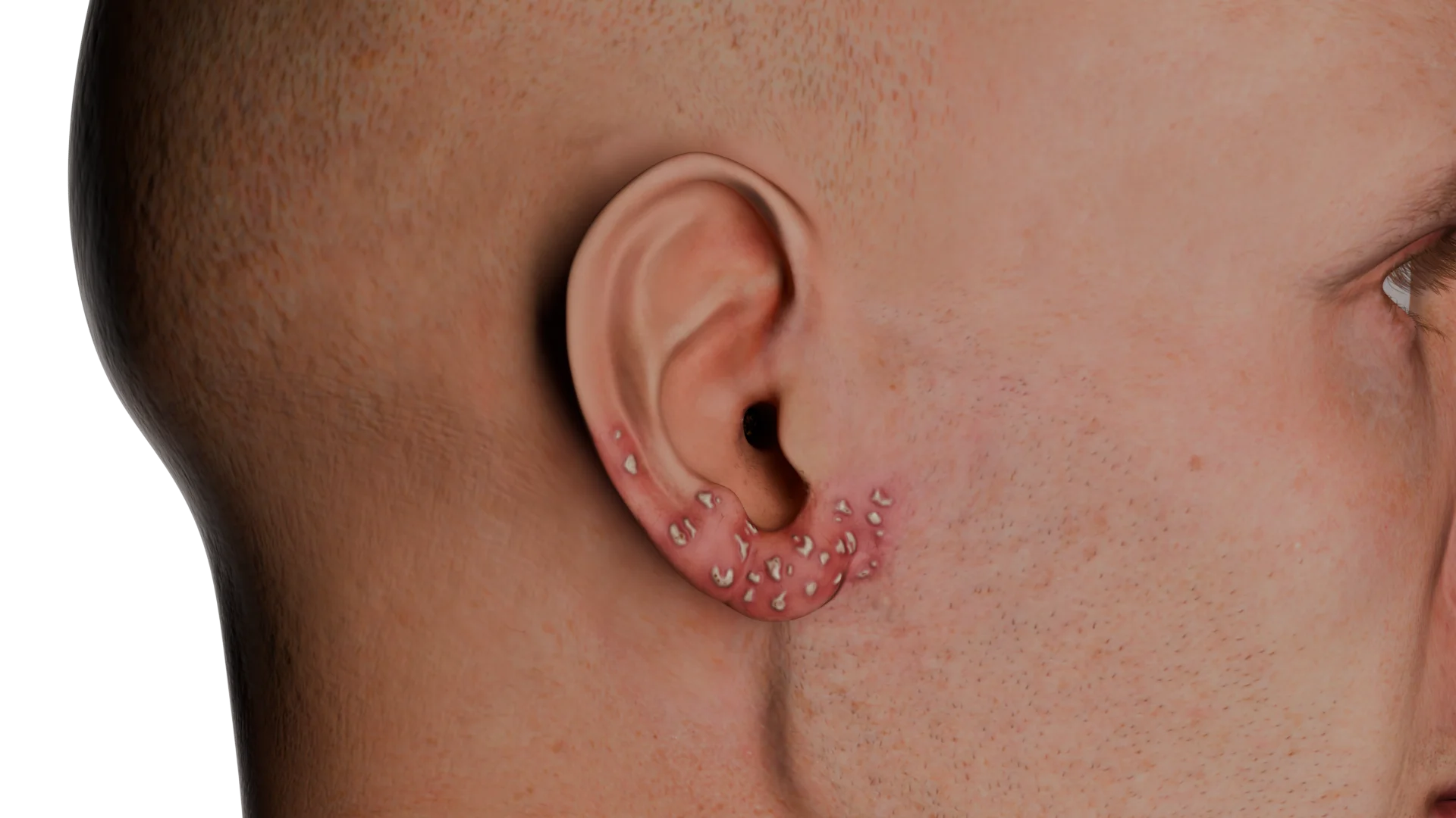 Herpetic infection of auricle
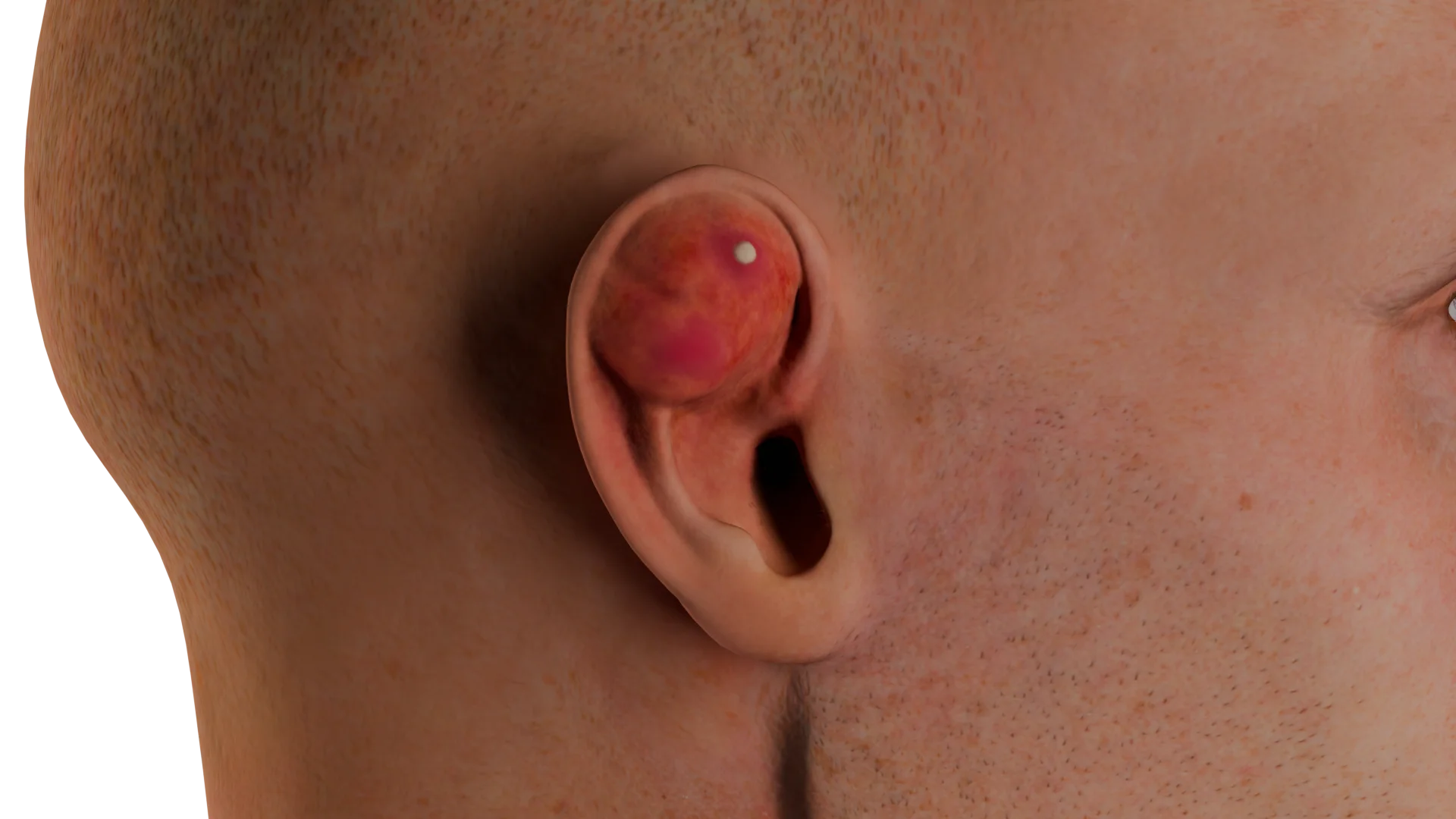 Auricular abscess
External ear infections can be caused by various microorganisms (bacteria, viruses, fungi, etc.) or their combinations (mixed infections). It is important to note that a disease can only develop if there are certain predisposing factors, such as:
The most common bacterial pathogens include:
The most severe infections are typically caused by:
Herpetic infections of the auricle are caused by herpes simplex virus type 1 (HSV-1) and human herpesvirus 3 (varicella-zoster virus; VZV).
Auricular erysipelas is an acute bacterial infection of the skin and the fatty layer of the subcutaneous tissue in the auricular region, including the earlobe.
The inflammation often extends beyond the auricle and continues to the neck, temporal, and buccal regions. It can be identified by a clear line of demarcation between involved and uninvolved tissue. The affected area is characterized by erythema and significant swelling, causing the skin to appear stretched and shiny. Moreover, regional lymph nodes may be involved, resulting in lymphadenitis.
In some cases, hemorrhagic and serous-filled blisters may appear, which subsequently burst and become covered with crusts.
Auricular perichondritis is a diffuse inflammation of the tissue covering the cartilage of the external ear (perichondrium). The skin of the auricle is also affected, while the earlobe remains intact.
Typically, this condition arises when an infectious pathogen enters the system through damaged skin or suppuration of an undrained auricular hematoma or seroma develops. In some cases, inflammation may develop as a complication of otitis externa.
The most common symptoms are local hyperemia and heterogeneous swelling of the auricle. Notably, the inflammatory process does not spread beyond the auricle and does not involve the earlobe.
Auricular abscess is a localized collection of pus between the perichondrium and the cartilage of the auricle. There may be a variety of causes for an auricular abscess. These include the lack or absence of proper treatment for perichondritis, an infection of an auricular hematoma, and immunodeficiency disorders.
It looks like a cavity filled with purulent fluid surrounded by inflamed auricular tissues. The skin around an abscess typically appears red, swollen, and stretched. Fluctuation can also be observed.
Herpetic infections of the auricle are a group of recurrent skin diseases caused by herpes viruses type 1 (HSV-1) and 3 (VZV).
After the initial infection, the herpes virus travels to the nerve ganglia, where it remains inactive for some time. This is called the latent (dormant) stage. The virus reactivates and spreads along the nerve fibers in response to a number of internal and external factors, including a weakened immune system, stress, UV radiation, exacerbation of chronic conditions, etc. This leads to an outbreak (exacerbation stage), which is characterized by typical skin changes.
The affected area becomes erythematous and swollen, with numerous small blisters on it. In 2–3 days, these vesicles rupture and crust over. Subsequently, the crusts fall off when the healing is complete. If scratched, a bacterial infection may develop, thus prolonging and complicating the course of the disease.
In HSV-1 infection, vesicles can spread randomly all over the skin. VZV, on the other hand, gives rise to two clinically distinct forms of disease. Chickenpox represents a primary infection that prevails in children. The other form of the disease is herpes zoster, also known as shingles (Herpes zoster oticus), which develops when the latent virus reactivates. It is characterized by the involvement of the dermatome, an area of skin innervated by a particular nerve. VZV typically affects the facial nerve, which can lead to its paresis or paralysis. The virus may also attack the geniculate ganglion of the facial nerve, resulting in Ramsay Hunt syndrome. Rarely, the infection may involve the trigeminal or vestibulocochlear nerves.
Since the infectious process spreads along the facial nerve, it can affect not only the skin of the auricle and external auditory canal but also the tympanic membrane. In severe cases, the central nervous system may be involved, leading to meningitis and encephalitis. Immunocompromised patients are at risk of developing a generalized herpetic infection because the virus can spread throughout the body. It is usually a recurrent disease that is extremely difficult to treat.
Auricular erysipelas is characterized by local skin changes and prominent systemic symptoms of intoxication. Fever may be accompanied by fatigue, headache, and, in some cases, vomiting.
On local examination, there is marked erythema and swelling of the auricle, including the earlobe. In some cases, inflammation extends beyond the auricle and the earlobe, continuing to the neck, retroauricular, and buccal regions. The affected area is clearly demarcated from the surrounding healthy tissue. The involved skin is tender and warm to touch and looks stretched and shiny. The regional lymph nodes become enlarged, tender, and elastic, not adhering to the surrounding tissues.
In auricular perichondritis, typical symptoms are erythema and heterogeneous swelling of the tissues covering the cartilage of the auricle. It is important to note that the inflammation does not spread to the earlobe and the surrounding areas, including the retroauricular region. Patients usually complain of severe aching or paroxysmal pain in this area. General intoxication symptoms are not typical, but a low-grade fever may occasionally be observed. If left untreated, the infection can involve deeper tissues and lead to an auricular abscess. Sometimes, the outcome of perichondritis is an ear deformity known as “cauliflower ear”, which may develop even with proper treatment.
As mentioned earlier, an auricular abscess typically results from a previous ear injury. Patients usually complain of general intoxication symptoms (fatigue, fever, chills, etc.). On examination, an abscess looks like a cavity filled with pus and surrounded by a capsule. It bulges above the tense and tender skin, and fluctuation is evident. The cavity may become so enlarged that the EAC can no longer be visualized. The deformity of the affected areas, most commonly the triangular and scaphoid fossae, can also be observed. Within 3–4 days, untreated abscesses may spontaneously rupture and drain, resulting in a purulent discharge.
HSV-1 infection is characterized by typical local changes of the skin. During an exacerbation, the affected area becomes hyperemic with multiple painless vesicles on it, which is accompanied by intense itching. The vesicles usually rupture within 2–3 days, resulting in small erosions that become covered with crusts. The healing process takes approximately 5–7 days, during which time the crusts completely disappear and the skin returns to its normal state. Most people do not experience systemic manifestations of infection. However, a low-grade fever and headache may occasionally be observed a few days before the rash develops. It is worth noting that recurrent outbreaks are common in this form of herpetic infection.
Herpes zoster, caused by VZV, differs clinically from HSV-1 infection. As stated above, this form of the disease results from the reactivation of a latent virus in individuals who have had chickenpox. It is more common in older adults and immunocompromised individuals.
A few days before the rash appears, there may be severe itching, burning sensations, and acute throbbing pain in the skin area prone to lesions. Patients may also have a fever, headache, and chills. Local changes manifest after a while, often within 2 to 3 days, when vesicles form on the hyperemic skin, spreading along the dermatome. The area of the lesion can be large and affect several adjacent dermatomes since the blisters can merge. It should be noted that the rash always distributes unilaterally. Intense pruritus and pain do not subside over time, which can sometimes lead to psychological issues such as anxiety and insomnia. If scratched, hemorrhagic crusts or secondary bacterial infections may develop.
The vesicles rupture within 5 to 7 days, forming erosions that eventually become crusted over. Subsequently, the crusts fall off after the healing process is complete. This area often remains depigmented. Postherpetic neuralgia (PHN) is the most common long-term complication of herpes zoster. Chronic pain along the affected nerve, which can persist for months or even years, is the main characteristic of this condition.
A comprehensive approach is required to diagnose infectious diseases of the external ear. It is crucial to take a detailed medical history and examine the structures of the ear (by means of otoscopy).
Laboratory tests include a complete blood count (CBC) and blood biochemistry (inflammatory markers, blood glucose, etc.). To identify the causative pathogen and its antimicrobial susceptibility, an ear discharge culture is performed.
Polymerase chain reaction (PCR), enzyme-linked immunosorbent assay (ELISA), and serological tests can be used to detect a viral pathogen. Some complications may necessitate a CT or MRI scan. Additional diagnostic options include blood culture and lumbar puncture.
Find more scientifically accurate content on our social media



Antibiotic therapy is the main treatment option for auricular infections since the principal causative pathogens are bacteria. Systemic antibiotics are administered orally or parenterally, at the discretion of a physician.
Penicillins (β-lactam antibiotics) are the first-line therapy and can be used in combination with fluoroquinolones. They can be used in combination with fluoroquinolones. Treatment can be subsequently adjusted based on the results of cultures and antimicrobial susceptibility.
Another crucial part of efficient therapy is applying antiseptic medications locally to the affected area. This helps dry the wound and speed up the healing process.
Patients with an auricle abscess should be treated surgically. One of the options is the ‘incision and drainage’ (I&D) procedure. This is when the abscess cavity is cut open to evacuate pus, and then a drain tube is placed.
After that, an aseptic dressing is applied. It should be changed on a daily basis until complete recovery. During the healing stage, topical medications for tissue repair and regeneration can be used.
Treatment of herpetic infections includes antiviral medications, namely acyclovir, valacyclovir, and famciclovir (treatment of choice). Symptomatic treatment is also provided and may include infusion therapy, antihistamines, nonsteroidal anti-inflammatory drugs (NSAIDs), and, in severe cases, glucocorticosteroids. In addition, antiseptic solutions are regularly applied to the affected areas of the skin.
Pain management is another important part of therapy, especially in postherpetic neuralgia. Medications used for this purpose include metamizole, gabapentin, pregabalin, and tricyclic antidepressants. In cases of intense pain, opioid analgesics (e.g., tramadol and morphine) can be administered.
1. What are the symptoms of an auricular infection?
2. What pathogens cause auricular infections?
3. What factors contribute to the development of auricular infections?
4. How are auricular infections diagnosed?
5. Can auricular infections cause complications?
6. How can auricular infections be prevented?
References
1.
VOKA 3D Anatomy & Pathology – Complete Anatomy and Pathology 3D Atlas. VOKA 3D Anatomy & Pathology.
Available from: https://catalog.voka.io/
2.
Sclafani AP, Dyleski RA, Pitman MJ, Schantz SP. Total otolaryngology—head and neck surgery. New York: Thieme Medical Publishers; 2015. ISBN: 978-1-60406-646-3.
3.
Behrbohm H, Kaschke O, Nawka T, Swift A. Bolezni ukha, gorla i nosa [Ear, nose, and throat diseases]. 2nd ed. Moscow: MEDpress-inform; 2016. 776 p. [In Russian.] ISBN 978-5-00030-322-1.
4.
Mitchell, S., Ditta, K., Minhas, S., & Dezso, A. (2015). Pinna abscesses: Can we manage them better? A case series and review of the literature. European Archives of Oto-Rhino-Laryngology. 2015 Nov;272(11):3163-7. https://doi.org/10.1007/s00405-014-3346-2 Epub 2014 Oct 28. PMID: 25348338.
5.
Spiller, W. H. S., & Aggarwal, R. (2019). Pathology clinic: Herpes simplex infection of the pinna. Ear, Nose & Throat Journal, 98(3), 134–135. doi: 10.1177/0145561318824510. Epub 2019 Jan 28. PMID: 30938246.
6.
Prasad, H. K., Sreedharan, S., Prasad, H. S., Meyyappan, M. H., & Harsha, K. S. (2007). Perichondritis of the auricle and its management. The Journal of Laryngology & Otology, 121(6), 530–534. 2007 Jun;121(6):530-4. https://doi.org/10.1017/S0022215107005877 Epub 2007 Feb 26. PMID: 17319983.
7.
Rivera-Morales, M. D., Rodríguez-Belén, J. L., Vera, A., & Anti Cureus. 12(10), Article e11141. doi: 10.7759/cureus.11141. PMID: 33251051; PMCID: PMC7686808.
Make VOKA your preferred source
See more VOKA articles in Google Search
Table of Contents









Summarize article with AI
Choose your preferable AI assistant:
Link successfully copied to clipboard
Thank you!
Your message is sent!
Our experts will contact you shortly. If you have any additional questions, please contact us at info@voka.io