Thermal Lesions of the External Nose: Burns and Frostbite
Danata A.Otorhinolaryngologist, MD
12 min read·April 28, 2025
This article is for informational purposes only
The content on this website, including text, graphics, and other materials, is provided for informational purposes only. It is not intended as advice or guidance. Regarding your specific medical condition or treatment, please consult your healthcare provider.
Thermal lesions of the nose – exposure to extremely low or high temperatures with damage to the skin and underlying tissues.
Nose burns
Etiology of nasal burns
Burns are caused by exposure of the nasal area to hot liquids, steam, and high doses of UV radiation (sunburns). As the temperature of the damaging liquid (vapor) and exposure time increase, the lesion volume increases accordingly.
Anatomyof nasal burns
Burns are categorized according to the depth of the injury.
In superficial burns (corresponding to the 1st degree), only the upper layer of the skin – the epidermis – is damaged.
In superficial incomplete layer burns (2nd degree), the epidermis and upper layers of the dermis (papillary layer) are damaged.
In deeper burns (3rd degree), the epidermis, subcutaneous fatty tissue and skin appendages (hair follicles, sweat glands) are affected.
In deep full-thickness burns (4th degree), in addition to the skin proper, necrosis of muscles, tendons and cartilage occurs, exposing bone tissue.
Healing of the first two degrees occurs by epithelization of the wound surface, 3-4 due to the growth of scar tissue.
Clinical manifestationsnasal burns
Burns of any degree produce severe pain at the site of the lesion.
Grade 1 is characterized by hyperemia of the skin. The burn site is painful on palpation, when pressing – pale.
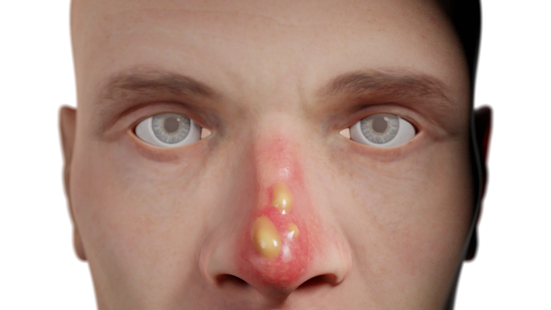
In 2nd degree burns, serous blisters with transparent contents are formed on hyperemic skin, the bottom is pink. Bubbles may form delayed within 24 hours of the burn. It is also characterized by soreness on palpation.
Grade 3 injuries are characterized by the formation of serous hemorrhagic blisters on hyperemic and infiltrated skin. There may also be dry necrosis. The area of the lesion is moderately painful or painless, and hair falls out.
Deep full-thickness burns of the 4th degree are characterized by the formation of black scab. The wound is painless, the wound bed is represented by bone tissue. It should be noted that isolated nasal burns are extremely rare and are usually accompanied by damage to other areas.
Classification of nasal burns by depth of injury
Degree
Depth of lesion
Local changes
Soreness
Healing
1st degree
Epidermis only
Hyperemia (redness) of the skin. On palpation, there is soreness. If you apply pressure, the area turns pale.
Moderate, when touched
Epithelialization, without scarring
2nd degree
Epidermis + upper dermis layers (papillary layer)
Serous blisters with transparent contents on hyperemic skin. The bottom of the bubbles are pink. Bubbles can appear for up to 24 hours
Serous hemorrhagic blisters, dry necrosis. The skin is hyperemic and infiltrated. The hair is easily removed
Reduced or absent
Scarring
4th degree
All layers of skin, muscle, tendons, cartilage, possibly bone
Black scab, the bottom of the wound is represented by bone tissue. Extensive necrosis
Absent
Gross scarring
Diagnosisnasal burns
Establishment of the diagnosis is based on collection of trauma history and otorhinolaryngologic examination. At the pre-hospital stage, it is mandatory to assess the patency of the respiratory tract. Subsequently, the nasopharyngeal mucosa is regularly reassessed for early detection of burns, reactive mucosal edema, and prevention of respiratory failure. It is necessary to assess the depth and volume of the lesion from the total body surface area to determine further treatment tactics. In case of a large area of damage to prevent the development of systemic complications perform biochemical study of blood, urine, ECG.
Treatment of nasal burns
It is imperative that the exposure to the damaging factor is stopped first. In case of smoke inhalation in the pre-hospital stage, insufflation of humidified oxygen is started.
If possible, it is necessary to cool the injured area of the body, adequately anesthetize the patient. For superficial burns, local therapy is recommended. Dressings with antibacterial ointments are prescribed . Small blisters should not be opened, while larger blisters should be dissected. Skin flap transplantation is indicated for patients with deep burns .
Extensive and deep burns require treatment in a burn hospital.
Nasal frostbite
Etiologyof nasal frostbite
Frostbite is caused by prolonged exposure of exposed areas of the body to low temperatures. With prolonged stay in unfavorable environmental conditions in the form of high humidity, wind frostbite can occur even at plus temperature.
Anatomyof nasal frostbite
Exposure to cold causes direct freezing of cells and subsequently celldeath. Also in frostbite there is a disturbance of blood circulation in the form of spasm of the distal parts of small blood vessels. Tissue ischemia occurs, which leads to cell death. The exact degree of frostbite in some cases can be assessed after several days, as characteristic changes appear delayed (blisters are formed after 4-8 hours, demarcation line at necrosis after several days).
In frostbite of the 1st degree, the superficial layers of the dermis are affected, there is pallor of the skin.
Grade 2 frostbite is characterized by the formation of serous blisters on pale skin.
Grade 3 frostbite has deep skin lesions. Blisters filled with hemorrhagic contents are formed.
The 4th degree of frostbite is characterized by necrosis of the entire thickness of the skin and underlying tissues. Dry gangrene is formed, distal areas are covered with black dry scab, in some cases self-amputation occurs. The skin is cyanotic, marbled. The lesions are combined with 2nd and 3rd degree manifestations, and the formation of blisters with various contents.
Clinical manifestationsnasal frostbite
Frostbite is initially characterized by burning and numbness of the affected area. The frostbitten area is pale, cold, hard, marbled. After warming, there is severe pain, the greater the degree of lesion, the more pronounced the pain syndrome. The skin becomes brightly hyperemic, edematous. Each of the degrees is characterized by local skin changes, which have been described above.
Classification of nasal frostbite by depth of injury
Degree
Depth of lesion
Local changes
Soreness
Healing
1st degree
Superficial layers of the dermis
Skin pallor, numbness, burning, cold area. After warming – hyperemia, swelling. No bubbles form
Moderate, worsens after warming up
Fast, no scarring
2nd degree
Epidermis + upper dermis layers (papillary layer)
Serous blisters with clear contents appear after 4-8 hours. Skin is pale, cold. Later, swelling, hyperemia.
Expressed
Complete, hyperpigmentation may occur
3rd degree
Deep layers of skin
Bubbles with hemorrhagic contents, the skin is marbled, cyanotic. The hair on the affected area falls out. Later, necrosis.
Strong, then decreased
Slow, with scarring
4th degree
All skin and underlying tissues (PJC, muscle, bone)
Cyanotic, marbled skin Dry gangrene, black dry scab, self-amputation. It is often combined with signs of grade 2-3. Demarcation after a few days
Absent due to nerve endings dying
Slow, with tissue loss and gross scarring
Diagnosisnasal frostbite
Diagnosis is based on the collection of history and complaints. The general condition and local changes are assessed.
Find more scientifically accurate content on our social media
Subscribe and don’t miss out the latest resources
Treatment of nasal frostbite
It is necessary to warm the injured area of the body. Under no circumstances should the skin be rubbed or open fires used, as there is a high risk of burns due to the lack of sensitivity. You can immerse the injured area in warm water or apply a dry warm bandage. Patients must be adequately anesthetized, and in some cases opioid analgesics are used. In general hypothermia of the body is recommended warm drinking,thermal blankets. Local treatment consists of treating the blisters with antibacterial ointments, applying sterile dressings. To improve tissue perfusion, anticoagulants, antiaggregants, vasodilators are prescribed. If dry gangrene is present, amputation is performed after a few days.
FAQ
1. Why is it not always possible to determine the degree of frostbite immediately?
Profound changes are not immediately apparent: blisters are formed after 4-8 hours, and the demarcation line after several days. The final evaluation of the degree is carried out in dynamics.
2. Why should I not rub my skin when I have frostbite?
Rubbing tissues that have lost sensitivity can cause mechanical damage and even thermal burns if an open heat source is used. Against the background of ischemia and cell death, this increases the risk of secondary complications and infections. Passive warming – using warm water or dry heat – is considered the correct way to warm up.
3 What is the difference between treating burns and frostbite?
In burns, it is important to eliminate the thermal factor, provide pain relief and prevent infection. Frostbite requires gradual warming, restoration of blood flow, and treatment of vascular disorders. Necrotized areas are sometimes surgically removed.
4. Is it possible to have a combined burn and frostbite injury at the same time?
In some clinical situations, especially when an inappropriate attempt is made to warm the frostbitten area with an open flame or hot objects, it is possible that thermal burns may be superimposed on tissue already damaged by the cold. This significantly worsens the course, increases the area of necrosis and increases the risk of complications.
5. What complications can develop in burns and frostbite of the nose?
Major complications include infection of the wound surface, gross scarring, and deformities of the external nose. Deep burns and frostbite can damage the cartilaginous skeleton, which can lead to persistent respiratory difficulties and impaired sense of smell. Also, when the nasal mucosa is affected, swelling often develops, complicating nasal breathing and creating a risk of secondary infection.
List of Sources
1.
VOKA Catalog.
https://catalog.voka.io/
2.
Total Otolaryngology—Head and Neck Surgery, Anthony P. Sclafani, Robin A. Dyleski, Michael J. Pitman, Stimson P. Schantz. Thieme Medical Publishers, Inc., 2015. ISBN 978-1-60406-646-3.
3.
Бербом Х. Болезни уха, горла и носа / Ханс Бербом, Оливер Кашке, Тадеус Навка, Эндрю Свифт; пер. с англ. – 2-е изд. – М. : МЕДпреcс-информ, 2016. – 776 с. : ил. ISBN 978-5-00030- 322-1.
Clark C, Ledrick D, Moore A. Facial Burns. [Updated 2023 Jul 3]. In: StatPearls [Internet]. Treasure Island (FL): StatPearls Publishing; 2025 Jan-. Available from:
https://www.ncbi.nlm.nih.gov/books/NBK559290/
6.
Basit H, Wallen TJ, Dudley C. Frostbite. [Updated 2023 Jun 26]. In: StatPearls [Internet]. Treasure Island (FL): StatPearls Publishing; 2025 Jan-. Available from:
St. Petersburg FL 33702, 7901 4th St N STE 300, USA
Thank you!
Your message is sent! Our experts will contact you shortly. If you have any additional questions, please contact us at info@voka.io
Cookie Consent
We use cookies to enhance your browsing experience, analyze site traffic, and deliver content. Please choose whether you accept all cookies or wish to reject non-essential tracking.
Cookie Preferences
Manage your cookie preferences below:
Essential cookies enable basic functions and are necessary for the proper function of the website.
Name
Description
Duration
Geolocation Config
This cookie is used to store the consent settings based on the visitor's location.
30 days
Cookie Preferences
This cookie is used to store the user's cookie consent preferences.
30 days
Google reCAPTCHA helps protect websites from spam and abuse by verifying user interactions through challenges.
Name
Description
Duration
_GRECAPTCHA
Google reCAPTCHA sets a necessary cookie (_GRECAPTCHA) when executed for the purpose of providing its risk analysis.
179 days
Statistics cookies collect information anonymously. This information helps us understand how visitors use our website.
Google Analytics is a powerful tool that tracks and analyzes website traffic for informed marketing decisions.
ID used to identify users for 24 hours after last activity
24 hours
_gat
Used to monitor number of Google Analytics server requests when using Google Tag Manager
1 minute
_gac_
Contains information related to marketing campaigns of the user. These are shared with Google AdWords / Google Ads when the Google Ads and Google Analytics accounts are linked together.
90 days
__utma
ID used to identify users and sessions
2 years after last activity
__utmt
Used to monitor number of Google Analytics server requests
10 minutes
__utmb
Used to distinguish new sessions and visits. This cookie is set when the GA.js javascript library is loaded and there is no existing __utmb cookie. The cookie is updated every time data is sent to the Google Analytics server.
30 minutes after last activity
__utmc
Used only with old Urchin versions of Google Analytics and not with GA.js. Was used to distinguish between new sessions and visits at the end of a session.
End of session (browser)
__utmz
Contains information about the traffic source or campaign that directed user to the website. The cookie is set when the GA.js javascript is loaded and updated when data is sent to the Google Anaytics server
6 months after last activity
__utmv
Contains custom information set by the web developer via the _setCustomVar method in Google Analytics. This cookie is updated every time new data is sent to the Google Analytics server.
2 years after last activity
__utmx
Used to determine whether a user is included in an A / B or Multivariate test.
18 months
_ga
ID used to identify users
2 years
_gali
Used by Google Analytics to determine which links on a page are being clicked
30 seconds
Clarity is a web analytics service that tracks and reports website traffic.