Intraductal Breast Papilloma: Etiology, Clinical Presentation, Diagnosis and Treatment
Svetlana D.Surgical oncologist, MD
9 min read·October 15, 2025
This article is for informational purposes only
The content on this website, including text, graphics, and other materials, is provided for informational purposes only. It is not intended as advice or guidance. Regarding your specific medical condition or treatment, please consult your healthcare provider.
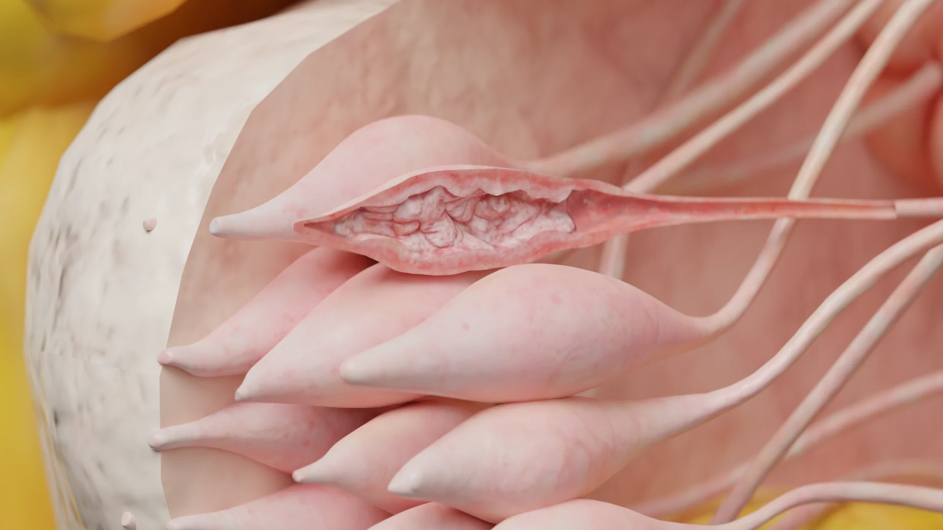
Intraductal papilloma is a benign mass located in the mammary ducts, which develops as a result of abnormal overgrowth of epithelial cells.
Intraductal papilloma: 3D model3D animation: intraductal papilloma
Intraductal papillomas may be solitary or multiple and located in any quadrant of the breast but are most commonly localized centrally behind the nipple, affecting the central duct.
Risk factors for developing intraductal papilloma:
Hormone replacement therapy with estrogen-containing agents;
Hereditary predisposition.
Etiology and pathogenesis of intraductal papilloma
The development of intraductal papilloma is based on hormonal imbalance, namely the increased sensitivity of breast tissue to estrogen and its hyperproduction.
Hyperestrogenaemia triggers proliferation of breast ductal epithelial cells, leading to hyperplasia of breast tissue and ductal dilation. As a result of these processes, papillary outgrowths are formed in the lumen of the duct, which are intraductal papilloma.
Morphological presentation
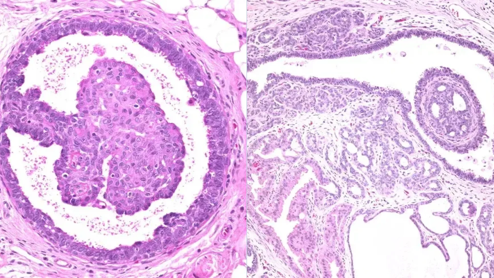
Intraductal papilloma can be central (usually solitary and large in size) and peripheral (small in size, may be multiple). Histologically, they are characterized by the presence of a fibrovascular rod covered with epithelial and myoepithelial cells (true papillae).
Morphological presentation of intraductal papilloma. Source: Webpathology. Intraductal Papilloma [8]
Intraductal papilloma may erode and sclerotize, but atypical proliferation, squamous cell or apocrine metaplasia with subsequent malignisation and development of ductal breast cancer is also possible.
3D animation: Intraductal papilloma with atypia
Clinical Manifestations
Clinical manifestations of intraductal papilloma depend on its size and localization.
Peripheral intraductal papillomas are usually asymptomatic and detected by screening ultrasound.
Central intraductal papillomas are characterized by the following symptoms:
A yellowish-transparent to red-brown (bloody) nipple discharge is the most common symptom;
Presence of a palpable mass in the areolar region behind the nipple.
Diagnosis of intraductal papilloma
Medical history, examination and palpation of breasts.
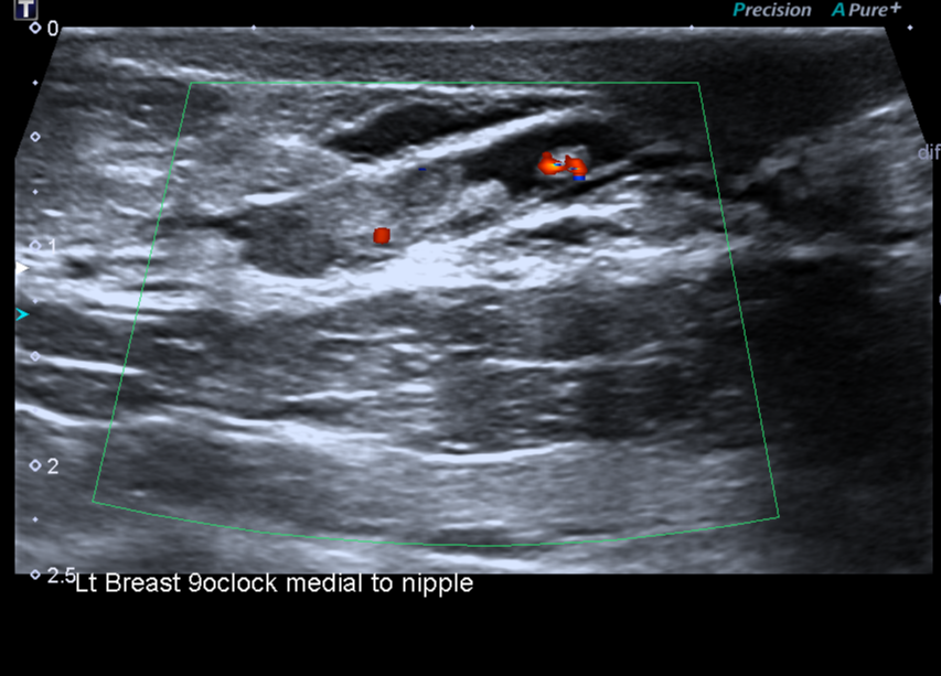
Breast ultrasound and peripheral lymph nodes: a mass protruding into the lumen of the duct or cystic cavity is visualized.
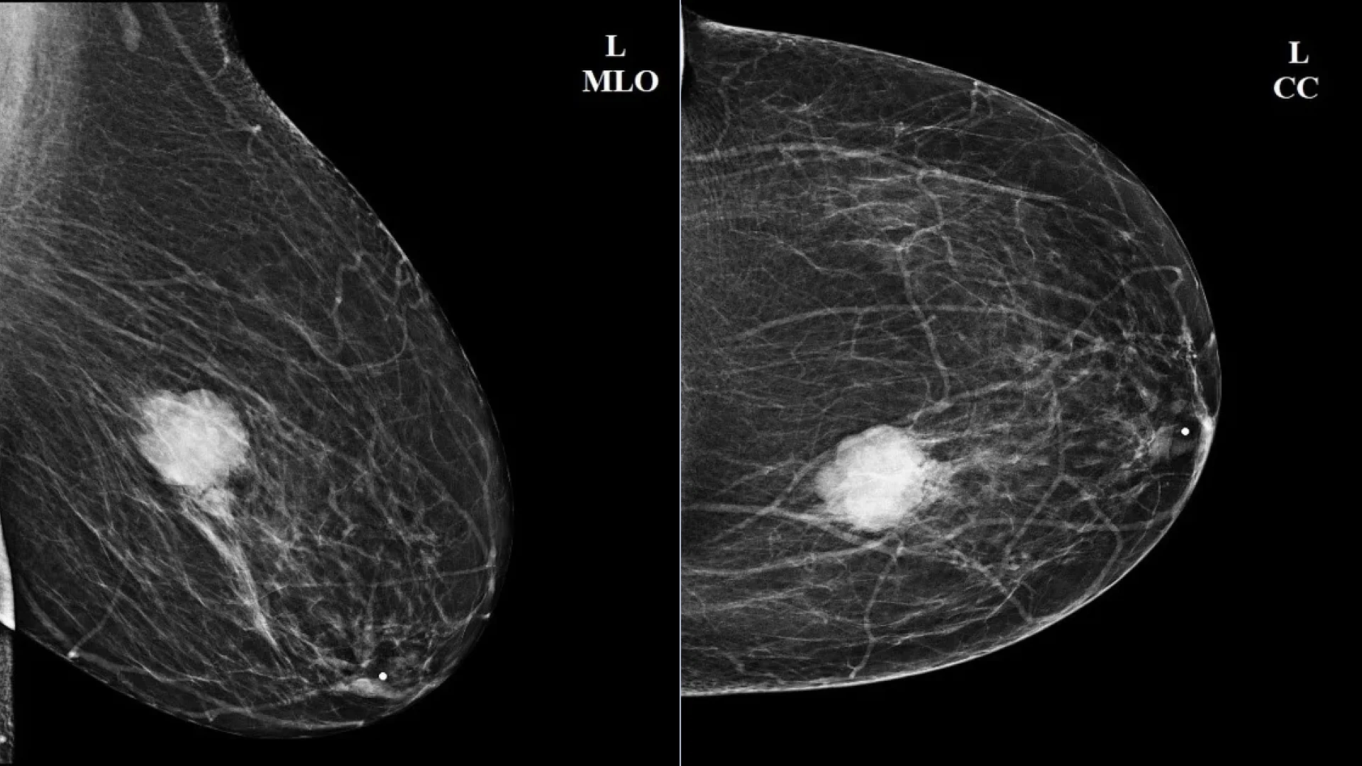
Mammography in most cases is uninformative and is used as a method of clarifying diagnosis when ductal breast cancer is suspected. Radiologically, an intraductal papilloma may be defined as a rounded mass with clear, even contours, similar to a cyst, but of higher density.
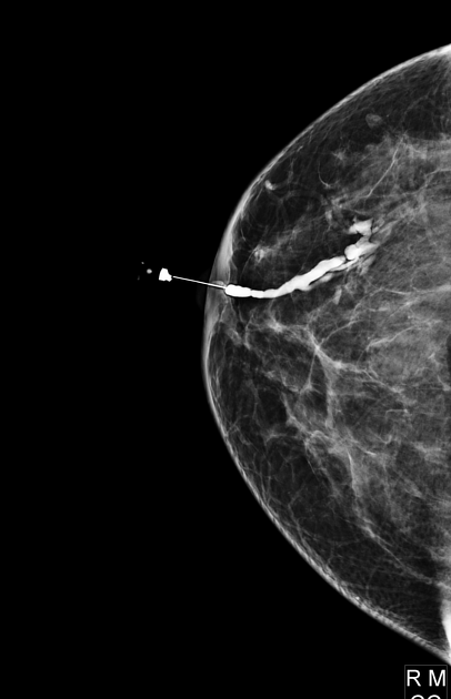
Ductography is used to clarify the diagnosis. On radiographs, after contrast agent injection, duct dilation and deformity, irregularities of contours with the presence of contrasted formations and filling defect (disruption in the contrast enhancement pattern) are determined.
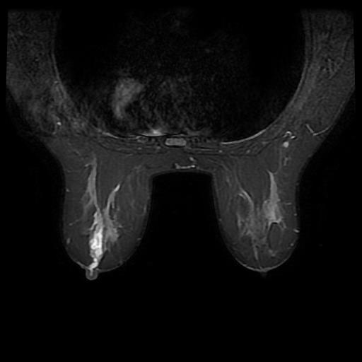
MRI is used for clarifying diagnosis. Intraductal papillomas are characterized by the presence of a well-defined rounded mass on MRI, isointense or slightly hypointense in T1-mode, and well accumulating contrast in T2-mode.
Cytological examination of nipple discharge.
Fine-needle aspiration biopsy of the tumor.
Core needle biopsy of the tumor.
Intraductal papilloma on ultrasound image. Author: Bruno Di Muzio. Source: Radiopaedia [11]Mammography of the left breast in two projections: intraductal papilloma. Author: Ammar Ashraf. Source: Radiopaedia [11]
You can see radiographs and MRI images illustrating an intraductal papilloma below. Authors: Mohammad Taghi Niknejad, Bahman Rasuli. Source: Radiopaedia [11]:
Ductography of the right breast: a filling defect typical of an intraductal papilloma is detected on the radiograph. Author: Mohammad Taghi Niknejad
MRI picture of intraductal papilloma in T2-weighted, fat-suppressed mode. Author: Bahman Rasuli
MRI picture of intraductal papilloma in T1-weighted mode. MRI for intraductal papilloma. Author: Bahman Rasuli
Find more scientifically accurate content on our social media
Subscribe and don’t miss out the latest resources
Treatment of intraductal papilloma
Intraductal papilloma should be treated surgically due to the high risk of malignization. A sectoral resection of the breast (lumpectomy) with subsequent histological examination of the removed material is indicated.
FAQ
1. What are the causes and risk factors for developing intraductal papilloma?
The main cause of development is hormonal imbalance, namely hyperestrogenaemia (increased estrogenic levels), which triggers abnormal proliferation of epithelial cells in the ducts. Key risk factors include any condition associated with this imbalance: endocrine diseases (obesity, diabetes mellitus), taking oral contraceptives or hormone replacement therapy, and hereditary predisposition.
2. What are the main symptoms of intraductal papilloma?
Clinical presentation depends on the localization. Peripheral papillomas are often asymptomatic. For central ones located near the nipple, the most typical symptom is nipple discharge. Discharges can range in color from yellowish-transparent to bloody (red-brown). Less frequently, a small mass in the areola region may be palpated.
3. Is an intraductal papilloma considered cancer, and what is the risk of malignization?
No, the intraductal papilloma itself is a benign mass. However, it is considered a precancerous condition or, more specifically, a mass with an increased risk of malignant transformation. Its cells can develop atypical proliferation, which is a precursor to ductal carcinoma in situ (DCIS). It is because of this risk of malignization that the management strategy for these patients is surgical.
4. What treatment is indicated for intraductal papilloma?
Intraductal papilloma is subject to mandatory surgical treatment. Non-surgical therapy and observation strategy are not indicated because of the risk of malignization. The standard surgery is sectoral resection of the breast (lumpectomy): excision of the duct together with the papilloma within healthy tissues. The removed material is necessarily sent for histological examination to exclude cancer.
5. Can an intraductal papilloma disappear or go away on its own without treatment?
Theoretically, the papilloma may undergo spontaneous necrosis and sclerosis (replacement by scar tissue). However, given that any papilloma can hide a focus of atypia or already developed cancer, the wait-and-see strategy is not applied. Relying on the mass to go away on its own is clinically unjustified and dangerous. A diagnosed intraductal papilloma is a direct indication for surgery.
6. What is the prognosis for intraductal papilloma after treatment?
After complete surgical removal, the prognosis is usually excellent. If histological study results show no atypia or signs of cancer in the removed material, the patient is considered healthy. However, having a history of papilloma may slightly increase the overall risk of developing breast cancer in the future, requiring regular follow-up with a mammologist.
References
1.
VOKA 3D Anatomy & Pathology – Complete Anatomy and Pathology 3D Atlas. VOKA 3D Anatomy & Pathology.
Available from: https://catalog.voka.io/
2.
Anisimov AV. BI-RADS system for ultrasound: description, classification, illustrations [Internet]. Moscow: Medison.ru. [In Russian.]
Available from: https://www.medison.ru/si/art434.htm
3.
Sencha AN. Ultrasound examination of the mammary glands. Step by step. From simple to complex. 2nd ed. Moscow: MEDpress-inform; 2019. 176 p. [In Russian.]
4.
Sencha AN, Bikeev YV. Ultrasound examination of the mammary glands. Atlas. Moscow: MEDpress-inform; 2016. 160 p. [In Russian.]
5.
Ternovoy SK, Abduraimov AB. Radiological mammology. Moscow: GEOTAR-Media; 2007. 128 p. [In Russian.]
50 Franklin Street, STE 203 Boston, MA 02110 United States
Thank you!
Your message is sent! Our experts will contact you shortly. If you have any additional questions, please contact us at info@voka.io
Cookie Consent
We use cookies to enhance your browsing experience, analyze site traffic, and deliver content. Please choose whether you accept all cookies or wish to reject non-essential tracking.
Cookie Preferences
Manage your cookie preferences below:
Essential cookies enable basic functions and are necessary for the proper function of the website.
Name
Description
Duration
Geolocation Config
This cookie is used to store the consent settings based on the visitor's location.
30 days
Cookie Preferences
This cookie is used to store the user's cookie consent preferences.
30 days
Google reCAPTCHA helps protect websites from spam and abuse by verifying user interactions through challenges.
Name
Description
Duration
_GRECAPTCHA
Google reCAPTCHA sets a necessary cookie (_GRECAPTCHA) when executed for the purpose of providing its risk analysis.
179 days
Statistics cookies collect information anonymously. This information helps us understand how visitors use our website.
Google Analytics is a powerful tool that tracks and analyzes website traffic for informed marketing decisions.
Used to monitor number of Google Analytics server requests
10 minutes
__utmb
Used to distinguish new sessions and visits. This cookie is set when the GA.js javascript library is loaded and there is no existing __utmb cookie. The cookie is updated every time data is sent to the Google Analytics server.
30 minutes after last activity
__utmc
Used only with old Urchin versions of Google Analytics and not with GA.js. Was used to distinguish between new sessions and visits at the end of a session.
End of session (browser)
__utmz
Contains information about the traffic source or campaign that directed user to the website. The cookie is set when the GA.js javascript is loaded and updated when data is sent to the Google Anaytics server
6 months after last activity
__utmv
Contains custom information set by the web developer via the _setCustomVar method in Google Analytics. This cookie is updated every time new data is sent to the Google Analytics server.
2 years after last activity
__utmx
Used to determine whether a user is included in an A / B or Multivariate test.
18 months
_ga
ID used to identify users
2 years
_gali
Used by Google Analytics to determine which links on a page are being clicked
30 seconds
_ga_
ID used to identify users
2 years
_gid
ID used to identify users for 24 hours after last activity
24 hours
_gat
Used to monitor number of Google Analytics server requests when using Google Tag Manager
1 minute
_gac_
Contains information related to marketing campaigns of the user. These are shared with Google AdWords / Google Ads when the Google Ads and Google Analytics accounts are linked together.
90 days
Clarity is a web analytics service that tracks and reports website traffic.