Brain Metastases: Classification, Etiology, Symptoms, Diagnosis, Treatment and Prognosis
Artemiy G.Neuro-oncological surgeon, MD
23 min read·December 23, 2025
This article is for informational purposes only
The content on this website, including text, graphics, and other materials, is provided for informational purposes only. It is not intended as advice or guidance. Regarding your specific medical condition or treatment, please consult your healthcare provider.
Metastases are the most common intracranial tumors in adults, occurring more frequently than primary brain tumors. In most cases, metastatic lesions of the brain are diagnosed following an already established systemic malignant process, but in some patients it may be the first manifestation of the tumor.
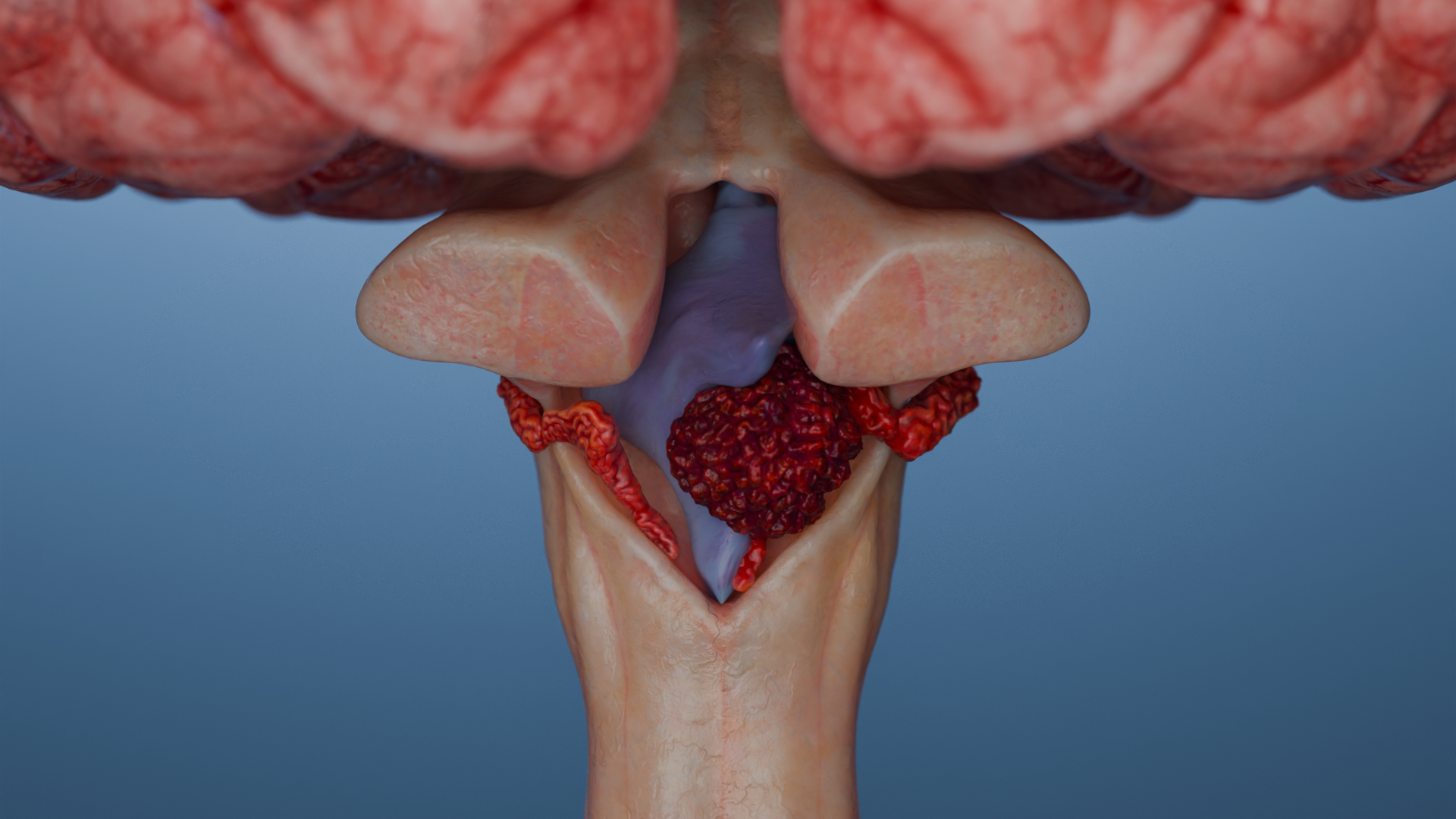
Kidney cancer metastasis in the left cerebral hemisphere: 3D model3D animation: kidney cancer metastasis in the left cerebral hemisphere
Etiology
5 primary tumors are responsible for ~80% of all brain metastases:
Lung cancer (30–60% of all brain metastases).
Breast cancer (10–30% of all metastases).
Melanoma (5–20% of all metastases). At the same time, melanoma has the highest individual rate of metastasis to the brain — 25–60% of patients with disseminated melanoma are found to have a brain lesion.
Kidney cancer (5–10% of all metastases).
Colorectal cancer (1–3% of all metastases).
Metastases in the brain are predominantly formed hematogenously; the cells penetrate the blood-brain barrier and linger at the gray-white matter interface due to the vascular features of this area.
At the time of initial diagnosis, brain metastases are found in approximately 2% of all patients with all tumor types and 12–15% of patients with already advanced (metastatic) disease.
During the course of the disease, brain metastases develop in 10–30% of patients with solid malignancies. For some types of neoplasms, the incidence can be as high as 40–50% in high-risk groups (HER2+ and triple-negative breast tumors, melanoma, non-small cell lung cancer).
Autopsy studies show that brain metastases are found in about 25% of patients with cancer, even if they were not detected during life.
Leptomeningeal metastases (along meninges) are more common in breast cancer, melanoma, lymphoma, and non-small cell lung cancer.
Anatomy
The majority (80%) of CNS metastases are located supratentorially, i.e. affect the hemispheres of the brain.
Up to 20% are located infratentorially (cerebellum, brainstem). Meninges are involved in up to 8% of cases.
The most common localization of metastases is the gray-white matter interface.
Classification of brain metastases
By number of metastatic foci
Solitary — 1 foci;
Oligometastases — 2–4;
Multiple — ≥5 foci (≥10 in some classifications).
This assessment is used when choosing between radiosurgery (SRS), whole brain radiation therapy (WBRT), and surgery.
By size
Small — <10 mm;
Medium — 10–30 mm;
Large — larger than 30 mm.
Small foci are available for stereotactic radiosurgery (SRS). Medium-sized foci (10–30 mm) usually imply dose constraints for single stereotactic radiosurgery (or require fractionation), and foci >30 mm are often considered for surgical removal or fractionated radiosurgery (large total dose is broken down and delivered in multiple steps), as single-fraction SRS in such cases carries a high risk of radionecrosis and edema.
By structure of metastasis
Solid metastasis is the typical form, parenchymatous foci (characteristic of lung, breast, kidney, and melanoma cancers).
Cystic metastasis presents as an almost entirely fluid (anechogenic or hypointense on T1 MRI) component with a thin wall and minimal solid area, usually indicating slow growth. This type of metastasis is most characteristic of papillary thyroid cancer and mucinous carcinomas of the gastrointestinal tract.
Cystic-solid metastasis is a mixed structure, contains both a distinct fluid component and a prominent solid area, often with uneven contrast accumulation (HER2+ breast cancer, colorectal cancer, EGFR+ non-small cell lung cancer, renal cancer).
Hemorrhagic metastasis: at high risk of hemorrhage (melanoma, kidney cancer, choriocarcinoma).
Leptomeningeal metastases: dissemination through the liquor (HER2+ and triple-negative breast cancer, non-small cell lung cancer, melanoma, medulloblastoma).
Hypervascular (hemorrhagic) metastasis of kidney cancer
Clinical significance of the structure of metastasis
Cystic and hemorrhagic metastases are more likely to cause cramping and edema, with an increased risk of brain herniation.
Large cystic metastases do not respond well to SRS and more often require surgery.
Hemorrhagic (melanoma, kidney cancer) may recur more often after SRS, have high risk of hemorrhage.
Clinical manifestations
Symptoms depend on the size, localization, growth rate and number of foci, as well as the involvement of liquor spaces and brain edema. The debut is usually acute or subacute.
Symptoms of metastatic brain lesions
Headache 35–70% (due to intracranial hypertension);
Seizures 15–35% (more common in cortical lesions, especially temporoparietal and frontal lobes; frequency is higher in melanoma and kidney metastases);
Cognitive impairment and behavioral changes 20–40% (associated with frontal foci, edema, elevated ICP);
Nausea and vomiting 15–30% (especially in the morning);
Coordination and gait disorders (ataxia) 10–25% (cerebellar and brain stem metastases);
Up to 20–30% of metastases are asymptomatic and are detected by imaging to clarify the diagnosis.
Acute and threatening symptoms
Acute impairment of consciousness / seizure status / severe vomiting + headache may indicate the following:
Dislocations (herniation) of the brain;
Decompensation with increasing cerebral edema;
Hemorrhage (typical in melanoma, kidney cancer, choriocarcinoma);
Obstructive hydrocephalus (with infratentorial tumors of the posterior cranial fossa).
These conditions require emergency measures (seizure control, anti-edema therapy, emergency imaging [CT/MRI with contrast] and probably emergency neurosurgical intervention).
Diagnosis of brain metastases
Main methods
MRI with contrast is the gold standard. The method is the most sensitive and allows detecting even small (5 mm) and asymptomatic metastases.
CT with contrast is an alternative when MRI is contraindicated. The method is less sensitive, but well detects large foci, hemorrhages, pronounced mass effect.
Additional methods
PET-CT, biopsy (in case of diagnostic doubts).
Assessment of the patient’s systemic status as a standard for staging and cancer screening (chest, abdominal and pelvic CT, PET-CT).
The molecular profile of the metastasis (EGFR, ALK, HER2, BRAF, etc.) is determined after obtaining histologic material and has a direct impact on the choice of systemic therapy and prognosis.
Differential diagnosis
Differential diagnosis is performed with other focal lesions of the brain. The key features suggestive of metastases are the presence of a known primary cancer history, multiple foci, their typical localization at the gray-white matter interface (corticosubcortical), severe perifocal edema, and clear annular enhancement on MRI with contrast.
The main conditions the metastases should be differentiated with:
Primary brain tumor
Gradual onset, focal neurologic symptoms, seizures, headache. On MRI, there is usually an infiltrative or solid mass (often solitary, as opposed to metastasis), edema, mass effect, and heterogeneous contrast accumulation.
Brain abscess
Often fever, leukocytosis, and a focus of infection (otitis media, sinusitis, bacteremia). Mimics the tumor on MRI (also has annular enhancement) but has clear diffusion restriction (DWI+) in the center. MR spectroscopy reveals specific amino acids/metabolites.
CNS lymphoma
On MRI, homogeneous enhancement and location in deep brain structures, especially in immunosuppressed patients. High diffusion restriction (DWI+), possible rapid regression after steroids, so it’s important to avoid them before biopsy.
Radiation necrosis
It occurs months to years after radiation therapy, may mimic a recurrence or new metastasis. Distinguished by MR perfusion and PET (necrosis means hypoperfusion, hypometabolism), may decrease on steroids.
Multiple sclerosis
(especially the tumor-like form). Young age, multifocal lesions in the white matter, FLAIR-hyperintense without mass effect or contrast; clinical course is recurrent, with remissions, often in the young.
Stroke
(especially in the subacute phase when there may be ring-shaped enhancement). Sudden onset, focal neurologic deficit, ischemic area visible on DWI/ADC MRI (diffusion restriction), more often localized across vascular basins.
Aneurysm
Sudden headache, on CT/MRI with contrast — vascular mass, on angiography — dilated artery, subarachnoid hemorrhage.
Encephalitis
Subacute onset, fever, confusion, and seizures — no focal mass. On MRI, changes are usually diffuse, often in the temporal lobes (e.g., in herpetic encephalitis), with no mass effect.
Cavernous sinus thrombosis
Acute onset with headache (often unilateral), eyelid swelling, ophthalmoplegia, and decreased vision. MRI/MR venography reveals thrombus in the venous sinuses. There is often infection of the facial, nasal, and sinus areas.
Cluster headache
Sharp pain around the eye, lacrimation, rhinorrhea, lasting minutes, recurring in series, not accompanied by neurologic deficit. No structural changes on the MRI.
Idiopathic hydrocephalus
Often in the elderly, a triad of symptoms (gait disturbance, dementia, urinary incontinence), there may be headache and nausea, on MRI ventricular dilation without focal masses.
Metabolic disorders
Causes diffuse rather than focal symptoms (confusion, seizures, tremors), with no focal mass on MRI. General tests (glucose, sodium, liver enzymes, creatinine, urea, etc.) help identify the cause.
Treatment of metastatic brain lesions
The main methods of treatment
Surgical removal or biopsy to clarify the diagnosis;
Stereotactic radiosurgery (SRS);
Whole Brain Radiation Therapy (WBRT);
Systemic (more often targeted) therapy for specific tumor types (e.g., EGFR+, ALK+, BRAF+, etc.).
General principles
Personalization of the strategy depending on the number of foci, patient functional status, systemic disease control, and molecular subtype of the tumor.
Symptomatic treatment: glucocorticoids, anticonvulsants, anticoagulants, etc.
Treatment for solitary metastases
Surgery: for large, symptomatic, superficial (i.e., available for removal), life-threatening foci. Increases life expectancy in patients with good performance status.
SRS (stereotactic radiosurgery) is an alternative for hard-to-reach or small foci (<3 cm), effective both after surgery and as a stand-alone method. Sometimes used before surgical removal to reduce the likelihood of recurrence.
WBRT (whole brain radiation therapy): as adjuvant (after surgery) and/or when SRS and surgical treatment are not possible.
Treatment for multiple metastases
Up to 4 foci (<3 cm each) — preferably SRS;
5–10 foci — SRS is acceptable in selected patients;
>10 foci or >3 cm — WBRT remains the standard.
WBRT (whole brain radiation therapy): effectiveness and side effects
The effect of whole brain radiation therapy (WBRT) is to control (stabilize) intracranial foci and reduce the incidence of intracranial recurrences.
It is used in multiple (>10) metastases and when SRS is not possible, and in leptomeningeal lesions.
Side effects:
Cognitive decline;
Leukoencephalopathy (diffuse lesion of the white matter of the brain);
Radiation necrosis (its likelihood increases with repeated radiation therapy).
Modern approaches: use of memantine (NDMA antagonist) and Intensity-Modulated Radiation Therapy (IMRT) with hippocampal exclusion reduce the risk of cognitive impairment.
The role of drug therapy
Challenges of drug treatment of brain metastases:
1. Hematoencephalicbarrier (BBB):
A physiologic filter that prevents most chemotherapy agents from penetrating brain tissue. Most classical cytotoxic agents (cisplatin, paclitaxel, docetaxel) have low permeability through the BBB.
It’s partially altered in the area of metastasis, but not always sufficient for effective drug concentration.
2. Heterogeneity of the tumor.
The molecular profile of metastases may differ from the primary tumor.
3. The immunoprivileged environment of the brain.
Less activity of T cells and antigen-presenting cells limits the efficacy of immunotherapy.
Targeted therapy shows encouraging results (intracranial control can reach more than 50–80%). It is used for tumors with potential sensitivity to therapy (e.g., lung cancer with EGFR/ALK, melanoma with BRAF, HER2+ breast cancer). The use of targeted therapies or immunotherapy may delay or replace radiation therapy.
Immunotherapy (PD-1/PD-L1 inhibitors) may be effective in metastatic non-small cell lung cancer, melanoma and HER2+ breast cancer.
CAR-T therapy targets tumors expressing certain antigens different from normal brain tissue and has shown encouraging results in clinical trials, but is not yet routinely used (the method is undergoing clinical trials and is currently experimental in CNS oncology; studies are limited due to the risk of systemic toxicity and unpredictable effects in the CNS).
Find more scientifically accurate content on our social media
Subscribe and don’t miss out the latest resources
Prognosis in brain metastases
The Disease-Specific Graded Prognostic Assessment (ds-GPA) scale is the most accurate prognostic tool for metastatic brain lesions. It considers biological and molecular characteristics of primary tumors (in addition to age, KPS functional status, number of brain metastases, and extracerebral metastases that are standard for GPA).
The scale is used for metastatic brain lesions in lung cancer, melanoma, breast cancer, kidney cancer, and colorectal cancer.
In practice, rigid and universal prognostic schemes should be avoided in metastatic brain lesions, preferring an individualized approach based on the type of primary tumor, molecular profile, functional status of the patient, and available treatments.
Modern treatment of metastatic brain lesions requires a multidisciplinary approach based on the type of primary tumor and molecular characteristics of the metastasis, the number and localization of foci, and the general condition of the patient. SRS and targeted therapies are becoming the treatment of choice in most patients with limited metastases and sensitive tumors.
FAQ
1. Which type of cancer most often metastasizes to the brain?
Lung cancer most commonly metastasizes to the brain, accounting for up to 60% of all cases. Breast cancer, melanoma, kidney cancer, and colorectal cancer are also common sources.
2. What do metastases look like on a brain MRI?
MRI with contrast is the primary method of diagnosis. Metastases are characterized by multiple foci of rounded shape with clear ring-shaped enhancement and marked swelling of the surrounding tissue. Typical localization of foci is the gray-white matter interface.
3. How fast do metastases in the head grow?
The growth rate of metastases is usually high, resulting in acute or subacute onset of clinical symptoms. This makes them different from many primary brain tumors, which may develop more slowly.
4. Can cancer be cured if it has metastasized to the brain?
Current treatment is aimed at controlling the disease, not a complete cure. A personalized strategy including surgery, stereotactic radiosurgery, and targeted therapy can control intracranial foci and improve quality of life.
5. What is the life expectancy for brain metastases?
Prognosis and life expectancy vary individually. They depend on the type of the primary tumor, its molecular characteristics, the number of metastases and the general condition of the patient, which is assessed using special prognostic scales.
References:
1.
VOKA Catalog. [Electronic resource].
https://catalog.voka.io/
2.
Vogelbaum MA, Brown PD, Messersmith H, Brastianos PK, Burri S, Cahill D, Dunn IF, Gaspar LE, Gatson NTN, Gondi V, Jordan JT, Lassman AB, Maues J, Mohile N, Redjal N, Stevens G, Sulman E, van den Bent M, Wallace HJ, Weinberg JS, Zadeh G, Schiff D. Treatment for Brain Metastases: ASCO-SNO-ASTRO Guideline. J Clin Oncol. 2022 Feb 10;40(5):492-516. doi: 10.1200/JCO.21.02314. Epub 2021 Dec 21. Erratum in: J Clin Oncol. 2022 Apr 20;40(12):1392. doi: 10.1200/JCO.22.00593. PMID: 34932393.
3.
Brenner AW, Patel AJ. Review of Current Principles of the Diagnosis and Management of Brain Metastases. Front Oncol. 2022 May 24;12:857622. doi: 10.3389/fonc.2022.857622. PMID: 35686091; PMCID: PMC9171239.
4.
Aizer AA, Lamba N, Ahluwalia MS, Aldape K, Boire A, Brastianos PK, Brown PD, Camidge DR, Chiang VL, Davies MA, Hu LS, Huang RY, Kaufmann T, Kumthekar P, Lam K, Lee EQ, Lin NU, Mehta M, Parsons M, Reardon DA, Sheehan J, Soffietti R, Tawbi H, Weller M, Wen PY. Brain metastases: A Society for Neuro-Oncology (SNO) consensus review on current management and future directions. Neuro Oncol. 2022 Oct 3;24(10):1613-1646. doi: 10.1093/neuonc/noac118. PMID: 35762249; PMCID: PMC9527527.
5.
Habbous S, Forster K, Darling G, Jerzak K, Holloway CMB, Sahgal A, Das S. Incidence and real-world burden of brain metastases from solid tumors and hematologic malignancies in Ontario: a population-based study. Neurooncol Adv. 2020 Dec 22;3(1):vdaa178. doi: 10.1093/noajnl/vdaa178. PMID: 33585818; PMCID: PMC7872008.
6.
Sener U, Wilcox JA, Boire AA. Leptomeningeal Disease: Current Approaches and Future Directions. Curr Neurol Neurosci Rep. 2025 Mar 18;25(1):25. doi: 10.1007/s11910-025-01412-y. PMID: 40100294; PMCID: PMC11920312.
7.
Sperduto PW, Kased N, Roberge D, Xu Z, Shanley R, Luo X, Sneed PK, Chao ST, Weil RJ, Suh J, Bhatt A, Jensen AW, Brown PD, Shih HA, Kirkpatrick J, Gaspar LE, Fiveash JB, Chiang V, Knisely JP, Sperduto CM, Lin N, Mehta M. Summary report on the graded prognostic assessment: an accurate and facile diagnosis-specific tool to estimate survival for patients with brain metastases. J Clin Oncol. 2012 Feb 1;30(4):419-25. doi: 10.1200/JCO.2011.38.0527. Epub 2011 Dec 27. PMID: 22203767; PMCID: PMC3269967.
8.
Huntoon K, Elder JB, Finger G, Ormond DR, Redjal N, Linskey ME, Olson JJ. Congress of Neurological Surgeons Systematic Review and Evidence-Based Guidelines Update for the Role of Emerging Therapies in the Management of Patients With Metastatic Brain Tumors. Neurosurgery. 2025 Jun 1;96(6):1172-1177. doi: 10.1227/neu.0000000000003383. Epub 2025 Mar 17. PMID: 40094364.
9.
Jacob Sperber, Seeley Yoo, Edwin Owolo, Tara Dalton, Tanner J Zachem, Eli Johnson, James E Herndon, Annee D Nguyen, Harrison Hockenberry, Brandon Bishop, Nancy Abu-Bonsrah, Steven H Cook, Peter E Fecci, Paul W Sperduto, Margaret O Johnson, Melissa M Erickson, C Rory Goodwin, Validation of the graded prognostic assessment and recursive partitioning analysis as prognostic tools using a modern cohort of patients with brain metastases, Neuro-Oncology Practice, Volume 11, Issue 6, December 2024, Pages 763–771.
10.
Brozos-Vázquez EM, Rodríguez-López C, Cortegoso-Mosquera A, López-Landrove S, Muinelo-Romay L, García-González J, López-López R, León-Mateos L. Immunotherapy in patients with brain metastasis: advances and challenges for the treatment and the application of circulating biomarkers. Front Immunol. 2023 Nov 3;14:1221113. doi: 10.3389/fimmu.2023.1221113. PMID: 38022574; PMCID: PMC10654987.
50 Franklin Street, STE 203 Boston, MA 02110 United States
Thank you!
Your message is sent! Our experts will contact you shortly. If you have any additional questions, please contact us at info@voka.io
Cookie Consent
We use cookies to enhance your browsing experience, analyze site traffic, and deliver content. Please choose whether you accept all cookies or wish to reject non-essential tracking.
Cookie Preferences
Manage your cookie preferences below:
Essential cookies enable basic functions and are necessary for the proper function of the website.
Name
Description
Duration
Geolocation Config
This cookie is used to store the consent settings based on the visitor's location.
30 days
Cookie Preferences
This cookie is used to store the user's cookie consent preferences.
30 days
Google reCAPTCHA helps protect websites from spam and abuse by verifying user interactions through challenges.
Name
Description
Duration
_GRECAPTCHA
Google reCAPTCHA sets a necessary cookie (_GRECAPTCHA) when executed for the purpose of providing its risk analysis.
179 days
Statistics cookies collect information anonymously. This information helps us understand how visitors use our website.
Google Analytics is a powerful tool that tracks and analyzes website traffic for informed marketing decisions.
Used to monitor number of Google Analytics server requests
10 minutes
__utmb
Used to distinguish new sessions and visits. This cookie is set when the GA.js javascript library is loaded and there is no existing __utmb cookie. The cookie is updated every time data is sent to the Google Analytics server.
30 minutes after last activity
__utmc
Used only with old Urchin versions of Google Analytics and not with GA.js. Was used to distinguish between new sessions and visits at the end of a session.
End of session (browser)
__utmz
Contains information about the traffic source or campaign that directed user to the website. The cookie is set when the GA.js javascript is loaded and updated when data is sent to the Google Anaytics server
6 months after last activity
__utmv
Contains custom information set by the web developer via the _setCustomVar method in Google Analytics. This cookie is updated every time new data is sent to the Google Analytics server.
2 years after last activity
__utmx
Used to determine whether a user is included in an A / B or Multivariate test.
18 months
_ga
ID used to identify users
2 years
_gali
Used by Google Analytics to determine which links on a page are being clicked
30 seconds
_ga_
ID used to identify users
2 years
_gid
ID used to identify users for 24 hours after last activity
24 hours
_gat
Used to monitor number of Google Analytics server requests when using Google Tag Manager
1 minute
_gac_
Contains information related to marketing campaigns of the user. These are shared with Google AdWords / Google Ads when the Google Ads and Google Analytics accounts are linked together.
90 days
__utma
ID used to identify users and sessions
2 years after last activity
Clarity is a web analytics service that tracks and reports website traffic.





.webp)