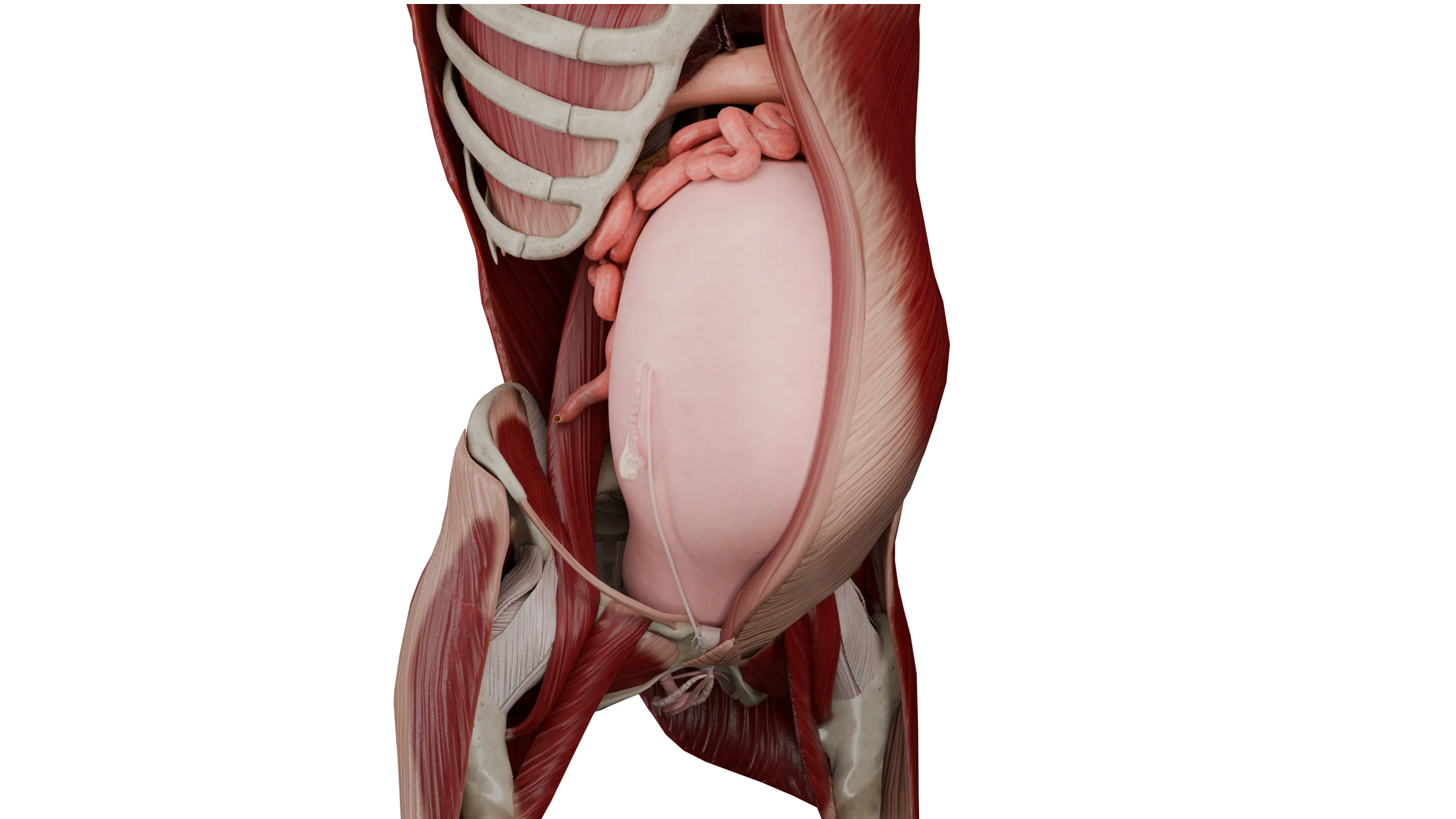
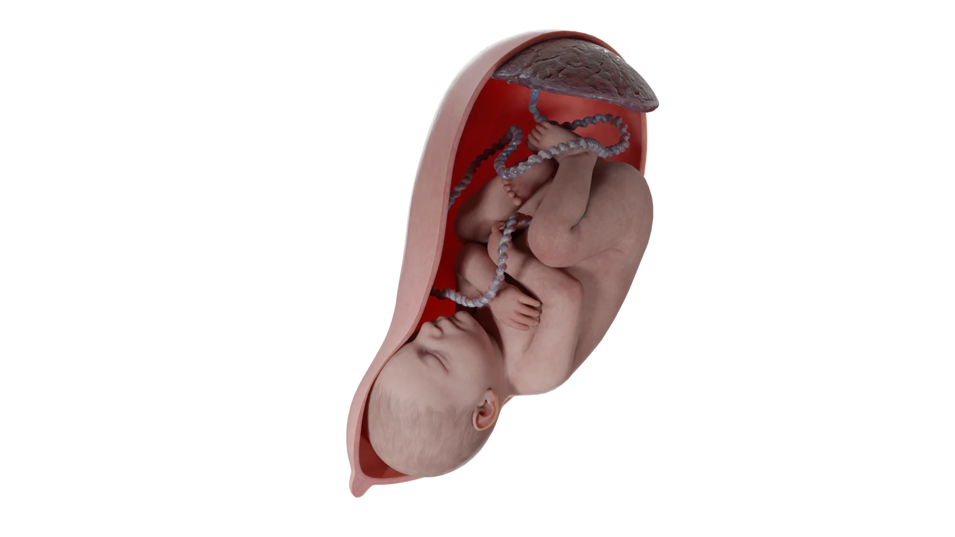
Placenta Previa: Classification, Diagnosis, Risks, and Management of Pregnancy
Daria G.Gynecologist, fertility specialist, MD
15 min read·June 11, 2025
This article is for informational purposes only
The content on this website, including text, graphics, and other materials, is provided for informational purposes only. It is not intended as advice or guidance. Regarding your specific medical condition or treatment, please consult your healthcare provider.
Placenta previa is a condition in which the placenta is implanted in the lower uterine segment, which is poorly vascularized. In such cases, the placenta covers the cervical os. This condition occurs in approximately 0.3 % to 2 % of pregnancies during the third trimester. In earlier gestations, a similar condition is defined as chorion previa, the outer germinal membrane from which the placenta later forms (up to 16 weeks), but this condition is not a pathology in early pregnancy.
Etiology
The fundamental cause of the development of placenta previa has not been established. However, its development has been associated with endometrial injury caused by various etiological factors.
Therefore, the following risk factors have been identified that increase the chances of developing placenta previa:
The mother’s age is more than 35 years old;
Multiple births;
History of pregnancy termination;
Surgical interventions in the uterine cavity (curettage, etc.);
Large uterine leiomyoma;
Utilization of in vitro fertilization technologies;
A history of uterine surgery;
History of placenta previa;
Smoking;
Race (is a controversial risk factor, some studies show that risk is increased in Asian and African women).
Implantation requires an environment with an adequate blood supply. The trophoblast cells of the outer layer of the blastocyst develop into the placenta and fetal membranes. The trophoblast attaches to the decidua, a specialized mucosal lining of the uterus. Pathological mechanisms behind the risk factors can adversely affect placental vessels, disrupting placental blood flow and leading to placentation abnormalities.
Classification of placenta previa
Placenta previa is diagnosed when the placental edge is located less than 20 mm from the cervical os. The condition may be classified into central (complete) when the cervical canal is completely obstructed and and lateral (partial) when ⅔ of the cervical canal is blocked — 3D Models:
Complete (Central) Placenta Previa: Side View (Left) and Ventral View (Right)
Partial (Lateral) Placenta Previa
By contrast, there is also a condition known as a low-lying placenta. In this case, the edge of the placenta is 20 to 35 mm away.
Types of placenta previa
Types of Placenta Previa
Distance between placental edge and cervical os
Degree of cervical canal obstruction
Low placentation
20–35 mm
Cervical os not obstructed
Placenta previa
< 20 mm
Partial or complete obstruction
Partial (lateral) placenta previa
< 20 mm
≈ 2/3 of cervical os obstructed
Complete (central) placenta previa
< 20 mm
Complete (central) placenta previa
3D Animation: Complete (Central) Placenta Previa
3D Animation: Partial (Lateral) Placenta Previa
Symptom of placenta previa
Painless vaginal bleeding at any gestational age is a typical sign of placenta previa. Such bleeding may also be linked to sexual intercourse or pelvic examination; sometimes, the exact cause may remain unclear.
Diagnosis of placenta previa
During a speculum examination, the placenta may be visualized if the cervix is dilated. Note that pelvic and speculum examinations should be performed in an operating room.
Placenta previa may be identified in a timely manner if ultrasound is conducted during routine prenatal screening. Not only transabdominal but also transvaginal examination should be performed. Transvaginal sonography has been found to be safe and more accurate for the diagnosis of placenta previa. The condition is typically diagnosed between Weeks 28 and 32 of gestation, with precise measurement of the distance from the placental edge to the cervical canal. It should be stressed that transvaginal examination must be performed with caution, as it may provoke bleeding.
It is also important to emphasize that the majority of placentas identified as low-lying in early pregnancy are no longer classified as such by the end of gestation. The placenta itself does not move, but grows towards the increased blood supply in the uterine fundus. The change in the location of the placenta is the result of the formation of the growing lower segment of the uterus.
Abnormal placentation in a poorly vascularized lower uterine segment induces compensatory placental growth and an increase in surface area in response to reduced placental perfusion. This process leads to characteristic histopathological changes, including coagulative necrosis of chorionic villi and fibrin deposition in the intervillous space. Morphologic changes occurring in the placenta previa may play an important role in maintaining adequate perfusion, which may prevent adverse neonatal outcomes.
Differential diagnosis is typically performed to distinguish among conditions that may cause vaginal bleeding at any gestational age during pregnancy. During the 1st and 2nd trimesters, potential causes of this complication include:
Subchorionic hematoma;
Threatened abortion;
Ectopic pregnancy;
Cervical cancer.
In the third trimester, vaginal bleeding is most commonly caused by threatened preterm labor. However, in approximately 1 % of cases, it may result from premature placental abruption, and more rarely, from vasa previa.
It is important to note that women with placenta previa or a low-lying placenta are at a higher risk of preterm labor throughout their pregnancy. Moreover, pregnant women with placenta previa are more prone to preterm labor compared to those with a low-lying placenta.
Progesterone, cervical pessaries, and/or cervical cerclage may be quite effective in preventing unfavorable outcomes, but to date, there is no conclusive data regarding their safety and effectiveness in this patient population.
Placental abruption also presents with vaginal bleeding accompanied by severe pain.
Vasa previa is an abnormal attachment of the umbilical vessels to the fetal membranes. It occurs in 1 in 5,000 pregnancies. Vessel integrity may be compromised if the membranes rupture, whether spontaneously or due to medical intervention.
Complication of placenta previa
Placental invasion (placenta accreta spectrum) is a complication of placenta previa and should be ruled out during ultrasound examination. This condition requires in most cases a hysterectomy due to massive bleeding, but it is possible to save the uterus with routine high-tech surgery, or a technique of delayed placenta separation can be applied until the placental bed is devascularized so that the remaining placental tissue can be more safely removed. MRI is indicated if the deviation is suspected.
Radionuclide imaging and arteriography may also yield valuable retrospective data.
Treatment
Tocolytic therapy may be considered in cases of mild bleeding occurring prior to Week 36 of pregnancy. Special medications are also administered to prevent fetal respiratory distress and to provide neuroprotective support.
Hemostatic therapy: To halt bleeding, hemostatic agents, blood products, plasma substitutes, and fibrinolysis inhibitors are used.
Compliance: Patients are advised to maintain bed rest, limit physical activity, and abstain from sexual intercourse.
Anemia management: Recurrent bleeding increases the risk of anemia, requiring medical therapy with iron supplements.
Transfusion availability: Inpatient care requires immediate access to compatible blood and blood components
Childbirth: peculiarities of cesarean section surgery
The primary delivery mode is an elective cesarean section at Week 37 of pregnancy. However, some patients may experience complications necessitating an emergency cesarean section at an earlier gestational age. This is usually caused by massive bleeding of more than 250 ml.
A vertical skin incision ensures optimal surgical access during a cesarean section. If the placenta is located within and occupies the lower uterine segment, or if the lower uterine segment is underdeveloped, a vertical uterine incision is required. After delivery of the fetus, the placenta should be separated spontaneously; manual separation of the placenta is prohibited. Women should be informed about possible uterine artery embolization or hysterectomy. Neuroaxonal analgesia is recommended, and general anesthesia is used only in emergency cases.
Natural childbirth
Patients diagnosed with a low placenta may qualify for physiologic management of labor, provided that the pregnancy is premature and amniotomy is performed early, but they have an increased risk of bleeding in the postpartum period, and delivery may end with surgery. It is worth noting that there are no data on the benefits of methotrexate.
Prognosis for the fetus
Neonatal mortality and morbidity rates are 3–4 times higher in cases of placenta previa, primarily due to premature birth.
Find more scientifically accurate content on our social media
Subscribe and don’t miss out the latest resources
Prognosis for the mother
If the placenta is located on the anterior wall of the uterus, there is an increased likelihood of massive blood loss, which increases the risks of hysterectomy. This patient group is at elevated risk for the following complications:
Blood transfusions;
Damage to adjacent organs;
Septicemia;
Providing resuscitative measures;
Development of placenta previa in the next pregnancy;
Death.
FAQ
1. How does placenta previa manifest?
The primary clinical manifestation of placenta previa is painless vaginal bleeding, which may occur suddenly at any stage of pregnancy. Bleeding often follows sexual intercourse or a gynecological examination, though it may also occur spontaneously. In cases of complete placenta previa, bleeding is typically more profuse and associated with an earlier onset compared to partial placenta previa cases.
2. When is complete placenta previa diagnosed?
Complete placenta previa is diagnosed via ultrasound when the placenta entirely covers the cervical os. A definitive diagnosis is typically not feasible until after Week 28 of pregnancy, as placental migration may still occur earlier in gestation. Transvaginal ultrasound is the most accurate diagnostic modality for assessing placental location.
3. What should be avoided in diagnosed placenta previa?
Patients with placenta previa must abstain from sexual activity, avoid physical exertion (including lifting objects weighing more than 3 kg), and undergo vaginal examinations only in an operating room setting. Due to the risk of bleeding, long-distance travel and air flights are contraindicated. Special attention should be paid to the patient’s psychoemotional state, as stress may trigger hemorrhagic complications. Any bloody discharge from the genital tract requires immediate medical attention.
4. What are the risks of marginal placenta previa?
Marginal placenta previa may provoke sudden bleeding, which may lead to anemia and premature birth. Although in such instances the placenta only partially covers the cervical os, an emergency cesarean section may be required if the bleeding becomes severe or life-threatening.
5. What are the clinical recommendations for placenta previa management?
Placenta previa necessitates an elective cesarean section at Weeks 36–37 in cases of complete placenta previa, mandatory hospital admission in the event of bleeding, and regular ultrasound monitoring throughout pregnancy. Watchful waiting and complete bed rest may be an option in cases of mild bleeding during early pregnancy.
References
1.
VOKA 3D Anatomy & Pathology – Complete Anatomy and Pathology 3D Atlas. VOKA 3D Anatomy & Pathology.
Available from: https://catalog.voka.io/
2.
Ahn KH, Lee EH, Cho GJ, Hong SC, Oh MJ, Kim HJ. Anterior placenta previa in the mid-trimester of pregnancy as a risk factor for neonatal respiratory distress syndrome. PLoS One. 2018;13(11):e0207061.
3.
Ryu JM, Choi YS, Bae JY. Bleeding control using intrauterine continuous running suture during cesarean section in pregnant women with placenta previa. Arch Gynecol Obstet. 2019 Jan;299(1):135-139.
Jing L, Wei G, Mengfan S, Yanyan H. Effect of site of placentation on pregnancy outcomes in patients with placenta previa. PLoS One. 2018;13(7):e0200252.
6.
Carusi DA. The Placenta Accreta Spectrum: Epidemiology and Risk Factors. Clin Obstet Gynecol. 2018 Dec;61(4):733-742.
Wing DA, Paul RH, Millar LK. Management of the symptomatic placenta previa: a randomized, controlled trial of inpatient versus outpatient expectant management. Am J Obstet Gynecol. 1996 Oct;175(4 Pt 1):806-11.
9.
Riveros-Perez E, Wood C. Retrospective analysis of obstetric and anesthetic management of patients with placenta accreta spectrum disorders. Int J Gynaecol Obstet. 2018 Mar;140(3):370-374.
10.
Jansen C, de Mooij YM, Blomaard CM, Derks JB, van Leeuwen E, Limpens J, Schuit E, Mol BW, Pajkrt E. Vaginal delivery in women with a low-lying placenta: a systematic review and meta-analysis. BJOG. 2019 Aug;126(9):1118-1126.
11.
American College of Obstetricians and Gynecologists; Society for Maternal-Fetal Medicine. Obstetric Care Consensus No. 7: Placenta Accreta Spectrum. Obstet Gynecol. 2018 Dec;132(6):e259-e275.
50 Franklin Street, STE 203 Boston, MA 02110 United States
Thank you!
Your message is sent! Our experts will contact you shortly. If you have any additional questions, please contact us at info@voka.io
Cookie Consent
We use cookies to enhance your browsing experience, analyze site traffic, and deliver content. Please choose whether you accept all cookies or wish to reject non-essential tracking.
Cookie Preferences
Manage your cookie preferences below:
Essential cookies enable basic functions and are necessary for the proper function of the website.
Name
Description
Duration
Geolocation Config
This cookie is used to store the consent settings based on the visitor's location.
30 days
Cookie Preferences
This cookie is used to store the user's cookie consent preferences.
30 days
Google reCAPTCHA helps protect websites from spam and abuse by verifying user interactions through challenges.
Name
Description
Duration
_GRECAPTCHA
Google reCAPTCHA sets a necessary cookie (_GRECAPTCHA) when executed for the purpose of providing its risk analysis.
179 days
Statistics cookies collect information anonymously. This information helps us understand how visitors use our website.
Google Analytics is a powerful tool that tracks and analyzes website traffic for informed marketing decisions.
Used to monitor number of Google Analytics server requests
10 minutes
__utmb
Used to distinguish new sessions and visits. This cookie is set when the GA.js javascript library is loaded and there is no existing __utmb cookie. The cookie is updated every time data is sent to the Google Analytics server.
30 minutes after last activity
__utmc
Used only with old Urchin versions of Google Analytics and not with GA.js. Was used to distinguish between new sessions and visits at the end of a session.
End of session (browser)
__utmz
Contains information about the traffic source or campaign that directed user to the website. The cookie is set when the GA.js javascript is loaded and updated when data is sent to the Google Anaytics server
6 months after last activity
__utmv
Contains custom information set by the web developer via the _setCustomVar method in Google Analytics. This cookie is updated every time new data is sent to the Google Analytics server.
2 years after last activity
__utmx
Used to determine whether a user is included in an A / B or Multivariate test.
18 months
_ga
ID used to identify users
2 years
_gali
Used by Google Analytics to determine which links on a page are being clicked
30 seconds
_ga_
ID used to identify users
2 years
_gid
ID used to identify users for 24 hours after last activity
24 hours
_gat
Used to monitor number of Google Analytics server requests when using Google Tag Manager
1 minute
_gac_
Contains information related to marketing campaigns of the user. These are shared with Google AdWords / Google Ads when the Google Ads and Google Analytics accounts are linked together.
90 days
Clarity is a web analytics service that tracks and reports website traffic.