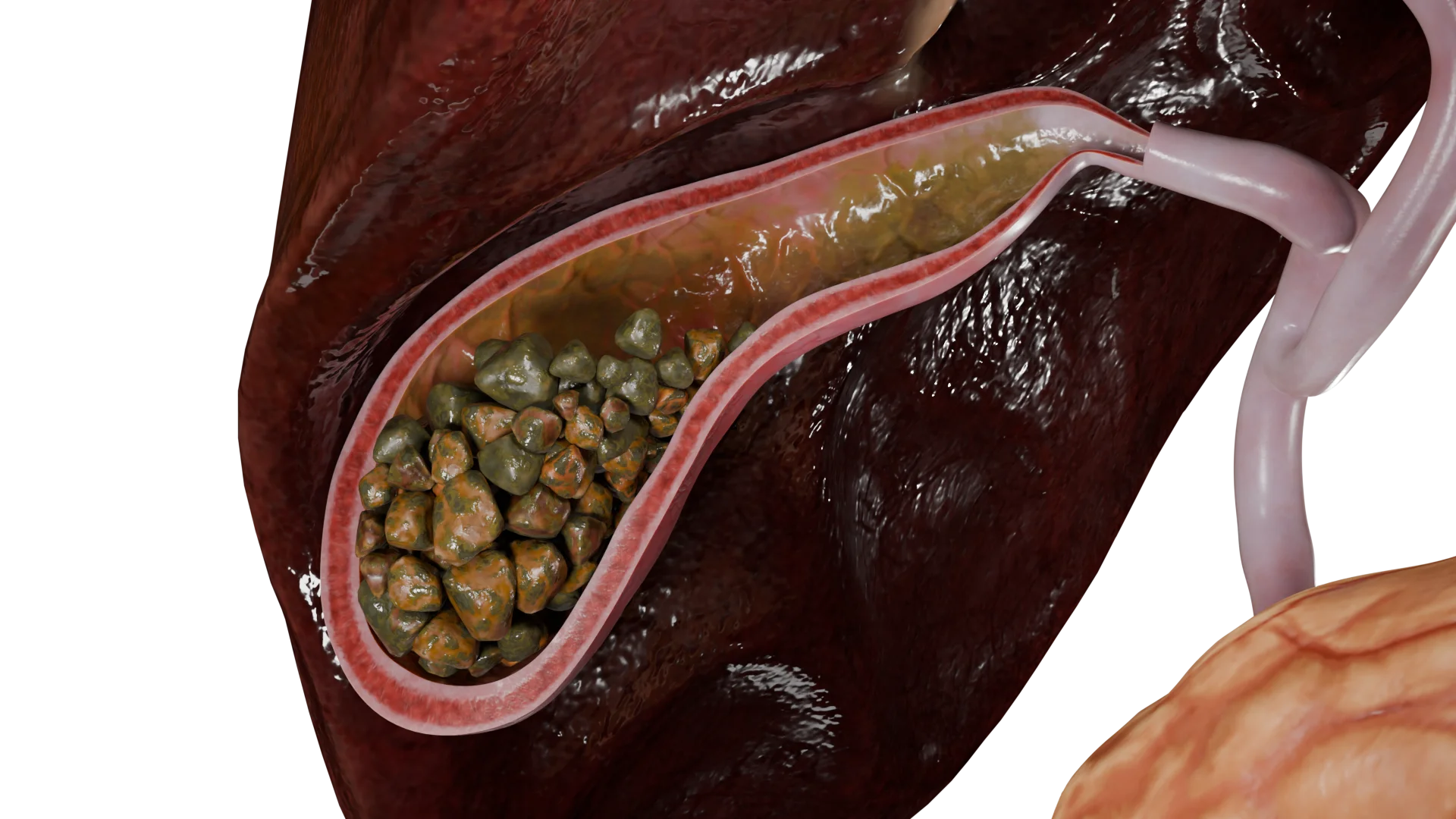
Liver Сirrhosis: Etiology, Clinical Manifestations, Diagnosis and Treatment
Aleksandr F.Abdominal surgeon, MD
12 min read·July 29, 2025
This article is for informational purposes only
The content on this website, including text, graphics, and other materials, is provided for informational purposes only. It is not intended as advice or guidance. Regarding your specific medical condition or treatment, please consult your healthcare provider.
Hepatic cirrhosis (also known as liver cirrhosis) is a diffuse liver disease characterized by parenchymal necrosis, diffuse fibrosis, and abnormal areas of hepatic tissue regeneration. These changes disrupt the normal internal and external architecture of the liver and impair its function.
Classification of Hepatic Cirrhosis
Hepatic cirrhosis is classified according to structural changes and stages of compensation.
By structural changes:
Micronodular cirrhosis (nodules measuring 3 mm to 1 cm in diameter);
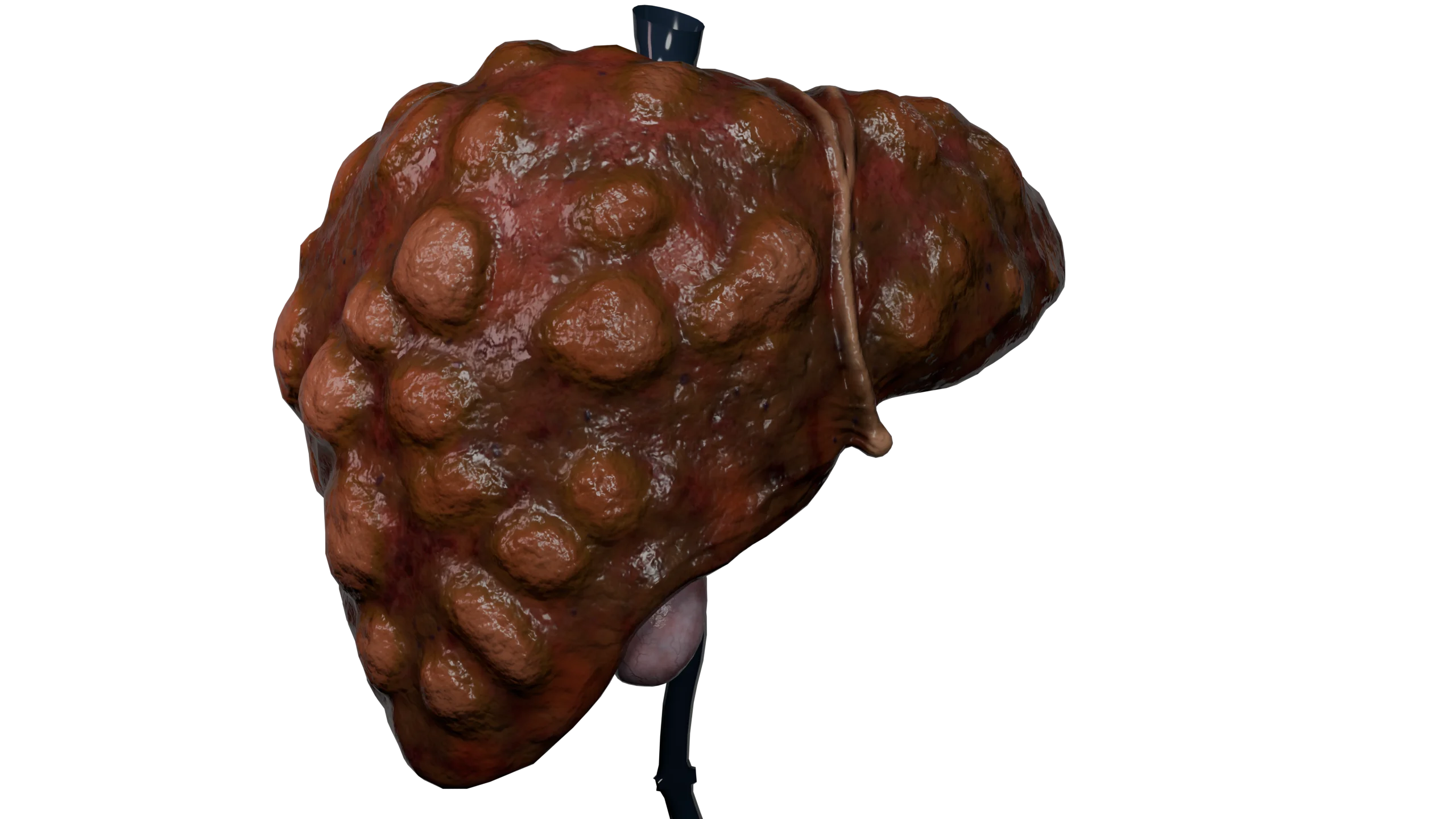
Macronodular cirrhosis (nodules larger than 1 cm in diameter);
Mixed cirrhosis;
Biliary cirrhosis.
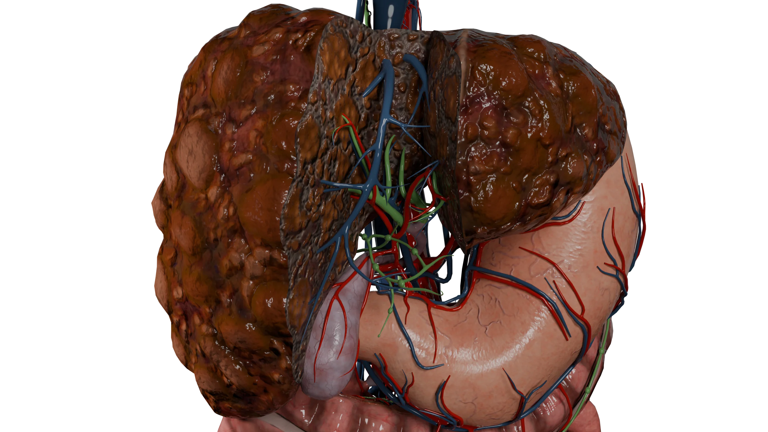
3D Models of hepatic cirrhosis forms by structural changes:
Micronodular Hepatic Cirrhosis
Macronodular Hepatic Cirrhosis
Mixed Hepatic Cirrhosis
Biliary Hepatic Cirrhosis
By stage of compensation:
Compensated;
Subcompensated;
Decompensated;
Terminal.
Another widely used classification is the Child–Pugh score, which considers clinical and laboratory parameters to assess the severity of cirrhosis.
Classification of Hepatic Cirrhosis According to the Child–Pugh Score
Clinical parameter
1 point
2 points
3 points
Ascites
no
Mild (responsive to conservative treatment)
Severe (refractory to conservative treatment)
Serum albumin (g/L)
>35
28-35
< 28
INR (International Normalized Ratio)
< 1,7
1,7-2,3
>>2.3
Total bilirubin (µmol/L)
< 34
34-51
>51
Hepatic encephalopathy
no
Grade I–II
Grade III–IV
Outcomes:
5–6 points: Compensated cirrhosis (class A);
7–9 points: Subcompensated cirrhosis (class B);
10–15 points: Decompensated or terminal hepatic cirrhosis (class C).
Etiology of Hepatic Cirrhosis
According to a report by the European Association for the Study of the Liver, approximately 40% of liver transplantations in Europe are performed for cirrhosis of viral etiology, associated with hepatitis B, C, or D virus infection. Alcohol-related cirrhosis accounts for 33% of cases, and 5% are due to a combination of these etiologic factors.
Autoimmune diseases, as well as the toxic effects of certain medications and systemic toxins that damage liver tissue, may also contribute to biliary cirrhosis. This form is associated with impaired bile flow through the intrahepatic and extrahepatic bile duct systems.
Pathogenesis
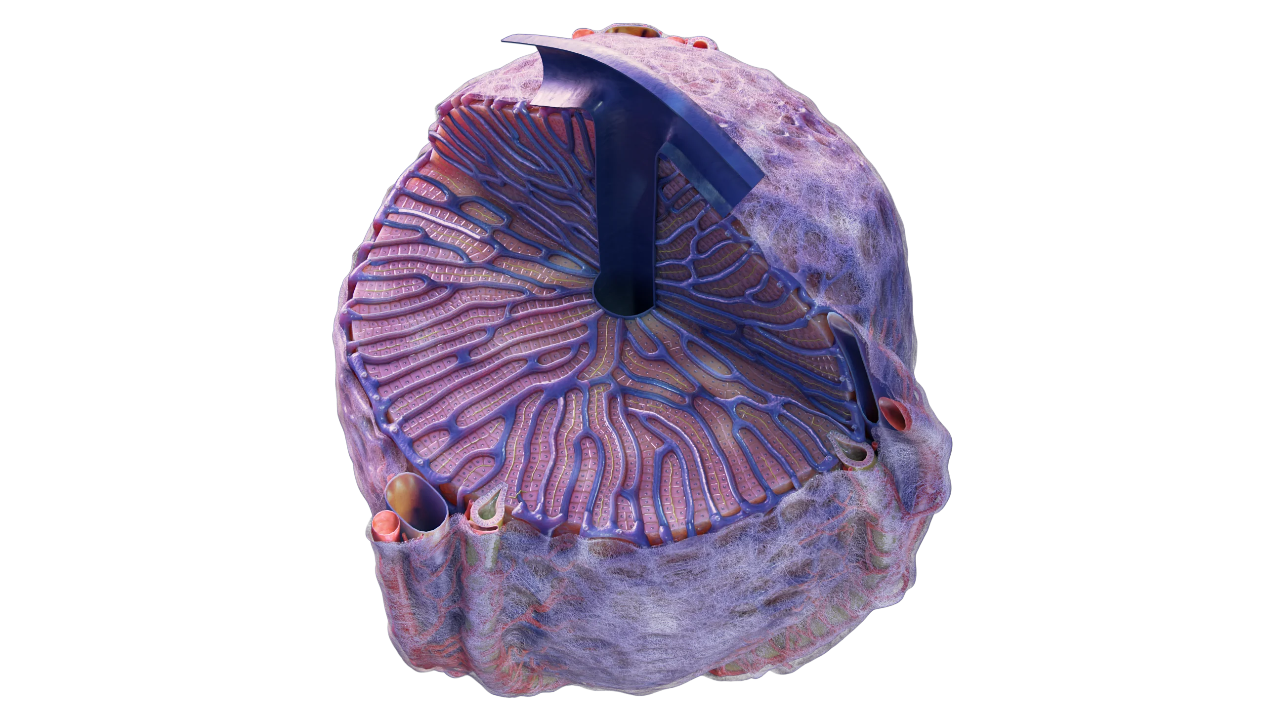
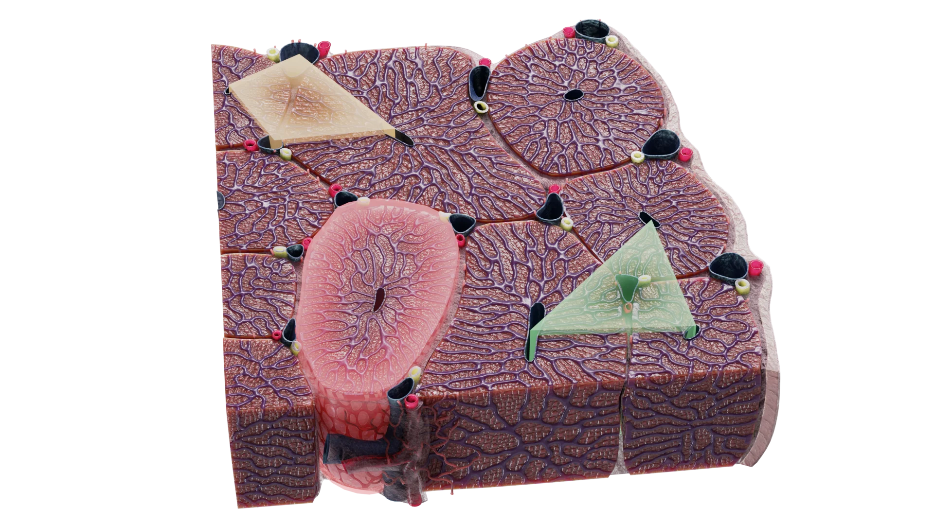
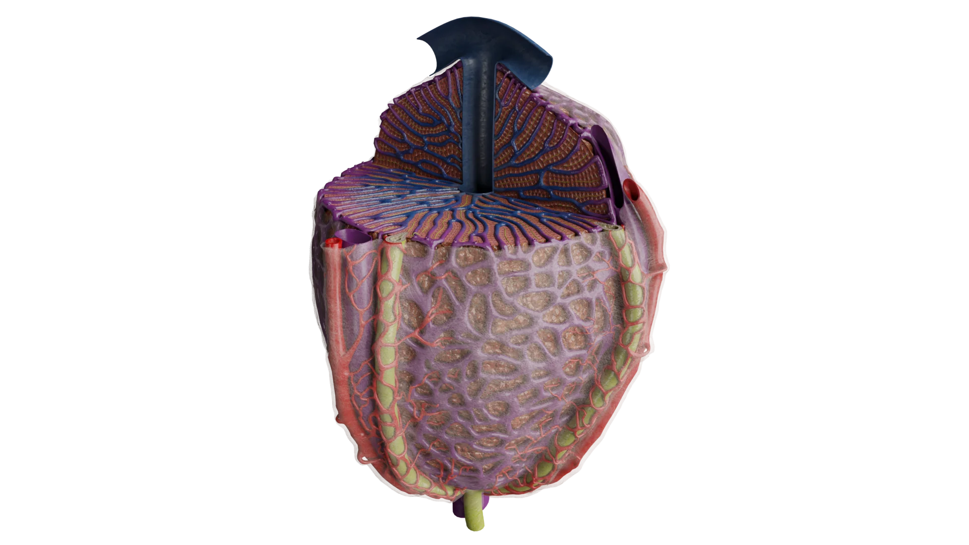
The development of hepatic cirrhosis is based on direct injury to the hepatic lobule, the structural and functional unit of the liver, caused by one of the etiologic factors.
The damaging process is prolonged and may last from several months to several years. As a result, areas of hepatic parenchymal necrosis develop. Around these necrotic areas, zones of regeneration and chronic inflammation form and subsequently progress to fibrosis.
In turn, regenerative nodules compress the bile ducts. This leads to congestion in the hepatic venous system and the development of portal hypertension.
Structure of Liver Tissue: Lobules and Periportal Tracts — 3D Model
Clinical Manifestations of Hepatic Cirrhosis
In the early stages, the disease may be asymptomatic.
As the disease progresses, the following syndromes may develop:
General symptoms, including weakness, headache, nausea, fever, arthralgia, a bitter taste in the mouth, asthenia, and intermittent abdominal pain.
Jaundice syndrome, which develops as a result of intrahepatic cholestasis, meaning impaired bile outflow, increased blood levels of direct bilirubin, and increased bile acid concentrations. Elevated bile acid levels are associated with pruritus.
Hepatomegaly and splenomegaly.
Portal hypertension syndrome, which develops at the stage of decompensation. It is characterized by increased pressure in the portal venous system. Clinical manifestations of portal hypertension include ascites, esophageal varices, rectal varices, and dilated veins of the anterior abdominal wall. Bleeding often occurs against the background of varices.
Appearance of the Abdomen in a Patient With Hepatic Cirrhosis: Ascites and Dilated Veins of the Anterior Abdominal Wall — 3D Model
Pain syndrome associated with biliary dyskinesia.
Hepatic encephalopathy, which develops in the setting of prolonged liver dysfunction and accumulation of neurotoxic substances. These substances may cross the blood–brain barrier and damage brain cells.
Impairment of the synthetic and detoxifying functions of the liver.
It is important to understand that hepatic cirrhosis is a staged disease, and its clinical manifestations change as the condition progresses. In the decompensated and terminal stages, the pathologic process involves not only the hepatobiliary system but also other organ systems, including the urinary, digestive, nervous, and cardiovascular systems.
Diagnosis of Hepatic Cirrhosis
Diagnosis is based on clinical manifestations, identification of the etiologic factor, and findings from instrumental and laboratory studies.
Instrumental Studies
Ultrasound, including liver elastography, and Doppler ultrasound to assess blood flow in the portal venous system;
Computed tomography (CT) and magnetic resonance imaging (MRI);
Liver biopsy;
Esophagogastroduodenoscopy (EGD);
Plain abdominal radiography.
Laboratory Tests
Testing for antibodies to hepatitis B, C, D, and E viruses;
Measurement of serum alpha-fetoprotein (AFP);
Biochemical blood analysis, including the liver profile;
Complete blood count (CBC);
Coagulogram;
Urinalysis;
Assessment of electrolyte levels.
Find more scientifically accurate content on our social media
Subscribe and don’t miss out the latest resources
Treatment of Hepatic Cirrhosis
Treatment of hepatic cirrhosis should be comprehensive and include the following measures:
Complete avoidance of alcohol;
A medically supervised diet for liver disease, correction of electrolyte disturbances, including sodium, potassium, and chloride, and correction of protein imbalance;
Restriction of medications with hepatotoxic potential.
Medical therapy
Infusion therapy, if symptoms of intoxication are present;
Ursodeoxycholic acid preparations;
S-adenosyl-L-methionine preparations;
Glucocorticoids;
Prevention of hemorrhagic complications;
Diuretics;
Treatment aimed at eliminating hepatotropic viruses;
Treatment of concomitant conditions.
Surgical methods cover paracentesis (also known as laparocentesis or abdominal paracentesis) and liver transplant.
Indications for liver transplantation include:
An irreversible pathologic process with an estimated life expectancy of less than 12 months;
Lack of response to conservative therapy;
Progressive hepatic cirrhosis with life expectancy expected to be shorter without transplantation than after transplantation.
FAQ
1. What is hepatic cirrhosis in simple terms?
Hepatic cirrhosis is the end result of various chronic liver diseases. In this condition, normal liver cells are replaced by fibrous scar tissue, which disrupts all major functions of the organ.
2. How does hepatic cirrhosis manifest?
Hepatic cirrhosis may cause symptoms such as increased fatigue, nausea, a bitter taste in the mouth, and abdominal pain, most often in the right upper quadrant. Other manifestations include jaundice, meaning yellowing of the skin and eyes; ascites, or fluid accumulation in the abdomen, which causes abdominal enlargement; and enlargement of the liver and spleen. In the later stages, more severe symptoms develop, including hepatic encephalopathy, bleeding, and progressive liver failure. These complications may be life-threatening.
3. Can hepatic cirrhosis be cured?
Hepatic cirrhosis cannot be completely cured. However, in the early stages, appropriate diet and treatment can slow disease progression and improve both quality of life and life expectancy.
4. How is hepatic cirrhosis diagnosed?
Diagnosis of hepatic cirrhosis is based on clinical manifestations, laboratory tests, and instrumental studies. These may include blood tests, such as a complete blood count, biochemical blood analysis, and blood tests for viral hepatitis; abdominal ultrasound or CT; and, in unclear cases, liver biopsy.
5. What is the difference between hepatic cirrhosis and liver cancer?
Hepatic cirrhosis is a chronic liver disease associated with scarring of liver tissue and impaired liver function. Liver cancer, or hepatocellular carcinoma, is a malignant tumor.
6. What are the stages of hepatic cirrhosis?
Hepatic cirrhosis is divided into four stages depending on the extent of liver damage and organ function: compensated, subcompensated, decompensated, and terminal.
7. Are there differences between hepatic cirrhosis in women and men?
Hepatic cirrhosis causes similar symptoms in women and men. However, hormonal disturbances may appear earlier in women, while signs of portal hypertension may be more prominent in men. In terms of etiology, cirrhosis in women more often develops in the setting of toxic exposure, including alcohol, whereas in men it is more often associated with viral hepatitis or alcohol use disorder.
8. Why is hepatic cirrhosis dangerous?
Hepatic cirrhosis can cause a number of serious complications, including liver failure, portal hypertension, ascites, variceal bleeding, and a substantially increased risk of hepatocellular carcinoma. In the terminal stages, complications of hepatic cirrhosis may lead to death.
References
1.
VOKA 3D Anatomy & Pathology – Complete Anatomy and Pathology 3D Atlas. VOKA 3D Anatomy & Pathology.
Available from: https://catalog.voka.io/
2.
Aprosina, Z. G. (1996). Chronic diffuse liver diseases: Current trends. Clinical Pharmacology and Therapy, 5(1), 14–18. [In Russian.]
3.
Bakulin, I. G., & Varlamicheva, A. A. (2014). Hepatorenal syndrome: Practical recommendations for diagnosis and treatment. Almanac of Clinical Medicine, 33, 23–31. [In Russian.]
4.
Ivashkin, V. T., & Lapina, T. L. (Eds.). (2015). Gastroenterology. National guidelines. Moscow: GEOTAR-Media; 2015. 480 p. [In Russian.]
5.
Mekhtiev, S. N., Grinevich, V. B., Chepur, S. V., & Gancho, V. Y. (2004). Portal hypertension in patients with chronic hepatitis and liver cirrhosis. S. Bresta. 320 p. [In Russian.]
6.
Nikitin, I. G., & Storozhakov, G. I. (2001). Chronic hepatitis C: Current issues of diagnosis and treatment. Clinical Perspectives in Gastroenterology and Hepatology, (3), 7–11. [In Russian.]
7.
Pavlov, C. S., Damulin, V. D., & Ivashkin, V. T. (2016). Hepatic encephalopathy: Pathogenesis, clinical features, diagnosis, and therapy. Russian Journal of Gastroenterology, Hepatology, Coloproctology, 26(1), 44–53. [In Russian.]
8.
Pimanov, S. (2016). Decompensated liver cirrhosis: Treatment considering international recommendations. Prakticheskaya Meditsina. 88 p. [In Russian.]
9.
Schiff, E. R., Sorrell, M. F., & Maddrey, W. C. (Eds.). (2012). Schiff’s diseases of the liver: Liver cirrhosis and its complications. Liver transplantation. Moscow: GEOTAR-Media; 2012. 592 p. [In Russian.]
10.
Arroyo, V., Moreau, R., Jalan, R., & Ginès, P., for the EASL-CLIF Consortium CANONIC Study. (2015). Acute-on-chronic liver failure: A new syndrome that will re-classify cirrhosis. J Hepatol. 62(Suppl. 1), S131–S143.
11.
European Association for the Study of the Liver. (2015). EASL clinical practice guidelines: Wilson’s disease. J Hepatol. 2012 Mar;56(3):671-685.
12.
European Association for the Study of the Liver. (2015). EASL clinical practice guidelines on the management of hepatic encephalopathy. Journal of Hepatology, J Hepatol. 77(3), 807–824.
13.
European Association for the Study of the Liver. (2015). EASL clinical practice guidelines: Autoimmune hepatitis. J Hepatol. 2015;63(4):971-1004.
14.
Sanyal, A., Mullen, K., & Bass, N. (2010). The treatment of hepatic encephalopathy in the cirrhotic patient. Gastroenterology & Hepatology, 6(4 Suppl. 8), 1–12.
50 Franklin Street, STE 203 Boston, MA 02110 United States
Thank you!
Your message is sent! Our experts will contact you shortly. If you have any additional questions, please contact us at info@voka.io
Cookie Consent
We use cookies to enhance your browsing experience, analyze site traffic, and deliver content. Please choose whether you accept all cookies or wish to reject non-essential tracking.
Cookie Preferences
Manage your cookie preferences below:
Essential cookies enable basic functions and are necessary for the proper function of the website.
Name
Description
Duration
Geolocation Config
This cookie is used to store the consent settings based on the visitor's location.
30 days
Cookie Preferences
This cookie is used to store the user's cookie consent preferences.
30 days
Google reCAPTCHA helps protect websites from spam and abuse by verifying user interactions through challenges.
Name
Description
Duration
_GRECAPTCHA
Google reCAPTCHA sets a necessary cookie (_GRECAPTCHA) when executed for the purpose of providing its risk analysis.
179 days
Statistics cookies collect information anonymously. This information helps us understand how visitors use our website.
Google Analytics is a powerful tool that tracks and analyzes website traffic for informed marketing decisions.
Used to monitor number of Google Analytics server requests
10 minutes
__utmb
Used to distinguish new sessions and visits. This cookie is set when the GA.js javascript library is loaded and there is no existing __utmb cookie. The cookie is updated every time data is sent to the Google Analytics server.
30 minutes after last activity
__utmc
Used only with old Urchin versions of Google Analytics and not with GA.js. Was used to distinguish between new sessions and visits at the end of a session.
End of session (browser)
__utmz
Contains information about the traffic source or campaign that directed user to the website. The cookie is set when the GA.js javascript is loaded and updated when data is sent to the Google Anaytics server
6 months after last activity
__utmv
Contains custom information set by the web developer via the _setCustomVar method in Google Analytics. This cookie is updated every time new data is sent to the Google Analytics server.
2 years after last activity
__utmx
Used to determine whether a user is included in an A / B or Multivariate test.
18 months
_ga
ID used to identify users
2 years
_gali
Used by Google Analytics to determine which links on a page are being clicked
30 seconds
_ga_
ID used to identify users
2 years
_gid
ID used to identify users for 24 hours after last activity
24 hours
_gat
Used to monitor number of Google Analytics server requests when using Google Tag Manager
1 minute
_gac_
Contains information related to marketing campaigns of the user. These are shared with Google AdWords / Google Ads when the Google Ads and Google Analytics accounts are linked together.
90 days
Clarity is a web analytics service that tracks and reports website traffic.