Cholecystitis: Etiology, Classification, Clinical Manifestations, Diagnosis, and Treatment
Aleksandr F.Abdominal surgeon, MD
10 min read·October 30, 2025
This article is for informational purposes only
The content on this website, including text, graphics, and other materials, is provided for informational purposes only. It is not intended as advice or guidance. Regarding your specific medical condition or treatment, please consult your healthcare provider.
Cholecystitis is an inflammation in the gallbladder caused by impaired bile outflow. The condition may develop due to a mechanical obstruction by gallstones (calculous cholecystitis) or for other reasons (acalculous cholecystitis).
Classification of Cholecystitis
By type of the disease:
Acute,
Chronic.
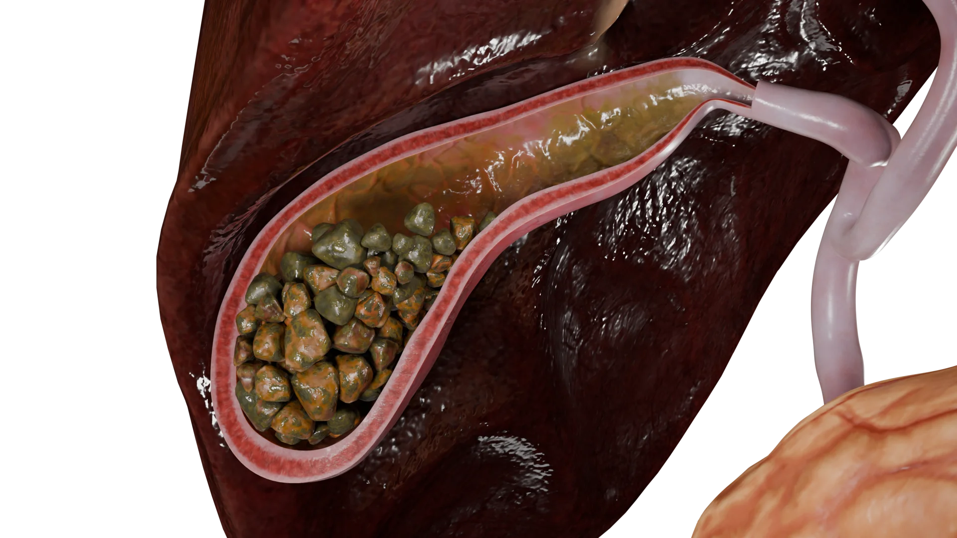
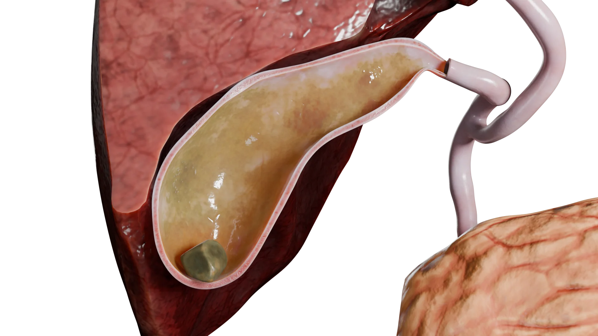
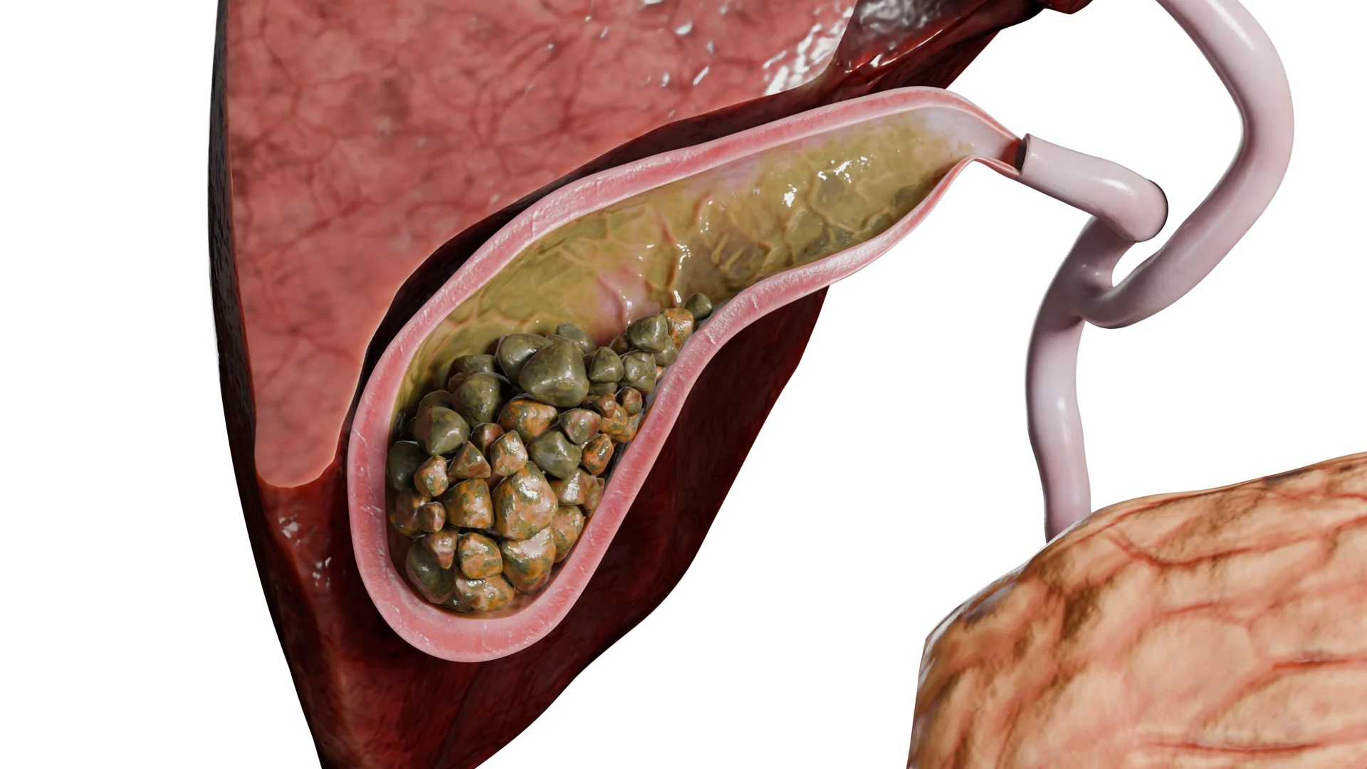
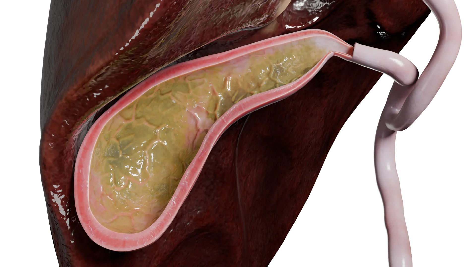
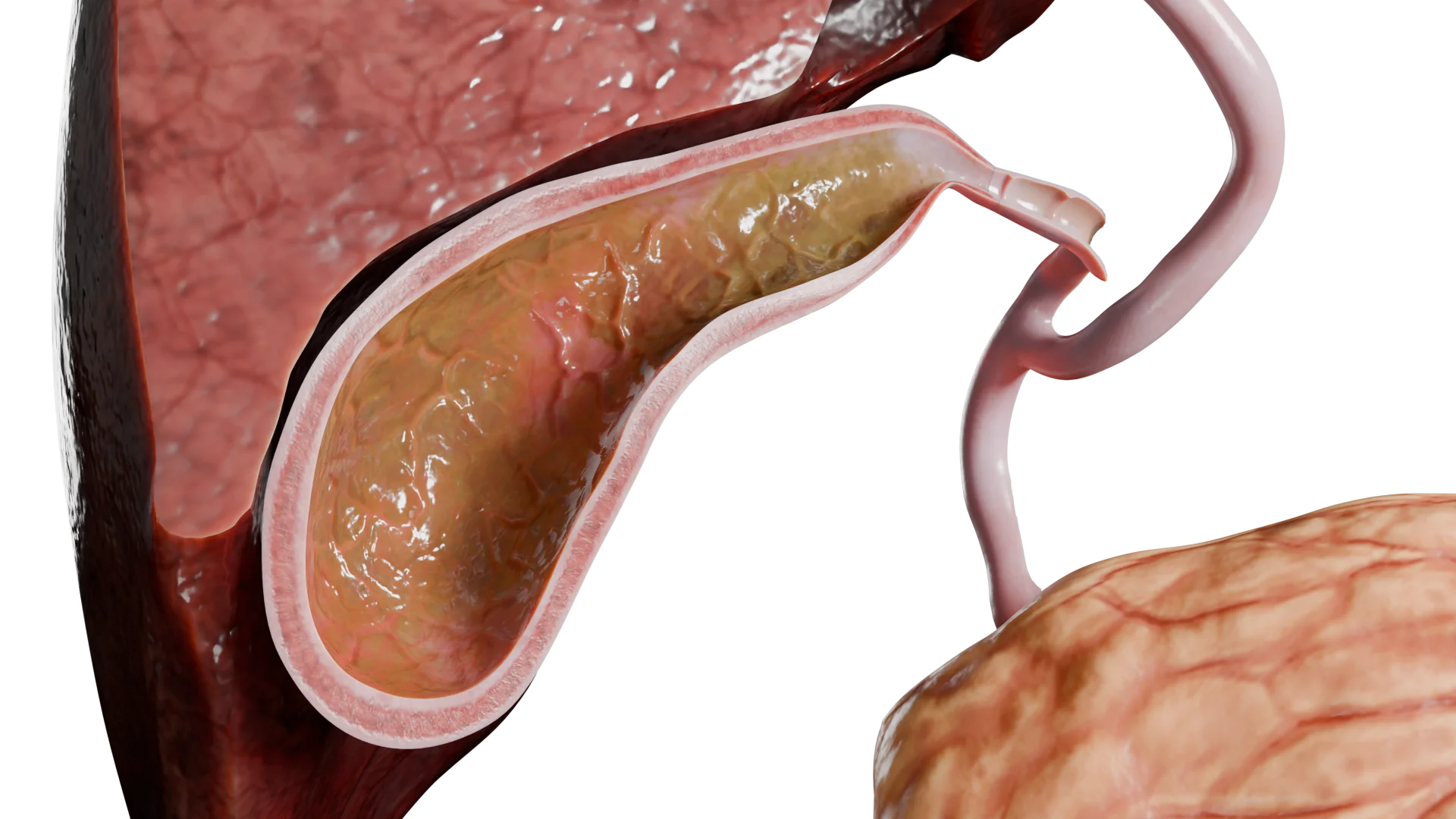
Acute Catarrhal Cholecystitis Associated with Gallstones — 3D ModelChronic Cholecystitis Associated with Gallstones, without Acute Exacerbation — 3D Model
By etiology:
Calculous (associated with gallstones),
Acalculous (not caused by gallstones).
Acute Calculous Catarrhal Cholecystitis — 3D ModelAcute Catarrhal Cholecystitis without Gallstones — 3D Model
By clinical and morphological features:
Catarrhal,
Phlegmonous,
Gangrenous.
3D models of cholecystitis according to clinical and morphological features:
Acute Catarrhal Cholecystitis
Acute Phlegmonous Cholecystitis
Acute Gangrenous Cholecystitis
By severity:
Mild: limited gallbladder inflammation;
Moderate: gallbladder inflammation persists for more than 72 hours, and its walls bear signs of destructive changes;
Severe: total gallbladder destruction accompanied by signs of multiple organ failure.
Etiology and Pathogenesis
Inflammation of the gallbladder wall may arise due to certain damaging factors.
Based on the cause, cholecystitis is differentiated into its calculous and acalculous forms.
Calculous cholecystitis
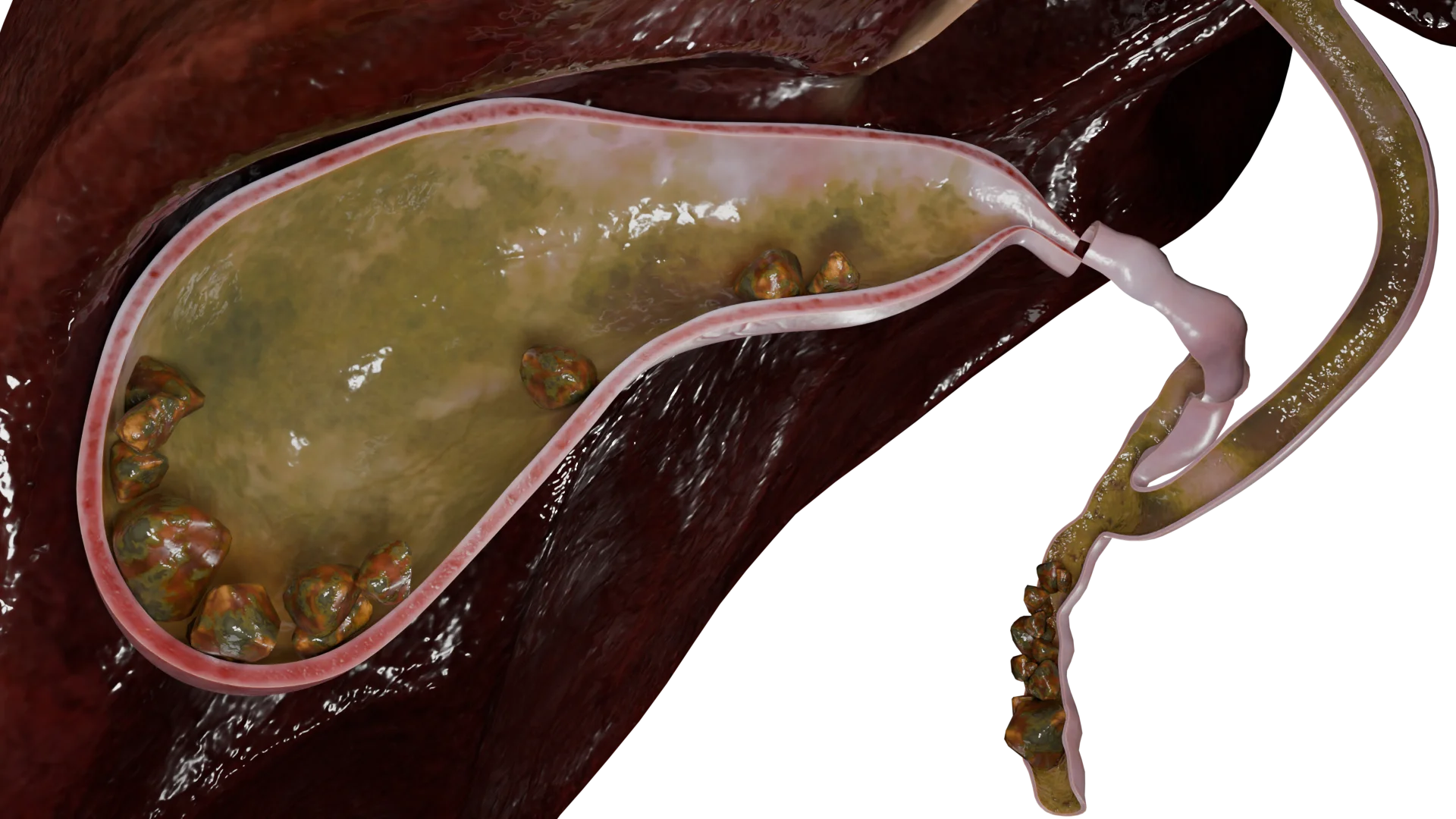
Cholecystitis develops secondary to gallstones (cholelithiasis) when a solitary or multiple calculi obstruct the cystic duct or gallbladder neck. The obstruction increases intraluminal gallbladder pressure, damages its mucosa, and causes local ischemic changes in the gallbladder wall. This triggers inflammation and the release of biologically active substances and inflammatory mediators.
Multiple Gallstones in Gallbladder and Cystic Duct — 3D Model
Only 50–75% of cholecystitis cases secondary to cholelithiasis have a bacterial nature. The most commonly identified gram-negative bacteria are: E. coli, Klebsiella spp., Pseudomonas spp., Enterobacter spp. Among gram-positive bacteria, Enterococcus spp. and Streptococcus spp. are most frequent. Anaerobes include Clostridium spp.
Acalculous cholecystitis
Unlike the calculous form,acalculous cholecystitis is caused by a variety of factors, such as impaired gallbladder contractility, hematogenous infection, systemic conditions, intestinal infections, or congenital bile duct disorders (e.g., choledochal cyst). However, all of these causes follow the same inflammation mechanism in the gallbladder wall that is typically observed in calculous cholecystitis.
Clinical Manifestations
Cholecystitis is associated with a range of signs and symptoms, such as:
Pain,
Dyspepsia,
Inflammation,
Peritoneal irritation,
Obstructive jaundice.
In general, pain in cholecystitis typically manifests as biliary colic. The episode of biliary colic may resolve spontaneously or with the help of antispasmodic drugs. The pain is intense. It is typically localized in the right upper quadrant (right hypochondrium) but may sometimes radiate to the adjacent regions (lumbar region, scapula, or shoulder). As the condition progresses, the pain tends to exacerbate and irritate the peritoneum.
Furthermore, dyspepsia may develop secondary to pain. This syndrome covers nausea, vomiting, and a bitter taste in the mouth. Vomiting, either single-episode or repeated, usually brings no relief.
Even during the first few days, patients often have a low-grade fever, which may gradually rise to a moderate fever as the inflammation progresses.
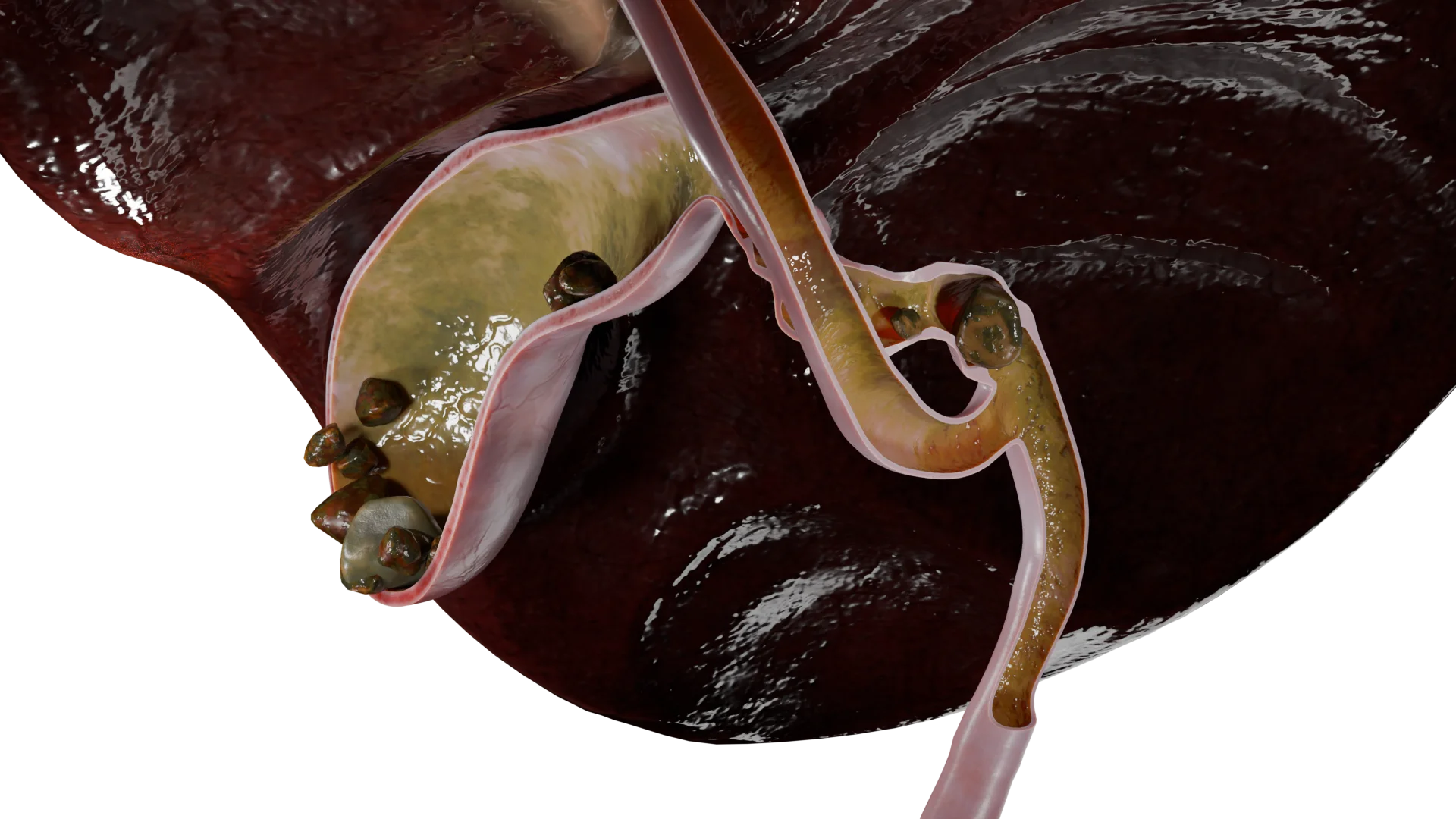
During examination, a healthcare professional should pay particular attention to signs of jaundice, including the yellow discoloration of the skin, sclerae, and mucous membranes. Dark urine and pale stools are quite common. The primary cause of these symptoms is a gallstone, either solitary or multiple, that obstructs (or blocks) the hepatobiliary system.
Multiple Gallstones in Gallbladder and Bile Duct — 3D Model
Diagnosis of Cholecystitis
Diagnosis of cholecystitis is based on clinical evaluation and investigations (both laboratory and instrumental).
Laboratory tests: complete blood count (CBC), urinalysis, and blood tests (liver function tests, inflammatory markers, electrolytes). In critically ill patients, the acid-base balance should also be assessed.
Investigations: abdominal ultrasound, plain abdominal and chest X-ray, computed tomography (CT), and esophagogastroduodenoscopy (EGD).
Find more scientifically accurate content on our social media
Subscribe and don’t miss out the latest resources
Treatment of Cholecystitis
Cholecystitis can be managed with medical or surgical approaches.
Medical Management
Medical therapy is aimed at stabilizing the acute episode and preparing the patient for potential surgery.
Pain relief: Nonsteroidal anti-inflammatory drugs (NSAIDs) and antispasmodics are used to control pain.
Infusion therapy: Intravenous fluids help to reverse any toxic effects of the condition and restore fluid and electrolyte balance.
Antimicrobial therapy: Antibiotics are administered when signs of a bacterial infection develop. The exact medications are chosen based on microbial sensitivity and the patient’s allergy history.
Surgical Management
Surgical removal of the gallbladder (cholecystectomy) is the definitive treatment for cholecystitis.
If conservative treatment has proven efficient and acute cholecystitis has no complications, elective cholecystectomy may be performed. However, when medications have failed and the condition progresses, emergency surgery is required. In this case, the intervention is most feasible within the first 24–72 hours after the onset.
There are two main surgical approaches:
Laparoscopic cholecystectomy: Removal of the gallbladder through small abdominal incisions; minimally invasive and considered the gold standard.
Open cholecystectomy: Performed through either an upper midline incision or an incision in the right hypochondriac region.
FAQ
1. What are calculous and acalculous cholecystitis, and what causes them?
Cholecystitis is an inflammation of the gallbladder. The most common type is calculous cholecystitis, which occurs secondary to gallstones. When these stones block the cystic duct, they can trigger an acute episode. In chronic cases, gallstones may be present without causing any obstruction. Acalculous cholecystitis is less common and is not related to gallstones. It often results from poor gallbladder motility, severe infections, or circulatory disorders.
2. What are the symptoms of cholecystitis, and how does an acute episode present?
One of the main symptoms is sudden, intense pain in the right upper quadrant (right hypochondrium), commonly known as biliary colic. The pain may be accompanied by nausea, vomiting, fever, and a bitter taste in the mouth. Chronic cholecystitis can flare up with similar symptoms, though they are usually milder. If bile flow becomes obstructed, jaundice may develop, along with dark urine and pale stools.
3. What complications can arise from cholecystitis, and why is it dangerous?
If left untreated, cholecystitis can lead to serious complications. The inflammation may progress to destructive forms, such as phlegmonous or gangrenous cholecystitis, which can cause gallbladder wall perforation and peritonitis. In severe cases, it can result in multiple organ failure and become life-threatening.
4. How is cholecystitis treated, and can it be cured permanently?
According to modern clinical practice, treatment usually begins with conservative therapy to relieve acute symptoms. This may include painkillers, antispasmodics, and antibiotics if a bacterial infection is suspected. However, the only definitive way to cure calculous cholecystitis and prevent recurrence and complications is surgical removal of the gallbladder (cholecystectomy).
5. What laboratory and imaging tests are used to diagnose cholecystitis?
Diagnosis combines laboratory tests and imaging studies. Abdominal ultrasound is considered the gold standard and can reveal gallbladder wall thickening, gallstones, and other typical signs of inflammation. In more complex or unclear cases, CT scans may be performed to provide a more detailed assessment.
6. Can an episode of cholecystitis resolve without treatment?
The pain of biliary colic may subside on its own, but this does not mean the inflammation has resolved. The underlying disease process usually persists, and without proper treatment, it may progress, increasing the risk of serious complications.
7. What diet is recommended for acute and chronic cholecystitis?
Diet plays a key role in treatment. During an acute episode, fasting is usually recommended first, followed by a strict therapeutic diet that excludes fatty, fried, spicy foods and alcohol. In chronic cholecystitis during remission, similar dietary principles apply but can be followed with greater flexibility. Meals should be small and frequent, ideally 5–6 times a day.
8. How does cholecystitis differ from cholestasis?
These are different conditions. Cholecystitis is inflammation of the gallbladder wall. Cholestasis, on the other hand, refers to a reduced or blocked bile flow from the liver. It can contribute to gallbladder inflammation, but it is not the same condition.
9. Can cholecystitis be prevented?
Yes. Preventive measures focus on reducing the risk of gallstone formation. A balanced diet low in fat and rich in fiber, maintaining a healthy weight, regular physical activity, and adequate hydration can help lower the risk of developing the disease.
10. How does cholecystitis present in children?
Cholecystitis is uncommon in children and is usually acalculous. It may develop as a complication after severe infections (such as scarlet fever or sepsis) or as part of systemic diseases. Symptoms are generally similar to those in adults. Treatment is based on antibiotic therapy and, when required, surgical intervention.
References
1.
VOKA 3D Anatomy & Pathology – Complete Anatomy and Pathology 3D Atlas. VOKA 3D Anatomy & Pathology.
Available from: https://catalog.voka.io/
2.
Dadvani SA, Vetshev PS, Shulutko AM, Prudkov MI. Zhelchnokamennaya bolezn’ [Gallstone disease]. Moscow: Vidar; 2000. 144 p. [In Russian.]
3.
Dorofeeva SG, Konoplya EN, Mansimova OV, Shelukhina AN, Anyushonkov OS. Zhelchnokamennaya bolezn’: sovremennye predstavleniya ob etiologii i patogeneze [Gallstone disease: current concepts of etiology and pathogenesis]. Integrativnye tendentsii v meditsine i obrazovanii [Integrative Trends in Medicine and Education]. 2020;2:21–25. [In Russian.]
4.
Ivashkin VT, Maev IV, Baranskaya EK. Rekomendatsii Rossiiskoi gastroenterologicheskoi assotsiatsii po diagnostike i lecheniyu zhelchnokamennoi bolezni [Recommendations of the Russian Gastroenterological Association for the diagnosis and treatment of gallstone disease]. RZhGGK. 2016;3:64–80. [In Russian.]
5.
European Association for the Study of the Liver (EASL). EASL Clinical Practice Guidelines on the prevention, diagnosis and treatment of gallstones. J Hepatol. 2016;65:146–181.
6.
Leishner U. Prakticheskoe rukovodstvo po zabolevaniyam zhelchnykh putei [Practical guide to biliary tract diseases]. Moscow: GEOTAR-Meditsina; 2001. 264 p. [In Russian.]
7.
Litovskii IA, Gordienko AV. Zhelchnokamennaya bolezn’, kholetsistity i nekotorye assotsiirovannye s nimi zabolevaniya: voprosy patogeneza, diagnostiki, lecheniya [Gallstone disease, cholecystitis and some associated diseases: issues of pathogenesis, diagnosis, treatment]. Saint Petersburg: SpetsLit; 2019. 358 p. [In Russian.]
8.
Podoluzhnyi VI. Oslozhneniya zhelchnokamennoi bolezni [Complications of gallstone disease]. Fundamentalnaya i Klinicheskaya Meditsina [Fundamental and Clinical Medicine]. 2017;2(1):102–114. [In Russian.]
9.
Saveliev VS, Kirienko AI. Khirurgicheskie bolezni [Surgical diseases]. Vol. 1. Moscow: GEOTAR-Media; 2005. p. 179-183. 185–201. [In Russian.]
10.
Skvortsov VV, Khalilova UA. Diagnostika i lechenie zhelchnokamennoi bolezni [Diagnosis and treatment of gallstone disease]. Eksperimentalnaya i Klinicheskaya Gastroenterologiya [Experimental and Clinical Gastroenterology]. 2018;157(9):142–150. [In Russian.]
11.
Uspenskii YuP, Fominykh YuA, Nadzhafova KN. Lipidnyi obmen u lits s zhelchnokamennoi bolezn’yu [Lipid metabolism in patients with gallstone disease]. University Therapeutic Journal. 2020;2(1):74–75. [In Russian.]
12.
Khimich SD. Analiz morfo-funktsional’nykh osobennostei razvitiya i techeniya nekotorykh ostrykh khirurgicheskikh zabolevanii organov bryushnoi polosti u lits s ozhireniem: avtoreferat dissertatsii na soiskanie uchenoi stepeni doktora meditsinskikh nauk [Analysis of morphofunctional features of the development and course of certain acute surgical diseases of the abdominal organs in obese patients: dissertation abstract]. Moscow; 2002. 38 p. [In Russian.]
13.
Barak O., Elazary R., Appelbaum L., Rivkind A., Almogy G. Conservative treatment for acute cholecystitis: clinical and radiographic predictors of failure. Israel Medical Association Journal, 11, 11. 739–743.
14.
Chen, L. Y., Qiao, Q. H., Zhang, S. C., Chen, Y. H., Chao, G. Q., & Fang, L. Z. (2012). Metabolic syndrome and gallstone disease. World Journal of Gastroenterology, 18, 4215–4220.
15.
Cotton P.B., Elta G.H., Carter C.R., Pasricha P.J., Corazziari E.S. Rome IV. Gallbladder and Sphincter of Oddi Disorders. Gastroenterology, pii: S0016-5085(16)00224-9.
16.
McGlone E.R., Bloom S.R. Bile acids and the metabolic syndrome. Annals of Clinical Biochemistry, 56(3), 56(3). 326–337.
17.
Wang Y., Qi M., Qin C., Hong J. Role of the biliary microbiome in gallstone disease. Expert Review of Gastroenterology & Hepatology, 12(12), 12(12). 1193–1205.
405 Lexington Avenue, 26th Floor New York, NY 10174 United States
Thank you!
Your message is sent! Our experts will contact you shortly. If you have any additional questions, please contact us at info@voka.io
Cookie Consent
We use cookies to enhance your browsing experience, analyze site traffic, and deliver content. Please choose whether you accept all cookies or wish to reject non-essential tracking.
Cookie Preferences
Manage your cookie preferences below:
Essential cookies enable basic functions and are necessary for the proper function of the website.
Name
Description
Duration
Geolocation Config
This cookie is used to store the consent settings based on the visitor's location.
30 days
Cookie Preferences
This cookie is used to store the user's cookie consent preferences.
30 days
Google reCAPTCHA helps protect websites from spam and abuse by verifying user interactions through challenges.
Name
Description
Duration
_GRECAPTCHA
Google reCAPTCHA sets a necessary cookie (_GRECAPTCHA) when executed for the purpose of providing its risk analysis.
179 days
Statistics cookies collect information anonymously. This information helps us understand how visitors use our website.
Google Analytics is a powerful tool that tracks and analyzes website traffic for informed marketing decisions.
Used to monitor number of Google Analytics server requests
10 minutes
__utmb
Used to distinguish new sessions and visits. This cookie is set when the GA.js javascript library is loaded and there is no existing __utmb cookie. The cookie is updated every time data is sent to the Google Analytics server.
30 minutes after last activity
__utmc
Used only with old Urchin versions of Google Analytics and not with GA.js. Was used to distinguish between new sessions and visits at the end of a session.
End of session (browser)
__utmz
Contains information about the traffic source or campaign that directed user to the website. The cookie is set when the GA.js javascript is loaded and updated when data is sent to the Google Anaytics server
6 months after last activity
__utmv
Contains custom information set by the web developer via the _setCustomVar method in Google Analytics. This cookie is updated every time new data is sent to the Google Analytics server.
2 years after last activity
__utmx
Used to determine whether a user is included in an A / B or Multivariate test.
18 months
_ga
ID used to identify users
2 years
_gali
Used by Google Analytics to determine which links on a page are being clicked
30 seconds
_ga_
ID used to identify users
2 years
_gid
ID used to identify users for 24 hours after last activity
24 hours
_gat
Used to monitor number of Google Analytics server requests when using Google Tag Manager
1 minute
_gac_
Contains information related to marketing campaigns of the user. These are shared with Google AdWords / Google Ads when the Google Ads and Google Analytics accounts are linked together.
90 days
Clarity is a web analytics service that tracks and reports website traffic.