Vulvitis: Predisposing Factors, Clinical Manifestations, Diagnosis, and Treatment
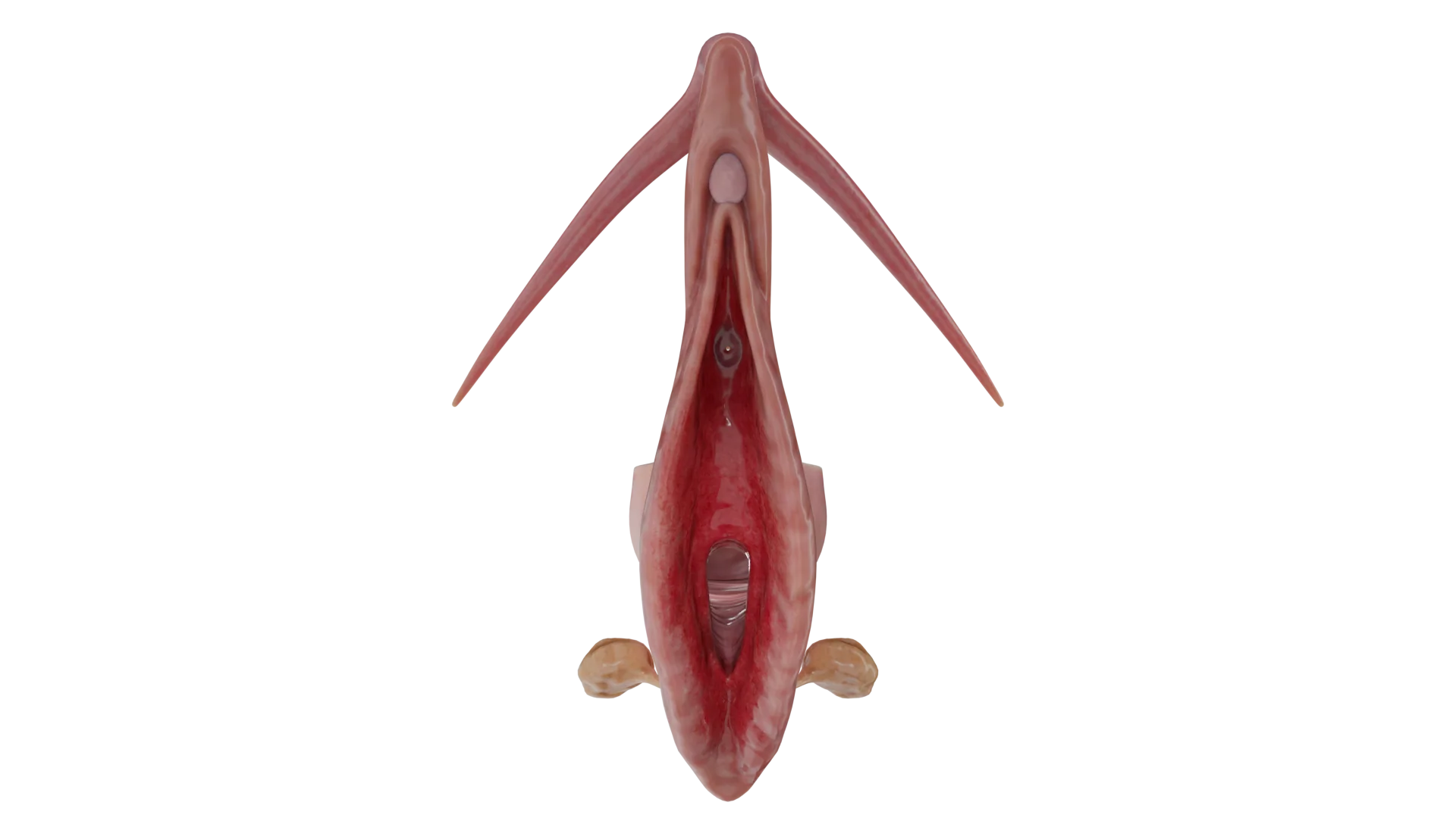
Vulvitis refers to vulvar inflammation affecting the labia, clitoris, mons pubis, and vestibule of the vagina. Clinical manifestations, diagnosis, and treatment.


Anesthesia
Pain management and sedation techniques

Angiology
Arterial and venous pathologies

Cardiology
Acquired and congenital heart diseases

Dentistry
Diseases of teeth, gums, and the oral cavity

Dermatology
Disorders of the skin and subcutaneous tissue

Embryology
Stages of embryogenesis and congenital anomalies

Endocrinology
Disorders of the glands and hormonal imbalance

Gastroenterology
Stomach, intestinal, and digestive diseases

Gynecology
Diseases of female reproductive organs

Hematology
Hematopoiesis and blood-related disorders

Hepatology
Liver, gallbladder, and biliary tract diseases

Histology
Microscopic tissue and cell structures

Infectious diseases
Bacterial, viral, and parasitic infections

Neurology
Brain, spinal cord, and peripheral nerve disorders

Obstetrics
Pregnancy complications and abnormal fetal positions

Oncology
Cancer types, benign and malignant tumors

Ophthalmology
Conditions affecting the eyes and vision

Orthopedics
Bone, joint, and soft tissue disorders

Otorhinolaryngology
Ear, nose, and throat diseases

Pediatrics
Child health, development, and clinical conditions

Physiology
Biological processes within organs and systems

Pulmonology
Lung and respiratory tract diseases

Traumatology
Acute injuries and musculoskeletal trauma

Urology
Urinary tract and male reproductive disorders
Anesthesia
Pain management and sedation techniques
Angiology
Arterial and venous pathologies
Cardiology
Acquired and congenital heart diseases
Dentistry
Diseases of teeth, gums, and the oral cavity
Dermatology
Disorders of the skin and subcutaneous tissue
Embryology
Stages of embryogenesis and congenital anomalies
Endocrinology
Disorders of the glands and hormonal imbalance
Gastroenterology
Stomach, intestinal, and digestive diseases
Gynecology
Diseases of female reproductive organs
Hematology
Hematopoiesis and blood-related disorders
Hepatology
Liver, gallbladder, and biliary tract diseases
Histology
Microscopic tissue and cell structures
Infectious diseases
Bacterial, viral, and parasitic infections
Neurology
Brain, spinal cord, and peripheral nerve disorders
Obstetrics
Pregnancy complications and abnormal fetal positions
Oncology
Cancer types, benign and malignant tumors
Ophthalmology
Conditions affecting the eyes and vision
Orthopedics
Bone, joint, and soft tissue disorders
Otorhinolaryngology
Ear, nose, and throat diseases
Pediatrics
Child health, development, and clinical conditions
Physiology
Biological processes within organs and systems
Pulmonology
Lung and respiratory tract diseases
Traumatology
Acute injuries and musculoskeletal trauma
Urology
Urinary tract and male reproductive disorders
Specialties
AnesthesiaAngiologyCardiologyDentistryDermatologyEmbryologyEndocrinologyGastroenterologyGynecologyHematologyHepatologyHistologyInfectious diseasesNeurologyObstetricsOncologyOphthalmologyOrthopedicsOtorhinolaryngologyPediatricsPhysiologyPulmonologyTraumatologyUrology
This article is for informational purposes only
The content on this website, including text, graphics, and other materials, is provided for informational purposes only. It is not intended as advice or guidance. Regarding your specific medical condition or treatment, please consult your healthcare provider.
Atrophic vaginitis is a complication of genitourinary syndrome of menopause, which is a constellation of symptoms and clinical signs associated with declining levels of sex steroids that result in atrophic changes affecting the labia, clitoris, vaginal vestibule, vagina, urethra, and bladder.
A decline in sex steroid hormone levels, particularly estrogen, leads to atrophic changes in the vaginal mucosa and reduced glycogen deposition within the vaginal epithelium. Glycogen is metabolized by the local vaginal microbiota to produce organic acids that are essential for protecting the genital tract. As a result, the concentration of Lactobacillus decreases leading to an increase in vaginal pH, which promotes the growth of pathogenic bacteria and contributes to the development of vaginitis.
Androgens (i.e. dehydroepiandrosterone, androstenedione, and testosterone) play an important role in vaginal mucosal metabolism because they serve as precursors for estrogen biosynthesis. In healthy premenopausal women, androgen production is significantly higher than estrogen production. Androgen receptors are widely distributed throughout the urogenital tract. Androgen-dependent protein products have trophic effects on various tissues of the urogenital organs (vaginal vestibule, clitoris, urethra, vagina, bladder, and pelvic floor muscles/ligaments). In addition to the cessation of estrogen production during menopause, the age-related decline in androgen levels may also contribute to the development of the signs and symptoms of genitourinary syndrome.
Patients are bothered by genital dryness, burning and irritation. Sexual symptoms such as lack of discharge, decreased lubrication, discomfort, pain during intercourse or vaginal bleeding associated with sexual activity. Urinary symptoms may include dysuria and recurrent urinary tract infections. It is worth saying that these symptoms in postmenopause have a negative impact on sexual interest, intimacy and relationship with a partner, mood and self-esteem.
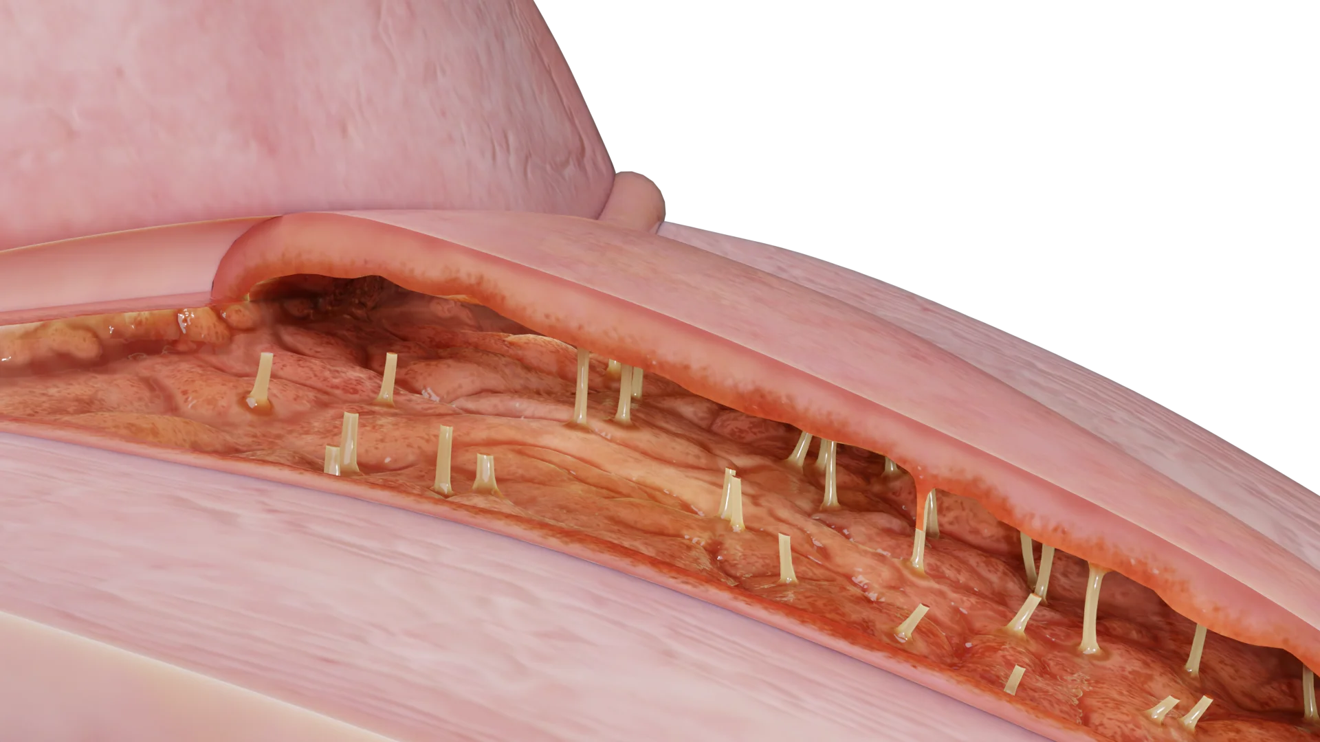
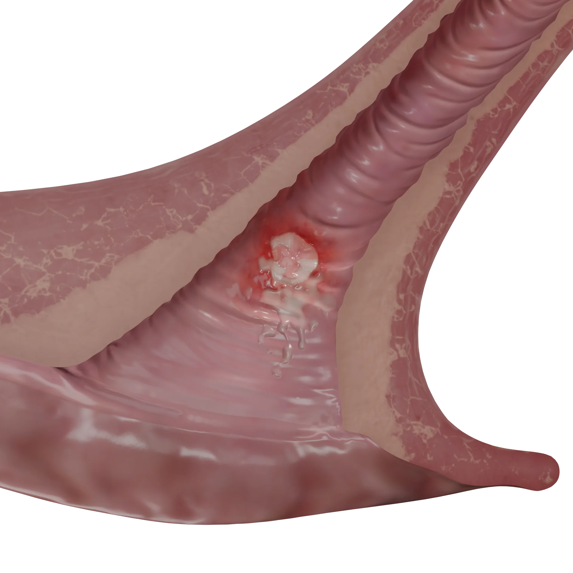
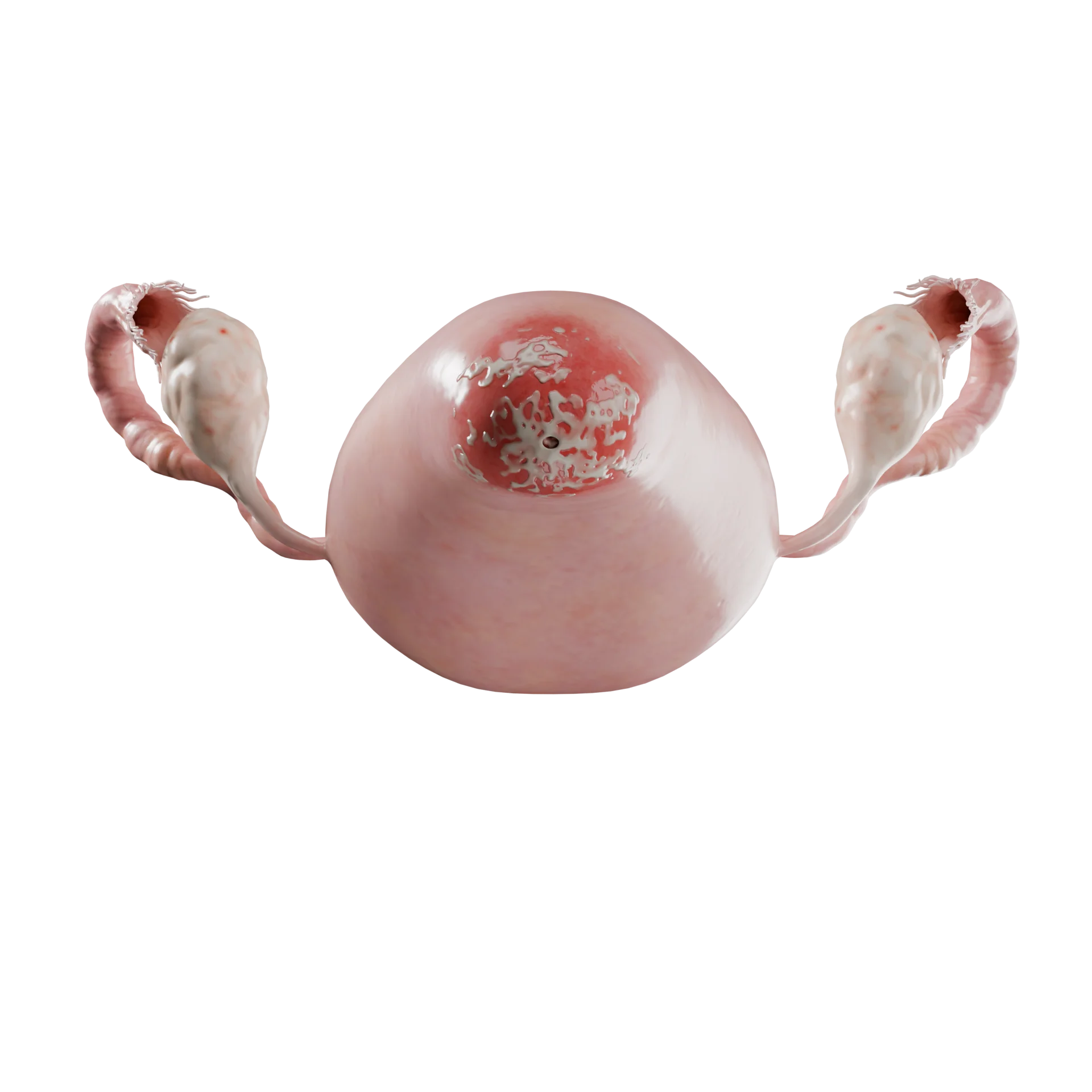
During a speculum examination, the vaginal mucosa appears thin and smooth, with erythema, absence of vaginal discharge, and the presence of localized petechiae or ulcerative lesions.

The Vaginal Maturation Index (VMI) reflects the proportional relationship among superficial, intermediate, and parabasal cells within the vaginal tissue. Estrogen deficiency is associated with an increased proportion of parabasal cells, resulting in a decreased VMI.
Histologic examination diagnoses a decrease in superficial squamous cells and an increase in parabasal cells. The hypoestrogenic state leads to loss of collagen, elastin fibers and blood vessels. These changes result in decreased elasticity and vascularization. Decreased vascularization in response to low estrogen levels leads to thinning of the vaginal mucosa and decreased discharge.
Vaginal therapy is the first-line pharmacologic treatment recommended by the International Menopause Society. Women should be started on the lowest dose and frequency that effectively manages their symptoms.
Vaginal estrogen is the treatment of choice. This therapy is appropriate for women with isolated vaginal symptoms because vaginal estrogen preparations contain lower doses of estrogen than systemic hormone therapy. Progestagen is generally not indicated for vaginal therapy.
Endometrial surveillance is also not required unless there is postmenopausal bleeding that requires diagnostic evaluation.
Vaginal therapy increases estrogen concentrations in the vaginal epithelium, uroepithelium, and helps reduce atrophic changes while minimizing systemic exposure.
A Cochrane systematic review found no evidence of differences in symptom improvement among women treated with the following vaginal estrogen formulations: estrogen vaginal ring versus estrogen cream, estrogen vaginal ring versus estrogen tablets, or estrogen tablets versus estrogen cream.
Estriol –is a natural estrogen. A low dose of estriol vaginal gel (0.005%) has been shown to significantly increase vaginal cell maturation index and decrease vaginal pH compared to a control group in postmenopausal women.
Find more scientifically accurate content on our social media



1. What is atrophic vaginitis?
2. What symptoms are characteristic of atrophic vaginitis?
3. What complications can develop if atrophic vaginitis is left untreated?
4. What preventive measures are recommended?
5. Which age groups are most commonly affected by atrophic vaginitis?
References
1.
VOKA 3D Anatomy & Pathology – Complete Anatomy and Pathology 3D Atlas. VOKA 3D Anatomy & Pathology.
Available from: https://catalog.voka.io/
2.
Brotman RM, Shardell MD, Gajer P, Fadrosh D, Chang K, Silver MI, et al. Association between the vaginal microbiota, menopause status, and signs of vulvovaginal atrophy. Menopause. 2018 Nov;25(11):1321-1330.
3.
Cheng, R. Interpretation on the 2023 Chinese Menopause Symptom Management and Menopausal Hormone Therapy Guidelines. Med. J. Peking Union Med. Coll. Hosp. 2023, 14, 514–519.
4.
Partridge L, Deelen J, Slagboom PE. Facing up to the global challenges of ageing. Nature. 2018 Sep;561(7721):45–56. doi:10.1038/s41586-018-0457-8.
5.
Valadares ALR, Kulak Junior J, Paiva LHSC, Nasser EJ, da Silva CR, Nahas EAP, et al. Genitourinary syndrome of menopause. Rev Bras Ginecol Obstet. 2022;44:319-324.
Make VOKA your preferred source
See more VOKA articles in Google Search









Summarize article with AI
Choose your preferable AI assistant:
Link successfully copied to clipboard
Thank you!
Your message is sent!
Our experts will contact you shortly. If you have any additional questions, please contact us at info@voka.io