Infective endocarditis is a potentially life-threatening disease associated with inflammation of the endocardium, predominantly the valve apparatus of the heart, caused by microbial invasion. Despite the development of antibiotic therapy and modern imaging techniques, the mortality rate of the total number of patients remains high.3D animation – Infective endocarditis of the left side of the heart3D animation – infective endocarditis of the right heart tract
Epidemiology
The incidence of infective endocarditis ranges from 3 to 15 cases per 100,000 population per year. The incidence increases with age, peaking in those over 60 years of age due to the accumulation of risk factors such as valve prostheses, cardiac implants, chronic diseases and frequent medical interventions. Men are about twice as likely to have the disease as women.
In recent decades, there has been a shift in the epidemiologic profile: instead of rheumatic heart disease and intravenous drug users, elderly patients, often with implanted valves, prostheses, and pacemakers, now predominate.
Etiology
The cause of IE can be caused by a variety of microorganisms, including:
Staphylococcus aureus is the leader in frequency, especially in hospital-acquired IE and in drug addicts.
Viridans streptococci are classic causative agents in native valve lesions in patients without obvious risk factors.
Enterococcus spp. – significant in elderly patients, often associated with urogenital interventions.
Coagulase-negative staphylococci are frequent causative agents in prosthetic valve infections.
Less frequently – HACEK-group, fungi, gram-negative bacilli, etc.
Risk factors include the presence of artificial valves, pacemakers, previous IE, heart defects, intravenous drug use, and chronic hemodialysis.
Pathogenesis
The pathogenesis of infective endocarditis involves several consecutive links:
Damage to valve endothelium (e.g. by turbulent blood flow) → exposure of extracellular matrix.
Adhesion of platelets and fibrin → formation of sterile thrombus (non-bacterial thrombotic endocarditis).
Microbial invasion – in transient bacteremia, microorganisms colonize the clot.
Vegetation formation – dense clusters of fibrin, inflammatory cells and bacteria protected from the immune response and antibiotics.
Destruction of valve structures and possible embolization → systemic complications, sepsis, acute heart failure. Immune mechanisms also contribute to the development of complications such as vasculitis and glomerulonephritis.
The key step is the formation of vegetations that promote persistence of infection and the development of embolic complications.
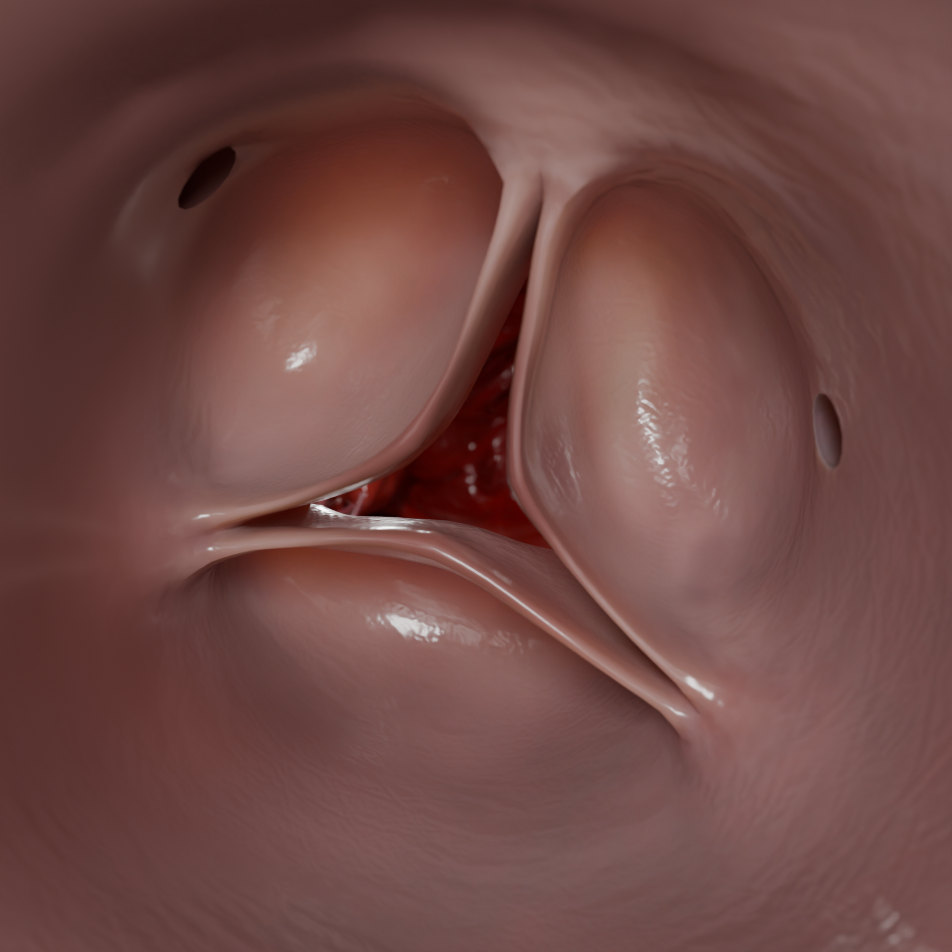
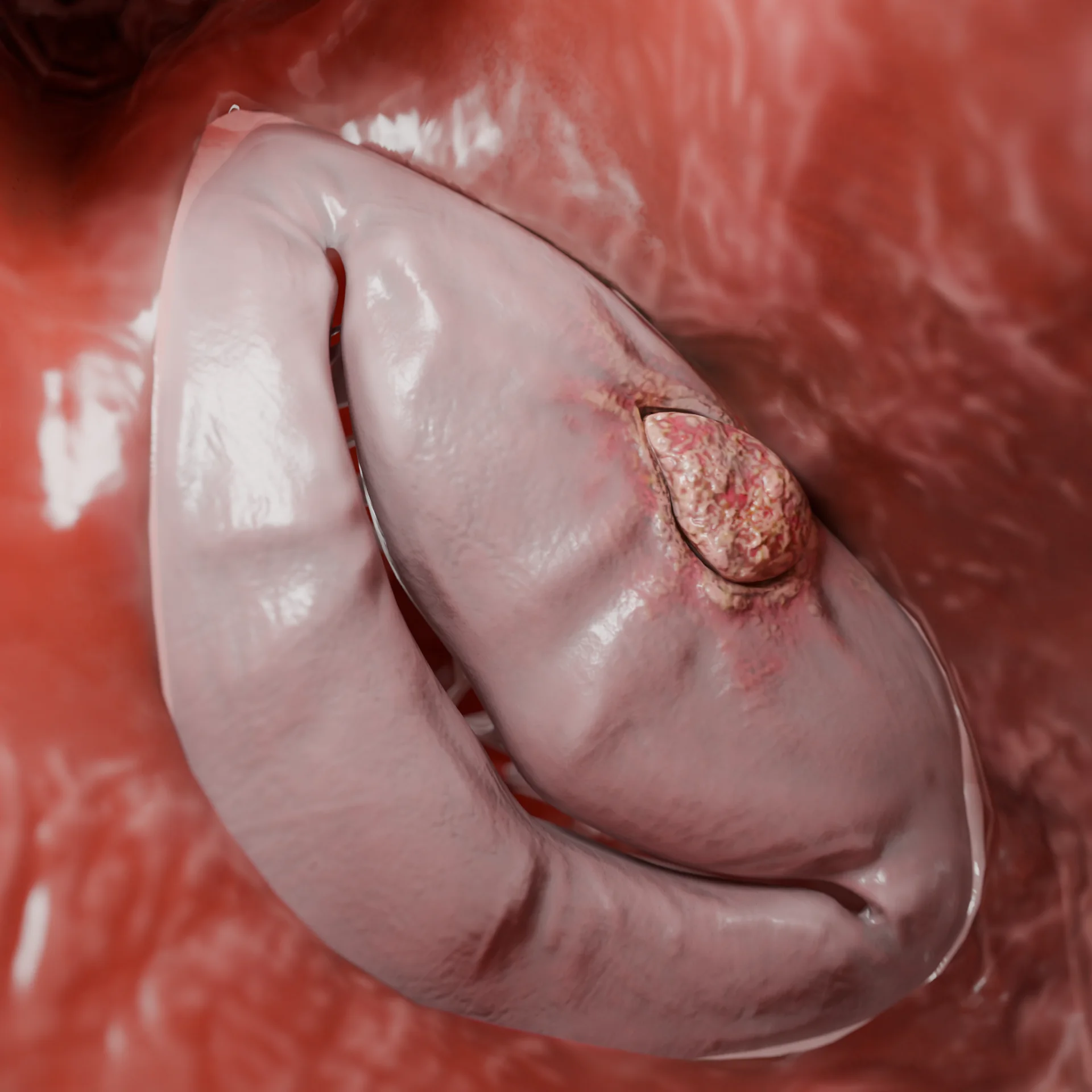
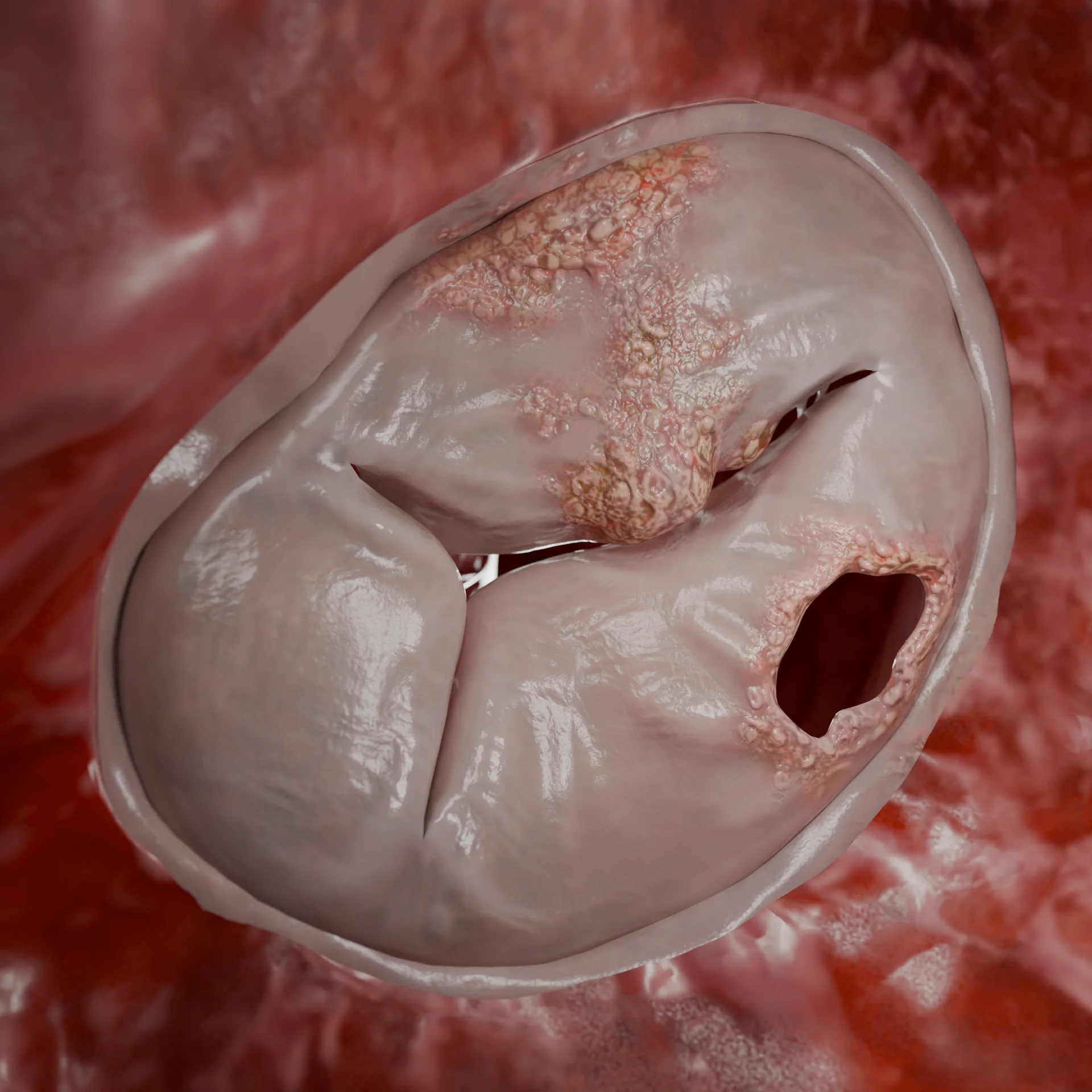
Vegetations on the mitral valveTricuspid valve leaflet perforation
Classification of infective endocarditis
By localization of the process: Left-sided endocarditis
Mitral and/or aortic valve damage (most common)
Right-sided endocarditis
Tricuspid and/or (less frequently) pulmonary valve damage (more common in injecting drug users, CVC patients)
Combined
Simultaneous lesions of the right and left divisions
Prosthetic endocarditis
Inflammation on a mechanical or biological valve prosthesis
Device endocarditis
Infection associated with pacemaker electrodes, ICDs, etc.
By etiology (causative agent): Gram-positive cocci
Candida spp., Aspergillus spp. (rare, more common in immunocompromised persons)
By clinical course: Acute
Onset: sudden Course: rapid, aggressive Pathogens more common: Staphylococcus aureus, β-hemolytic streptococci Features: rapid valve destruction, sepsis, high mortality rate
Subacute
Onset: gradual (weeks) Course: sluggish, with unclear symptomatology Pathogens more common: Streptococcus viridans, Enterococcus spp. Features: anemia, subfebrile, immune manifestations.
Chronic
Onset: prolonged (months) Course: latent or recurrent Pathogens more common: low virulence bacteria, culture-negative forms Features: prolonged inflammation, persistent valve changes, possible relapse after therapy
Left infective endocarditis (vegetations on valves and mitro-aortic junction) – 3D modelInfective right-sided endocarditis (vegetations and perforation on the tricuspid valve) – 3D model
Clinical Manifestations
The symptomatology of IE is variable, ranging from nonspecific to life-threatening:
Common symptoms of infective endocarditis include fever, sweating, fatigue, and weight loss.
Cardiac signs: new or altered murmur, signs of heart failure.
Embolic complications: stroke, limb ischemia, infarcts of internal organs.
Lesion of the conductive system: blockades, arrhythmias.
In severe course of IE may develop: septic shock, multi-organ failure.
In prosthetic endocarditis, auscultatory murmurs may be less pronounced, periannular infection is more likely to develop: abscesses, pseudoaneurysms, fistulas.
Diagnosis of infective endocarditis
Laboratory Methods:
General blood analysis: normocytic anemia, leukocytosis, thrombocytopenia.
Repeated emboli or large vegetations c episode of emboli (>10 mm), vegetations >15 mm, especially in left-sided IE even without emboli;
Prosthetic endocarditis;
Fungal IE or caused by highly resistant microorganisms.
Contraindications:
Decompensated general condition, multi-organ failure;
Recent massive stroke with hemorrhagic component.
Types of operations(in the vast majority of cases, operations are performed under artificial circulation):
Replacement (prosthetics) of the affected valve;
Removal of vegetations, sanitation of abscesses;
Reconstructive interventions (valve, valve ring, aortic root plasty): if the aortic root is involved, replacement with a conduit (artificial vascular prosthesis with artificial valve) or homograft (human donor valve with a section of ascending aorta) may be necessary;
Removal of infected devices (If electrodes or pacemakers are involved: mandatory removal of the entire system. In TAVI-endocarditis: surgery is often highly lethal, but is indicated if therapy is ineffective).
FAQ
1. What is infective endocarditis?
Infective endocarditis is an inflammatory disease of the inner lining of the heart (endocardium), most often involving the valves, caused by a bacterial or fungal infection.
2. What symptoms are most characteristic of IE?
Most common symptoms: fever, chills, weakness, heart murmurs, weight loss. Possible complications: strokes, embolisms, heart failure.
3. Which bacteria most commonly cause IE?
The most common pathogens are Staphylococcus aureus, Streptococcus viridans, and Enterococcus spp. Persons with prostheses or intravascular devices often have Staph. epidermidis .
4. What are the dangers of infective endocarditis?
IE can lead to valve destruction, heart failure, embolic complications (strokes, organ infarcts), abscesses and sepsis. The mortality rate reaches 20-30%.
5. Can IE be treated without surgery?
Yes, in some cases – especially with native valves and sensitive microorganisms – complete cure with antibiotics is possible. However, surgery may be required for complications.
6. When is surgery necessary for IE?
Surgery is indicated for: • Heart failure due to valve dysfunction; • Abscesses, ruptures, perforations; • Ineffective antibiotic therapy; • Fungal infection; • Recurrent emboli.
7. How long is antibiotic treatment given?
The duration of therapy is usually 4-6 weeks, depending on the causative agent, type of valve (native or prosthetic) and complications.
8. Can infective endocarditis be prevented?
Yes, antibiotic prophylaxis is recommended in at-risk patients (e.g., those with prosthetic valves) before certain dental or surgical procedures.
9. Who is at high risk for IE?
Individuals with prosthetic valves, pacemakers, previous IE, congenital heart disease, and patients on hemodialysis and drug users.
List of Sources
1.
VOKA Catalog.
https://catalog.voka.io/
2.
2023 ESC Guidelines for the management of endocarditis. Delgado V, Ajmone Marsan N, de Waha S, Bonaros N, Brida M, Burri H, Caselli S, Doenst T, Ederhy S, Erba PA, Foldager D, Fosbøl EL, Kovac J, Mestres CA, Miller OI, Miro JM, Pazdernik M, Pizzi MN, Quintana E, Rasmussen TB, Ristić AD, Rodés-Cabau J, Sionis A, Zühlke LJ, Borger MA; ESC Scientific Document Group. Eur Heart J. 2023 Oct 14;44(39):3948-4042. doi: 10.1093/eurheartj/ehad193.
3.
Infective endocarditis. Li M, Kim JB, Sastry BKS, Chen M. Lancet. 2024 Jul 27;404(10450):377-392. doi: 10.1016/S0140-6736(24)01098-5.
4.
Infective endocarditis: A contemporary update. Rajani R, Klein JL. Clin Med (Lond). 2020 Jan;20(1):31-35. doi: 10.7861/clinmed.cme.20.1.1.
5.
Management Considerations in Infective Endocarditis: A Review. Wang A, Gaca JG, Chu VH. JAMA. 2018 Jul 3;320(1):72-83. doi: 10.1001/jama.2018.7596.
6.
Infective Endocarditis in Adults: Diagnosis, Antimicrobial Therapy, and Management of Complications: A Scientific Statement for Healthcare Professionals From the American Heart Association. Baddour LM, Wilson WR, Bayer AS, Fowler VG Jr, Tleyjeh IM, Rybak MJ, Barsic B, Lockhart PB, Gewitz MH, Levison ME, Bolger AF, Steckelberg JM, Baltimore RS, Fink AM, O’Gara P, Taubert KA; American Heart Association Committee on Rheumatic Fever, Endocarditis, and Kawasaki Disease of the Council on Cardiovascular Disease in the Young, Council on Clinical Cardiology, Council on Cardiovascular Surgery and Anesthesia, and Stroke Council. Circulation. 2015 Oct 13;132(15):1435-86. doi: 10.1161/CIR.0000000000000296.
7.
Infective endocarditis. Epidemiology, pathophysiology and histopathology. Iung B. Presse Med. 2019 May;48(5):513-521. doi: 10.1016/j.lpm.2019.04.009.
8.
Native-Valve Infective Endocarditis. Chambers HF, Bayer AS. N Engl J Med. 2020 Aug 6;383(6):567-576. doi: 10.1056/NEJMcp2000400.
9.
Infective Endocarditis-Update for the Perioperative Clinician. Jain A, Subramani S, Gebhardt B, Hauser J, Bailey C, Ramakrishna H. J Cardiothoracic Vasc Anesth. 2023 Apr;37(4):637-649. doi: 10.1053/j.jvca.2022.12.030.

.webp)
.webp)