Rhinitis: Symptoms, Stages, Diagnosis, and Treatment
Afanasyeva D.Otorhinolaryngologist, MD
20 min read·April 14, 2025
This article is for informational purposes only
The content on this website, including text, graphics, and other materials, is provided for informational purposes only. It is not intended as advice or guidance. Regarding your specific medical condition or treatment, please consult your healthcare provider.
Rhinitis is inflammation of the nasal cavity mucosa. The disease has common symptoms, regardless of the causes, such as nasal congestion and rhinorrhea (nasal discharge), and specific symptoms characteristic of certain types of rhinitis.
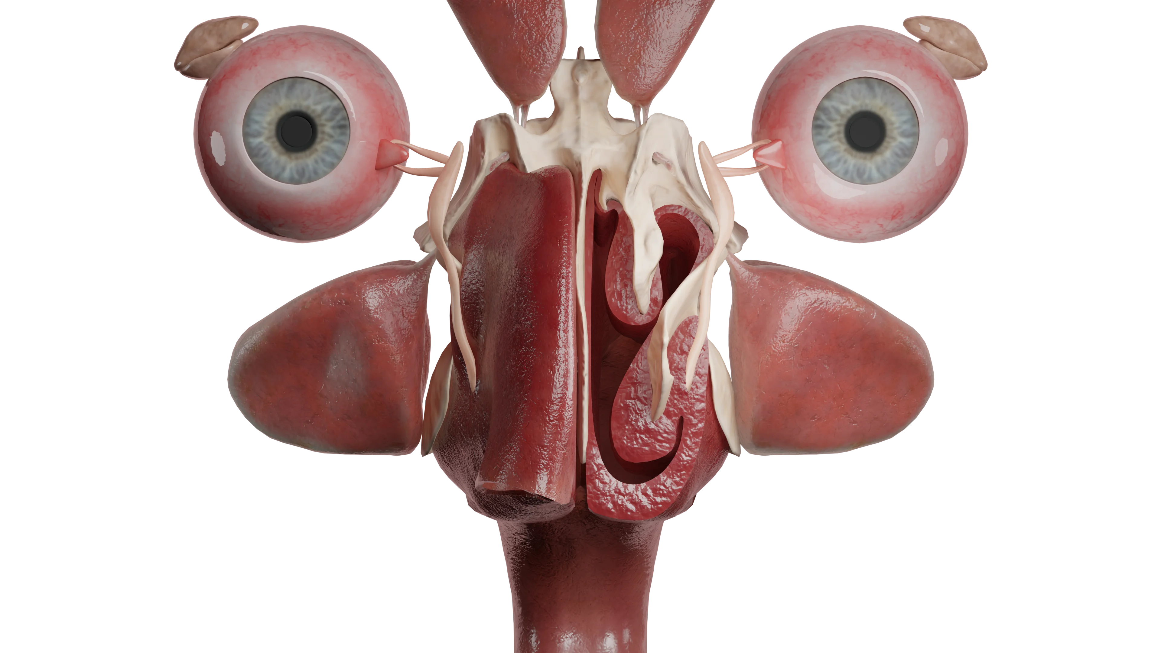
Nasal cavity (in cross-section)
Classification of Rhinitis
Acute rhinitis:
1st stage of dry irritation;
2nd stage of serous discharge;
3rd stage of serous-purulent discharge.
Chronic rhinitis:
Infectious;
Allergic;
Hypertrophic;
Vasomotor;
Atrophic;
Ozena.
Etiology
Acute rhinitis is inflammation of the nasal cavity mucosa lasting no more than 12 weeks, caused by viruses or bacteria that land on the epithelial surface and trigger a pathological reaction. This disease is non-specific. The viruses most commonly causing acute rhinitis are: adenovirus, rhinovirus, RS-virus, influenza, and parainfluenza viruses. Among bacteria causing nasal mucosa inflammation are streptococci, staphylococci, and pneumococci. Acute rhinitis can be an initial manifestation of specific diseases such as measles, scarlet fever, diphtheria, and meningococcal infection. For the development of the pathological process, in addition to the presence of pathogenic microflora, mucosal changes such as dryness and crusting, decreased general or local immunity, and the presence of chronic infections in the decompensation phase are necessary.
Chronic inflammation is considered to last over 12 weeks; if acute rhinitis persists beyond this period, it transitions to chronic infectious rhinitis, so the pathogens for these conditions are the same. This form of rhinitis can also accompany infectious diseases such as syphilis, tuberculosis, histoplasmosis, blastomycosis, leprosy, and others, which will be discussed in more detail in the corresponding sections.
Allergic rhinitis occurs when allergens affect the mucosa. The most common allergens are pet dander, dust mites, plant pollen, and mold spores. When allergens affect the mucosa, an IgE-mediated immune response develops with the release of inflammatory mediators that trigger pathological reactions. There is a hereditary predisposition and a general tendency towards atopy. Allergic rhinitis can also be a manifestation of helminth invasion, such as giardiasis, more common in children, pathogenetically explained by general sensitization of the body.
Hypertrophicrhinitis most often develops as a result of impaired nasal breathing of post-traumatic nature or chronic inflammation in the nasal cavity or paranasal sinuses.
Vasomotorrhinitis occurs when there is a disturbance in neuro-reflex processes, increasing hypersensitivity to various irritants.
Vasomotor rhinitis includes:
Medicamentous (due to prolonged use of nasal decongestants);
Hormonal (in pregnant women due to increased progesterone, in patients with thyroid diseases);
Professional (in contact with dust, wool, sawdust);
Emotional (during stress);
Nutritional (when consuming spicy, hot dishes, alcohol).
These forms of rhinitis have different etiologies but similar clinical and pathomorphological manifestations. The diagnosis is made based on a carefully collected medical history and identification of the causal factor.
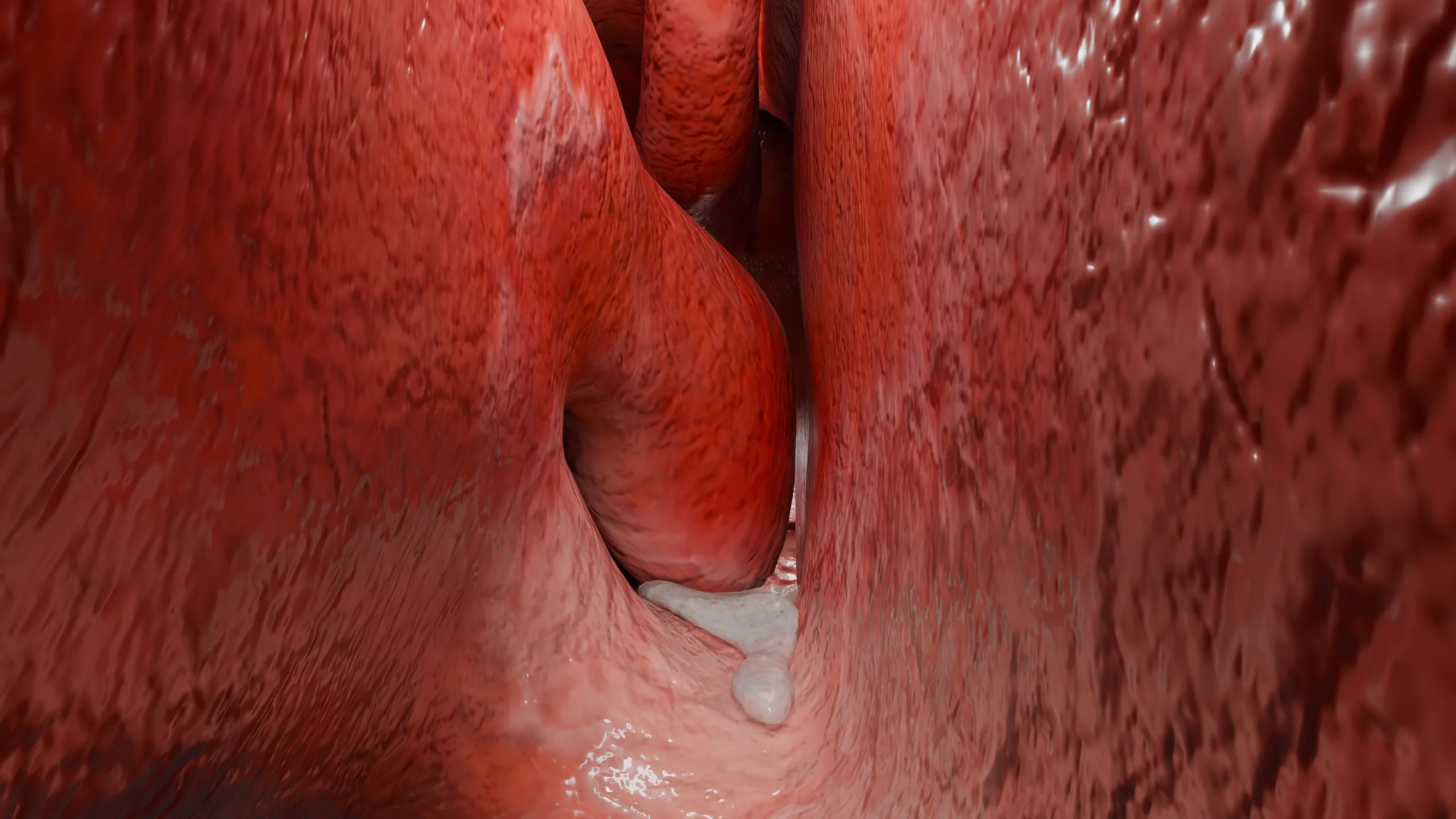
Vasomotor rhinitis (left nasal cavity in section) – 3D model
The etiology of atrophicrhinitis is not fully understood. Some authors attribute the causes of the disease to poor environmental conditions (dry air, dust), trauma or surgery in the nasal cavity, improper nasal hygiene, autoimmune diseases, hormonal changes (menopause, aging), and deficiencies in trace elements (particularly iron) and vitamins.
Ozena, or pruritic runny nose, is a special case of atrophic rhinitis. The causative agent is the bacterium Klebsiella ozaenae, but for the development of the disease in addition to pathogenic microflora, predisposing factors such as foci of chronic infection in the nasal cavity or perinasal sinuses, disturbance of aerodynamics, dryness and the presence of microcracks are necessary.
Anatomic Pathology
Acute rhinitis proceeds in 3 stages, successively changing each other. The 1st phase of dry irritation is characterized by hyperemia and dryness of the mucosa, this stage lasts from several hours (more often) to several days.
The stage of serous discharge is characterized by hyperemia and edema of the mucosa, its fullness and small foci of submucosal hemorrhages (petechiae), increased mucus production.
By days 4-5 from the onset of the disease, the discharge becomes mucopurulent due to the overproduction of lymphocytes and desquamated epithelium. With a favorable course, by the end of 7-10 days, the inflammation resolves.
Chronic infectious rhinitis is characterized by non-specific changes, such as hyperemia of the mucosa, congestion of the nasal turbinates, and hyperplasia of goblet cells with increased secretion production.
Allergic rhinitis is characterized by pale mucosa with a cyanotic tinge and pronounced edema of the nasal turbinates, with abundant discharge of clear mucous secretions. Allergic rhinitis is often combined with chronic polyposis rhinosinusitis, and in such cases, rhinoscopy reveals polyposis-altered mucosa or polyps.
In hypertrophic rhinitis, an excess of bone tissue in the inferior nasal turbinate is most often found along its entire length. Less common are vascular and fibrous forms, in which there is proliferation of blood vessels or connective tissue within the nasal turbinates.
Vasomotorrhinitis is morphologically characterized by congestion of the cavernous tissue vessels in the nasal turbinates, which become purple-bluish, thickened, with the nasal passages rarely narrowed, and an increase in the number of goblet cells. When the parasympathetic system is disrupted, there is hyperproduction of mucus, while disruption in the sympathetic system leads to edema and nasal congestion.
In atrophicrhinitis, a large number of crusts are found in the nasal cavity, the mucosa is pale pink, thinned, matte, ‘parchment-like’, with scant serous-mucous discharge. As the disease progresses, atrophic processes affect the olfactory nerve and blood vessels of the mucosa.
Ozena is characterized by the same changes as atrophic rhinitis, but as the process progresses, deep-lying tissues are destroyed, including the bone part of the nasal turbinates due to osteoclasts, blood vessels obliterate and scar. The number of goblet cells is sharply reduced, cilia are absent, resulting in non-functional mucociliary clearance, and nasal passages widen due to the deficit of nasal turbinate tissue. Tissue destruction contributes to the production of a foul odor. Rhinoscopy reveals a pathologically wide nasal cavity, with the posterior wall of the nasopharynx clearly visible. Gray-green crusts abundantly line the nasal cavity, forming so-called casts.
Symptoms of Rhinitis
Acute rhinitis begins with pronounced difficulty breathing, sneezing, burning in the nose, which corresponds to the 1st phase, there are general symptoms: headache, increased body temperature to subfebrile or febrile values. In the transition to the next phase, there are abundant mucous discharges, which in contact with the skin in the area of the nasolabial triangle cause its maceration, due to its chemical composition. Nasal congestion worsens, lacrimation appears, some patients note congestion in the ears.
3D animation – acute rhinitis: stage 2 serous discharge
When moving to stage 3 nasal discharge acquires yellow-green color, becomes thicker, stuffiness decreases. In any of the stages there may be pain in the projection of the sinuses due to the development of marked edema of the mucosa in the sinuses themselves and in the places of their exit into the nasal cavity.
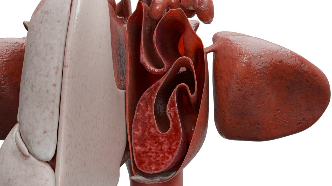
Acute rhinitis, stage 3 serous purulent discharge – 3D model
Chronic infectiousrhinitis is a slow-progressing disease in which patients note difficulty in nasal breathing and constant mucous or mucopurulent discharge with an unpleasant odor, sometimes accompanied by headaches or anosmia.
Allergicrhinitis is characterized by abundant serous discharge upon contact with an allergen, sneezing, itching in the nose and congestion, and may also include signs of allergic conjunctivitis with lacrimation and itching in the eyes. The aforementioned complaints manifest immediately upon contact with an allergen. Clinically, allergic rhinitis is subdivided into seasonal and perennial, persistent and intermittent, mild and moderate-severe. Seasonal rhinitis appears once or several times a year and is usually associated with the flowering of certain plants, while perennial rhinitis is constant and more often associated with household allergens (dust mites, pet hair, etc.). Intermittent rhinitis manifests up to 4 days a week or up to 4 weeks a year, while persistent rhinitis is characterized by continuity.
Mild rhinitis is not characterized by sleep disturbances or general activity impairment, while moderate-severe rhinitis, on the contrary, causes significant discomfort that disrupts the usual rhythm of life, negatively affecting sleep and work capacity. It is important to emphasize the close relationship between allergic rhinitis and bronchial asthma, due to a common pathogenetic mechanism. Allergic rhinitis is considered a risk factor in the development of bronchial asthma. It has been proven that without adequate treatment of allergic manifestations in the nose, the course of bronchial asthma significantly worsens. There is also a connection between this pathology and atopy. It usually manifests in childhood.
Hypertrophic rhinitis is characterized by persistent pronounced difficulty in nasal breathing, snoring, and less frequently, anosmia.
Vasomotorrhinitis is characterized by an intermittent nature of clinical manifestations, with periodic appearance of itching in the nose, sneezing, nasal congestion, and watery or mucous discharge, often dripping down the posterior wall. Patients note the appearance of complaints with changes in air temperature or humidity, body position in space (marked worsening when lying on the side), increased blood pressure, strong odors, etc. It usually manifests in adulthood.
Patients with atrophic rhinitis complain of dryness and itching in the nasal cavity, difficulty breathing despite pathologically wide nasal passages, the so-called “empty nose syndrome”, poorly detachable crusts, after removal of which no relief occurs, and in some cases, nosebleeds may occur. As the process progresses, the olfactory nerves are affected and anosmia develops, nasal septum perforation may form, and nosebleeds may develop. Since ozena is a subtype of atrophic rhinitis, all of the above complaints will be characteristic of it as well. A distinctive feature is the presence of a persistent foul odor from the nose, which the patients themselves do not sense, causing others to avoid interaction with them, which in turn affects the patients’ mental state. When attempting to remove crusts, they detach as casts, with minimal bleeding. General symptoms include headache, pronounced weakness, and fatigue.
Diagnosis of Rhinitis
To diagnose acute or chronic rhinitis, in most cases, a general examination (otorhinolaryngoscopy) is sufficient. The nature of complaints, condition of the mucosa, and discharge are assessed, and a thorough medical history is collected. In cases of prolonged course, lack of treatment effect, and presence of pain in the projection of the paranasal sinuses, an X-ray of the nasal sinuses is recommended.
For chronic infectious rhinitis, a bacteriological culture of nasal discharge is performed to identify the pathogen and determine sensitivity to antibacterial drugs.
For the diagnosis of allergic rhinitis, various tests are used depending on the clinic’s equipment. The rhinocytogram with quantitative determination of eosinophils in nasal mucus has lost its value today due to uncertain sensitivity, as the absence of eosinophils does not mean the absence of the disease, and their presence can also be found in patients with non-allergic rhinitis. The most common method is various skin tests (scarification, prick tests, etc.) where the allergen is applied to/under the skin, and the reaction at the contact site is recorded after a certain time. The “gold standard” for diagnosing allergic rhinitis is the determination of specific IgE to the most common allergens in blood serum.
Для выявления гипертрофического костного ринита выполняют анемизацию носовых раковин. Диагноз правомочен при отрицательной пробе.
Vasomotor and atrophic rhinitis are diagnosed after rhinoscopy, collection of complaints, and medical history.
Vasomotor rhinitis (nasal shells in section) – 3D model
In atrophic rhinitis and ozena, bacteriologic examination of the nasal cavity discharge is also performed. A blood test is performed to determine hemoglobin and serum iron levels. If perforation is present, a free edge biopsy with pathomorphologic examination is performed. If symptoms worsen rapidly, the patient should be evaluated for ANCA vasculitis. The diagnosis of ozaena is 100% when Klebsiella ozaenae is detected by microbiologic examination or blood examination with immunologic tests and antibody detection.
Find more scientifically accurate content on our social media
Subscribe and don’t miss out the latest resources
Treatment
Treatment of acuterhinitis is symptomatic. Nasal decongestants are prescribed (phenylephrine, xylometazoline, oxymetazoline), which reduce edema and mucous discharge, after which it is recommended to perform nasal toilet with saline solutions or seawater to evacuate pathologic contents from the nasal cavity. In the presence of pronounced general symptoms, it is possible to use NSAIDs (paracetamol, ibuprofen).
For the treatment of chronic infectiousrhinitis, it is necessary to prescribe antibiotic therapy locally or systemically, taking into account sensitivity. Regular nasal lavage with saline solutions or sea water is also prescribed.
In the treatment of allergicrhinitis, the most important thing is the elimination of the causative factor (allergen). Depending on the severity of the course, combinations of different drugs are prescribed. As therapy, intranasal decongestants are used for a short course of no more than 7-10 days. Antihistamines for local or systemic use are necessarily prescribed. In case of severe symptoms it is recommended to use glucocorticosteroids intranasally for a long time (at least 1 month) or antileukotriene drugs systemically. The majority of patients can achieve persistent remission with ASIT (allergen-specific immunotherapy), which is an etiotropic method (i.e., it fights the cause of the disease, not the symptoms). The essence of treatment is the long-term introduction of allergens into the patient’s body in minimal quantities (sublingually or subcutaneously). This results in the development of “immunity” to subsequent contacts with the allergen, minimizing unwanted reactions.
Treatment of hypertrophic bonyrhinitis is surgical. It involves performing a partial conchotomy, in which excess bone tissue is gently removed while preserving the anatomical landmarks and soft tissues of the nasal turbinates.
As therapy for vasomotorrhinitis, antihistamines of local or systemic action, topical hormonal preparations for a course of 1 month are used, and regular moisturizing of the mucosa with isotonic solutions is recommended. If conservative therapy is ineffective, surgical intervention is performed using various devices (laser coagulation/submucosal vasotomy/radiofrequency or ultrasonic destruction, etc.), during which the nasal turbinates are partially damaged from the inside and then scarred, reducing in size, while the mucous membrane remains intact and continues to perform its functions.
For atrophic rhinitis, treatment is aimed at moisturizing the mucosa. For this purpose, sprays based on isotonic solutions, sea water with the addition of dexpanthenol or hyaluronic acid are used. If there are no contraindications, lubricating the mucosa with iodine solutions is prescribed to irritate and stimulate goblet cells and increase the production of mucous secretion. A good effect is observed when treating the mucosa with oil solutions containing vitamins A, D, E, such as sea buckthorn, peach, or sesame oil, but they should be used in limited amounts as they impair the function of the ciliated epithelium. If pathogenic microorganisms are detected, topical antibacterial therapy is prescribed.
For the treatment of ozena, systemic (preferably parenteral) antibiotic therapy is necessary, based on sensitivity results. Locally, as with atrophic rhinitis, regular nasal irrigation using saline solution or sea water with the addition of iodine preparations is recommended, as well as moisturizing with oil solutions. To achieve a therapeutic effect, after softening the crusts, they must be regularly removed and then the nasal cavity irrigated with local antibacterial preparations.
FAQ
1. What are the main symptoms of rhinitis?
Common symptoms include: • Nasal congestion; • Nasal discharge (mucous or pus); • Sneezing, itchy nose; • Decreased sense of smell; • Headache, weakness (in acute rhinitis).
2. What stages of acute rhinitis are distinguished?
Acute rhinitis goes through three stages: 1. Dry irritation stage (dry, burning nose). 2. Serous discharge stage (copious watery discharge). 3. Stage of mucopurulent discharge (thick yellow-green discharge).
3. What complications can arise from rhinitis?
• Sinusitis (inflammation of the sinuses); • Otitis media (inflammation of the middle ear); • Anosmia (loss of sense of smell); • Nosebleeds.
4. How to distinguish rhinitis from sinusitis?
• Rhinitis – inflammation of the nasal mucosa, accompanied by stuffiness and nasal discharge; • Sinusitis – inflammation of the sinuses, accompanied by pain in the area of the sinuses, thick yellow-green discharge, fever.
5. What factors contribute to the development of rhinitis?
6. What complications can occur with improper treatment of rhinitis?
• Transition to a chronic form; • Development of sinusitis, otitis media; • Deterioration of quality of life (sleep disturbance, decreased efficiency).
List of Sources
1.
VOKA Catalog.
https://catalog.voka.io/
2.
Total Otolaryngology—Head and Neck Surgery, Anthony P. Sclafani, Robin A. Dyleski, Michael J. Pitman, Stimson P. Schantz. Thieme Medical Publishers, Inc., 2015. ISBN 978-1-60406-646-3.
3.
Бербом Х. Болезни уха, горла и носа / Ханс Бербом, Оливер Кашке, Тадеус Навка, Эндрю Свифт; пер. с англ. – 2-е изд. – М. : МЕДпреcс-информ, 2016. – 776 с. : ил. ISBN 978-5-00030- 322-1.
4.
Green RJ, Feldman C, Van Niekerk A, McDonald M, Friedman R, Richards GA. Treating acute rhinitis and exacerbations of chronic rhinitis – A role for topical decongestants? S Afr Fam Pract (2004). 2020 Mar 24;62(1):e1-e5. doi: 10.4102/safp.v62i1.5053. PMID: 32242436; PMCID: PMC8378128.
5.
Akhouri S, House SA. Allergic Rhinitis. 2023 Jul 16. In: StatPearls [Internet]. Treasure Island (FL): StatPearls Publishing; 2025 Jan–. PMID: 30844213.