Infectious Diseases of External Nose: Etiology, Clinical Manifestations, Diagnosis, and Treatment
Danata A.Otorhinolaryngologist, MD
10 min read·April 04, 2025
This article is for informational purposes only
The content on this website, including text, graphics, and other materials, is provided for informational purposes only. It is not intended as advice or guidance. Regarding your specific medical condition or treatment, please consult your healthcare provider.
Infectious lesions of the external nose refer to skin diseases of the external nose caused by bacterial infections that develop under favorable conditions. The affected area includes the skin, hair follicles, and subcutaneous adipose tissue, while the mucous membrane remains intact.
External nose
Classification
Furuncle of nose:
infiltration stage;
abscess stage.
3D Animation – Nasal Furuncle, Abscess Formation Stage
Other conditions:
sycosis of vestibule of nose;
eczema of nose;
eczema of nose;
Etiology
The aforementioned diseases are primarily caused by bacterial pathogens: staphylococci (S. aureus, S. epidermidis, S. saprophyticus) and streptococci (β-hemolytic group A). Infection develops due to factors such as damage to the integument (abrasions, cracks, macerations) and reduced overall body reactivity. In cases of metabolic disorders (coexistent diabetes mellitus) and immune deficiency, an extremely unfavorable course can be observed.
Nasal eczema develops due to a combination of several triggering factors:
maceration of the skin by pathologic discharge (in the presence of chronic infection of the nasal cavity or paranasal sinuses);
contact allergy;
concomitant systemic disorder (such as diabetes mellitus, atopic dermatitis, food allergies, or thyroid diseases).
Anatomy
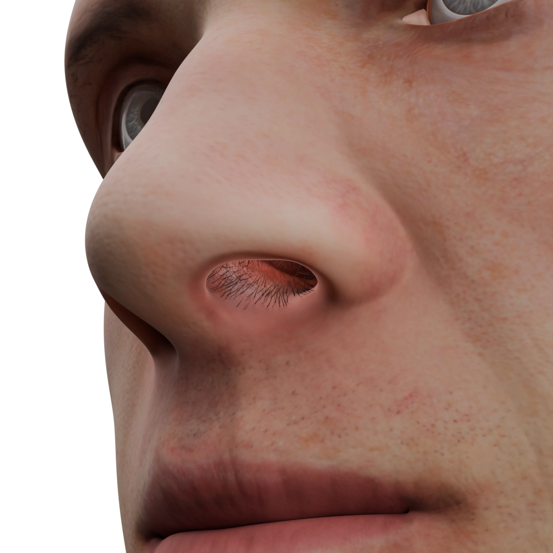
A nasal furuncle is a purulent-necrotic lesion of the hair follicle, sebaceous gland, and surrounding tissues (subcutaneous adipose tissue, skin). The tip, wings, or vestibule of the nose, as well as the upper lip area, may become involved in the pathological process. It should be noted that the process is strictly limited to the skin and never extends to the mucous membrane of the nasal cavity, due to the absence of hair follicles (in the latter).
Furuncle of the nose (infiltration stage) – 3D model
When multiple hair follicles in the same area are affected, the condition is referred to as a carbuncle.
A nasal furuncle follows a specific stage process: infiltration stage, abscess stage, and the healing stage. During the first stage, local infiltration, thickening, and hyperemia of the skin at the site of inflammation are noted, with a hair shaft located at the center of inflammation. Subsequently, the process progresses to the second stage in a few days, where purulent material forms with tissue liquefaction and fluctuation, which in rare cases may rupture spontaneously.
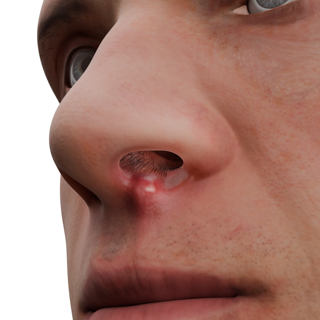
Furuncle of nasal vestibule (abscess stage) – 3D model
During the healing stage, the affected area is covered by connective tissue. Severe inflammation can lead to serious complications, including cavernous sinus thrombosis, brain abscess, and sepsis, which can be life-threatening. The pathogenesis involves infection along the venous drainage from the vestibule of the nose through the angular and ophthalmic veins to the cavernous sinus, where a thrombus subsequently forms.
Sycosis of the nasal vestibule (folliculitis) is characterized by damage to only the hair follicles (often in groups) in the nasal vestibule area or on the upper lip; underlying tissues remain intact. On hyperemic and infiltrated skin in the hair area, pustules with purulent content form, which open on the 2nd to 3rd day and subsequently become crusted.
Nasal eczema is a lesion of the epidermis characterized by a staged process. On hyperemic skin, papules and vesicles form, which rupture a few days later releasing serous content. Within 1–2 days, the affected area is covered by crusts and scales, which eventually fall off, sometimes leaving a depigmented area.
Erysipelas of the nose (erysipelas) is an infectious inflammation of the skin and subcutaneous adipose tissue with involvement of nearby lymphatic vessels and nodes. The skin becomes sharply hyperemic, edematous, hot; the boundaries of the lesion are well-defined. In some cases, serous or serous-hemorrhagic blisters form on the skin surface. Adjacent lymph nodes enlarge.
Clinical manifestations
For a nasal furuncle in the infiltration stage, significant tenderness at the inflammation site is characteristic (after 1–2 days, the pain becomes throbbing and intensifies). The skin is hyperemic, surrounding tissues are edematous. Signs of general intoxication may appear: weakness, headache, fever reaching febrile values, and enlargement of regional lymph nodes. Within 2–3 days, as the process transitions to the abscess stage, the patient experiences significant relief in general condition as tenderness decreases. As previously noted, the purulent core can rupture and be spontaneously expelled, resulting in abundant purulent or purulent-hemorrhagic discharge from the lesion area, while signs of general intoxication subside. In recovery, a small scar is formed.
If complications like cavernous sinus thrombosis arise, the clinical picture can vary. Symptoms of general intoxication always intensify. Patients experience intense pulsating headache and eye pain, nausea, vomiting, and vision impairment; seizures or loss of consciousness may occur. Local manifestations spread to the orbital area and are accompanied by exophthalmos, chemosis, and ptosis on the affected side.
Sycosis of the nasal vestibule is characterized by itching and burning at the site of the lesion; there may be a sensation of skin tension with slight tenderness. The skin is dimly hyperemic, with sparse pinpoint edemas. Subsequently, crusts form around hair shafts, which are frequently scratched, contributing to the chronicity of the process with periods of exacerbation and remission. In rare cases, general body intoxication worsens: fever rises to 38–38.5 °C, weakness appears, and regional lymph nodes enlarge.
Nasal eczema generally tends to be chronic. During exacerbations, severe itching and significant tenderness occur in the affected area. When vesicles rupture, the skin becomes moist, then gets covered with itchy crusts. The skin thickens, and the skin pattern becomes more pronounced. Itching is so bothersome to patients that it can lead to insomnia in some cases.
Erysipelas of the nose manifests as redness and swelling of the skin. The lesion area has clear boundaries (demarcation line), and sharp tenderness is noted upon touching; the skin feels hot to the touch. In some cases, the lesion takes on a ‘butterfly’ appearance, involving the skin of the nose and cheek areas on both sides. If serous or serous-hemorrhagic blisters are present, they rupture spontaneously, forming a moist surface that then becomes crusted. Acute intoxication phenomena are especially characteristic for erysipelas with fever increasing to 39–40 °C, chills, marked weakness, headache, and possible vomiting. On the 7th to 10th day, with adequate treatment, the process is stabilized. In some cases, the inflammation can be complicated by cavernous sinus thrombosis and sepsis.
Diagnosis
Diagnosis involves a clinical examination of the affected area. Laboratory indicators such as complete blood count (changes in leukocyte count and leukocyte formula), biochemical blood analysis (CRP, glucose level) are evaluated. Bacterial culture of wound discharge and tissues is also mandatory to identify the pathogen and determine antibiotic sensitivity. CT and MRI are performed in the event of complications. Additionally, blood culture for sterility and lumbar puncture may be prescribed. Allergy tests are performed in some cases to clarify the causes of nasal eczema.
Find more scientifically accurate content on our social media
Subscribe and don’t miss out the latest resources
Treatment Infectious lesions of the external nose
In mild conditions (furuncle and sycosis), local treatment is predominant. Ointments with NSAIDs and antibiotics are prescribed, along with antiseptic treatment of the foci for 5–7 days until symptoms resolve. Physical therapy, especially UV radiation, demonstrates a good effect.
Surgical intervention is indicated if the condition progresses to the abscess stage: it involves incision and cleansing of the suppurative focus at the site of maximum bulging with drainage installation and aseptic dressing application. Subsequently, regular dressings are carried out until complete healing.
In some cases, at the physician’s discretion (lack of local treatment effect, severe general intoxication, prolonged course), antibacterial agents are prescribed orally for a 5–7 day course, taking sensitivity into account.
In the recovery stage, wound healing and regenerating ointments are used.
Emergency hospitalization is indicated in the case of complications.
In the treatment of eczema, local therapy is fundamental. During the acute phase, ointments with glucocorticosteroids are used; in the case of integument infection, combined drugs with antibiotics are added. The skin is regularly treated with antiseptic solutions. To reduce itching and skin scratching, antihistamines are administered orally; in some cases, sedatives are prescribed. During the healing period (formation of crusts and scales) apply healing ointments and moisturizing lotions. During remission, the skin should be abundantly and regularly moisturized to prevent exacerbations. It is important to remember that eczema is a polyetiological disease, thus, in order to successfully treat skin manifestations, coexisting chronic diseases need to be managed.
For the treatment of erysipelas, antibiotic therapy is mandatory. The drugs of choice are penicillins, with adjustments made based on bacteriological culture results, administered orally or parenterally, depending on the severity of the condition, as the disease is prone to recurrence and has several serious complications. Locally, antiseptic treatment of the affected area is performed, along with physiotherapy using UV radiation.
FAQ
1. What is a nasal furuncle and why is it dangerous?
A nasal furuncle is a purulent-necrotic inflammation of a hair follicle and surrounding tissues. The danger lies in the risk of the infection spreading to the veins of the brain, which can lead to cavernous sinus thrombosis, brain abscess, or sepsis.
2. How does a furuncle differ from nasal vestibule sycosis?
A furuncle is a solitary purulent inflammation, while sycosis is a chronic affliction of hair follicles, often in groups, characterized by crust formation and a tendency for recurrence.
3. How to recognize nasal eczema?
Nasal eczema presents as skin redness, papule and vesicle formation, itching, oozing, and peeling. It often has a chronic course accompanied by periods of exacerbation.
4. What is nasal erysipelas?
Erysipelas is an acute infectious inflammation of the skin characterized by edema, redness, and increased temperature. The lesion has defined boundaries and may be accompanied by general symptoms of intoxication.
5. How is the diagnosis of infections of the external nose conducted?
Diagnosis includes clinical examination, complete and biochemical blood tests, bacteriological culture, and for complications, CT, MRI, and blood culture for sterility.
References
1.
VOKA 3D Anatomy & Pathology – Complete Anatomy and Pathology 3D Atlas. VOKA 3D Anatomy & Pathology.
Available from: https://catalog.voka.io/
2.
Sclafani AP, Dyleski RA, Pitman MJ, Schantz SP. Total otolaryngology—head and neck surgery. New York: Thieme Medical Publishers; 2015. ISBN: 978-1-60406-646-3.
3.
Behrbohm H, Kaschke O, Nawka T, Swift A. Ear, Nose, and Throat Diseases 2nd ed. Moscow: MEDpress-inform; 2016. 776 p. [In Russian.] ISBN 978-5-00030-322-1.
4.
Marra P, Colacurcio V, De Luca P, Bisogno A, Calvanese M, Scarpa A, et al. Nasal vestibulitis and vestibular furunculosis: a systematic review about two common nasal infections and considerations about correct diagnosis and management. Clin Ter. 2022 Nov-Dec;173(6):590-596. doi: 10.7417/CT.2022.2487. PMID: 36373460.
5.
Bakshi SS. Image Diagnosis: Nasal Furunculosis-A Dangerous Nose Infection. Perm J. 2018;22:17-076. doi: 10.7812/TPP/17-076. PMID: 29236652; PMCID: PMC5737918.
St. Petersburg FL 33702, 7901 4th St N STE 300, USA
Thank you!
Your message is sent! Our experts will contact you shortly. If you have any additional questions, please contact us at info@voka.io
Cookie Consent
We use cookies to enhance your browsing experience, analyze site traffic, and deliver content. Please choose whether you accept all cookies or wish to reject non-essential tracking.
Cookie Preferences
Manage your cookie preferences below:
Essential cookies enable basic functions and are necessary for the proper function of the website.
Name
Description
Duration
Geolocation Config
This cookie is used to store the consent settings based on the visitor's location.
30 days
Cookie Preferences
This cookie is used to store the user's cookie consent preferences.
30 days
Google reCAPTCHA helps protect websites from spam and abuse by verifying user interactions through challenges.
Name
Description
Duration
_GRECAPTCHA
Google reCAPTCHA sets a necessary cookie (_GRECAPTCHA) when executed for the purpose of providing its risk analysis.
179 days
Statistics cookies collect information anonymously. This information helps us understand how visitors use our website.
Google Analytics is a powerful tool that tracks and analyzes website traffic for informed marketing decisions.
ID used to identify users for 24 hours after last activity
24 hours
_gat
Used to monitor number of Google Analytics server requests when using Google Tag Manager
1 minute
_gac_
Contains information related to marketing campaigns of the user. These are shared with Google AdWords / Google Ads when the Google Ads and Google Analytics accounts are linked together.
90 days
__utma
ID used to identify users and sessions
2 years after last activity
__utmt
Used to monitor number of Google Analytics server requests
10 minutes
__utmb
Used to distinguish new sessions and visits. This cookie is set when the GA.js javascript library is loaded and there is no existing __utmb cookie. The cookie is updated every time data is sent to the Google Analytics server.
30 minutes after last activity
__utmc
Used only with old Urchin versions of Google Analytics and not with GA.js. Was used to distinguish between new sessions and visits at the end of a session.
End of session (browser)
__utmz
Contains information about the traffic source or campaign that directed user to the website. The cookie is set when the GA.js javascript is loaded and updated when data is sent to the Google Anaytics server
6 months after last activity
__utmv
Contains custom information set by the web developer via the _setCustomVar method in Google Analytics. This cookie is updated every time new data is sent to the Google Analytics server.
2 years after last activity
__utmx
Used to determine whether a user is included in an A / B or Multivariate test.
18 months
_ga
ID used to identify users
2 years
_gali
Used by Google Analytics to determine which links on a page are being clicked
30 seconds
Clarity is a web analytics service that tracks and reports website traffic.