


Acute Sinusitis (Acute Rhinosinusitis): Classification, Clinical Manifestations, Diagnosis, and Treatment
A detailed review of rhinosinusitis, including classification, symptoms, diagnostic approaches, and current treatment strategies.
Anesthesia
Pain management and sedation techniques
Angiology
Arterial and venous pathologies
Cardiology
Acquired and congenital heart diseases
Dentistry
Diseases of teeth, gums, and the oral cavity
Dermatology
Disorders of the skin and subcutaneous tissue
Endocrinology
Disorders of the glands and hormonal imbalance
Gastroenterology
Stomach, intestinal, and digestive diseases
Gynecology
Diseases of female reproductive organs
Hepatology
Liver, gallbladder, and biliary tract diseases
Neurology
Brain, spinal cord, and peripheral nerve disorders
Obstetrics
Pregnancy complications and abnormal fetal positions
Oncology
Cancer types, benign and malignant tumors
Ophthalmology
Conditions affecting the eyes and vision
Otorhinolaryngology
Ear, nose, and throat diseases
Pediatrics
Child health, development, and clinical conditions
Physiology
Biological processes within organs and systems
Pulmonology
Lung and respiratory tract diseases
Traumatology
Acute injuries and musculoskeletal trauma
Urology
Urinary tract and male reproductive disorders

This article is for informational purposes only
The content on this website, including text, graphics, and other materials, is provided for informational purposes only. It is not intended as advice or guidance. Regarding your specific medical condition or treatment, please consult your healthcare provider.
Infectious diseases of the external nose are bacterial infections of the external nose skin that develop under favorable or predisposing conditions. The skin, hair follicles, and the fatty layer of the subcutaneous tissue are most commonly affected, while the mucosa typically remains intact.
The conditions mentioned above tend to develop as a result of a staphylococcal (S. aureus, S. epidermidis, S. saprophyticus) or streptococcal (group A β-hemolytic) infection. These conditions may be triggered by a variety of factors, ranging from damaged integument (abrasions, cracks, macerations) to decreased body resistance. In cases of metabolic imbalance (such as concomitant diabetes mellitus) or immunodeficiency, the disease may be extremely severe.
Nasal eczema is typically set off by a combination of discharge-induced skin macerations (which may occur if a chronic infection of the nasal cavity or paranasal sinuses has developed), allergic contact dermatitis, and a concomitant systemic disorder (such as diabetes mellitus, atopic dermatitis, food allergies, or thyroid gland diseases).
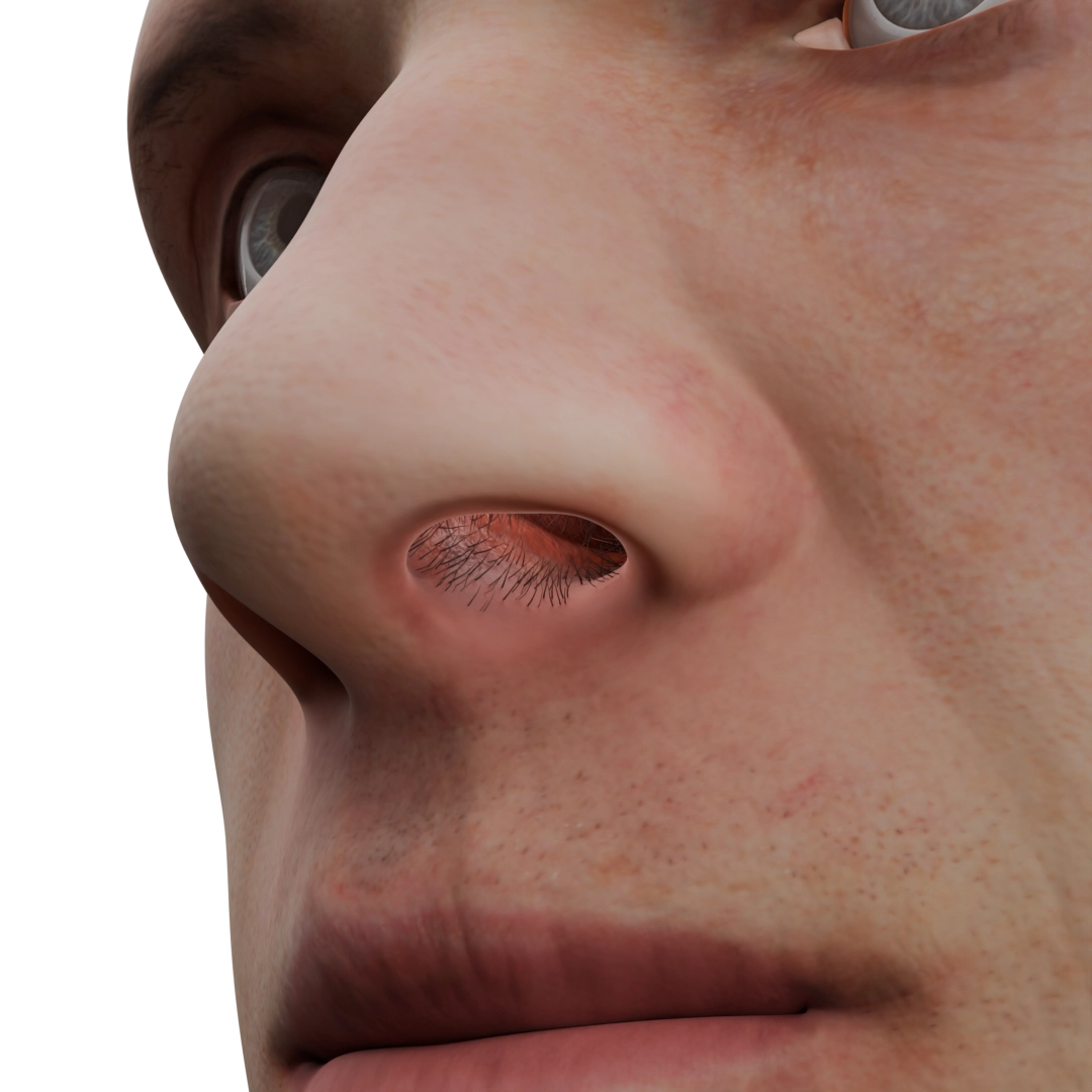
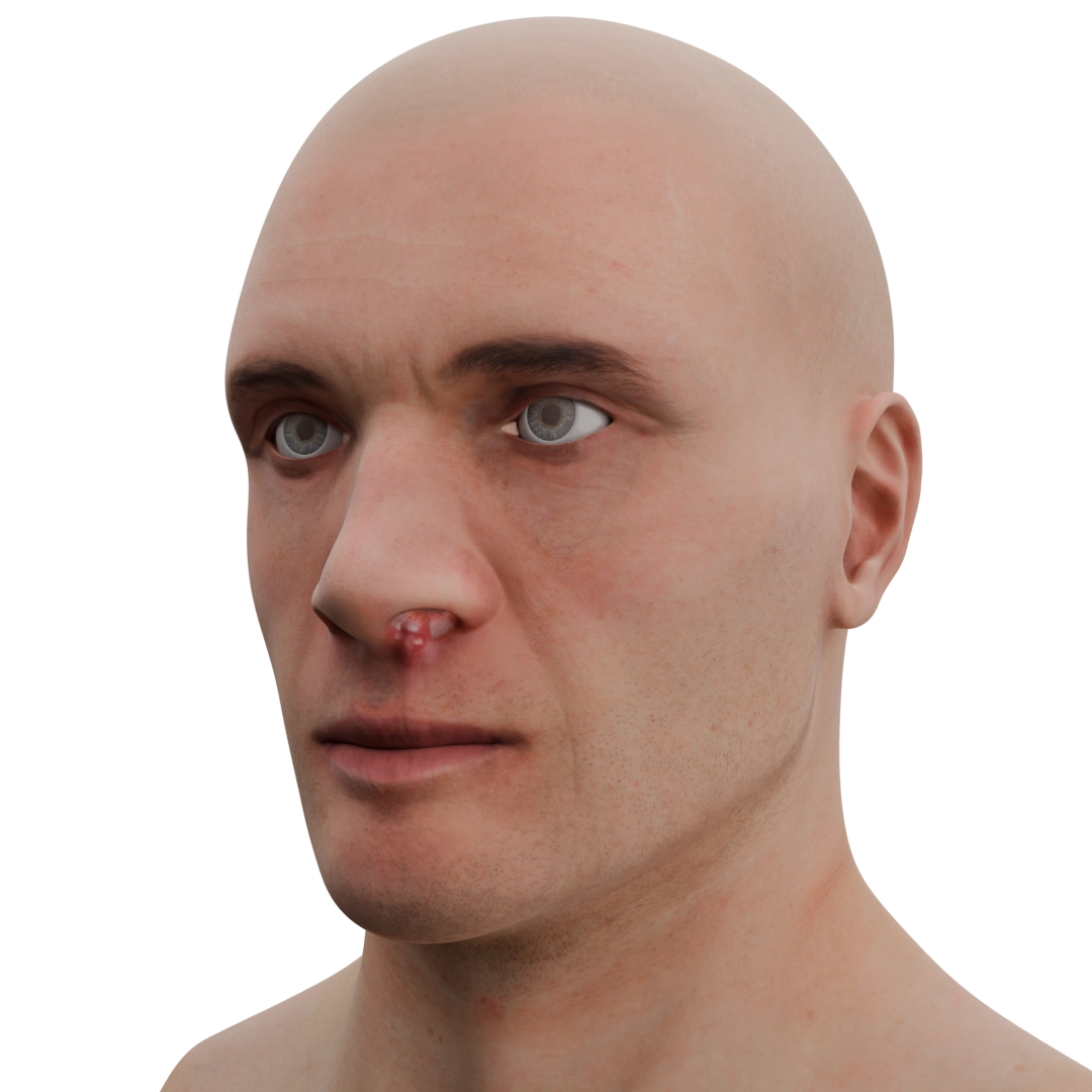
A furuncle of the nose is a purulent inflammation of the hair follicle, sebaceous gland, and surrounding tissues (the fatty layer of the subcutaneous tissue and skin). The pathological process may spread to the apex, ala, vestibule of the nose, or the upper lip. It should be noted that only the skin is affected; the mucosa of the nasal cavity, which does not contain any hair follicles, remains intact.
When several hair follicles within one area are involved, the condition is referred to as a carbuncle. The life cycle of a furuncle can be divided into three stages: infiltration, abscess, and healing. The first stage is characterized by local infiltration, induration, and hyperemic skin, with a hair shaft at the center. A few days later, the condition progresses to its second stage, during which necrosis occurs, resulting in the accumulation of pus and fluctuation. In rare cases, the furuncle may rupture spontaneously.
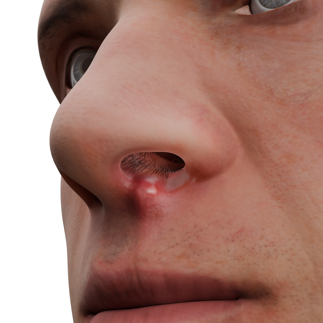
During the healing stage, the affected area is covered by connective tissue. Severe inflammation can lead to serious complications, including cavernous sinus thrombosis, intracranial abscess, and sepsis. These complications can, in turn, be life-threatening. The infection, which is the cause of the disease, spreads via the venous drainage system. It starts from the nasal vestibule, travels through the angular and ophthalmic veins all the way to the cavernous sinus, where a blood clot forms.
Nasal sycosis (folliculitis) is characterized by lesions only in the hair follicles, most often in a group in the area of the nasal vestibule or on the upper lip; the underlying tissues remain intact. Pustules with purulent content are formed on hyperemic and infiltrated skin in the hair area, which open on 2-3 days and are subsequently covered with crusts.
Nasal eczema is a lesion of the epidermis and is characterized by a staged process. On the affected hyperemic skin papules and vesicles are formed, which after a few days open and serous content is released. After 1-2 days, the affected area is covered with crusts, scales, which are then independently rejected, in some cases leaving a zone of depigmentation.
Rhinitis nasalis (Latin erysipelas) is an infectious inflammation of the skin and subcutaneous fatty tissue with involvement of nearby lymphatic vessels and lymph nodes. The skin becomes sharply hyperemic, edematous, hot, the borders of the lesion are clearly delineated, in some cases, serous or serous-hemorrhagic blisters are formed on the surface of the skin. Adjacent lymph nodes are enlarged.
Nasal furuncle in the infiltration stage is characterized by pronounced pain at the site of inflammation (after 1-2 days, the pain becomes throbbing, increases), the skin is hyperemic, the surrounding tissues are edematous, there may be signs of general intoxication, such as weakness, headache, fever to febrile values, enlargement of regional lymph nodes. After 2-3 days, when the process passes to the stage of abscission, the patient notes a significant relief of the general condition, pain decreases, as noted above, the purulent rod can be opened and rejected independently, in this case there is abundant purulent or purulent-hemorrhagic discharge from the lesion area, manifestations of general intoxication pass. In recovery, a small scar is formed.
When complications occur, such as cavernous sinus thrombosis, the clinic may vary, the symptoms of general intoxication always increase, patients note a pronounced throbbing headache and eye pain, nausea, vomiting, decreased vision, seizures or loss of consciousness may occur. Local manifestations extend to the ocular region, accompanied by exophthalmos, chemosis, ptosis on the affected side.
Sycosis of the nasal vestibule is characterized by itching and burning at the lesion site, there may be skin tension with slight soreness. The skin is dimly hyperemic, sparingly defined pitting wetting. Then crusts are formed around the hair shafts, which are often combed, which contributes to the chronicization of the process with periods of exacerbation and remission. In rare cases, general intoxication of the body increases, temperature rises to 38-38.5°C, weakness and enlarged regional lymph nodes.
Nasal eczema is more often characterized by a chronic course, in the period of exacerbation there is severe itching and marked soreness in the affected area, at the time of opening vesicles, the skin becomes moist and then covered with itchy crusts. The skin thickens and the skin pattern becomes more pronounced. Itching bothers patients so much that in some cases it leads to insomnia.
Nasal swelling is manifested by redness and swelling of the skin, the affected area has clear boundaries (demarcation line), when touching – sharp soreness, the skin is hot to the touch, in some cases presented in the form of a butterfly, with lesions of the skin of the nose and cheek area on 2 sides. If there are serous or serous-hemorrhagic blisters, they self-open and a wet surface is formed, which is then covered with a crust. Acute intoxication with fever up to 39-40°C, chills, marked weakness, headache, and vomiting may be characteristic of rheumatoid inflammation. On 7-10 days with adequate treatment the process is eliminated. In some cases, inflammation may be complicated by cavernous sinus thrombosis and sepsis.
Clinical examination of the affected area is used to make a diagnosis. Evaluation of laboratory indicators, such as a general blood count (changes in the number of leukocytes, leukocyte formula), biochemical blood count (CRP, glucose level) is performed. Also necessarily performed bacteriologic seeding of wound discharge and tissues to determine the causative agent and clarify sensitivity to antibiotics. If complications occur, CT and MRI are performed, additionally, blood culture for sterility and lumbar puncture may be prescribed. To clarify the cause of nasal eczema, allergy tests are performed in some cases.
Find more scientifically accurate content on our social media


With a mild course of diseases (furuncle and sycosis), local treatment prevails. Ointments with NSAIDs and antibiotics are prescribed, antiseptic treatment of foci for 5-7 days until resolution of symptoms. A good effect is noted with physical therapy, especially UV radiation.
In case of progression of the process (transition to the abscessed stage), surgical treatment is indicated – opening and sanation of the purulent focus in the place of the largest swelling with the installation of drainage and aseptic dressing. Subsequently, regular dressings are performed until complete healing.
In some cases, at the discretion of the doctor (in the absence of effect from local treatment, severe general intoxication, prolonged course) prescribe antibacterial drugs orally for a course of 5-7 days, taking into account sensitivity.
In the recovery stage, wound-healing and regenerating ointments are used.
In the presence of complications, emergency hospitalization is indicated.
In the treatment of eczema, the basis is also local treatment, in the acute period ointments with glucocorticosteroids are used, in case of infection of the skin, combined preparations with antibiotics are added. The skin is regularly treated with antiseptic solutions. To reduce itching and combing of the skin, antihistamines are prescribed orally, in some cases sedatives are prescribed. During the healing period (formation of crusts and scales) apply healing ointments and moisturizing lotions. During remission, the skin should be moisturized abundantly and regularly to prevent the development of exacerbations. It should be remembered that eczema is a polyetiologic disease and for the successful cure of skin manifestations it is necessary to treat concomitant chronic diseases.
For the treatment of swelling, antibiotic therapy is necessarily prescribed (drugs of choice – penicillin series, correction according to the results of bacteriological culture), orally or parenterally, depending on the severity of the condition, since the disease is prone to recurrence and has a number of formidable complications. Local antiseptic treatment of the affected area, physiotherapy with UV radiation is performed.
1. What is a nasal furuncle and how dangerous is it?
2. What is the difference between a furuncle and sycosis of the nasal vestibule?
3. How to recognize nasal eczema?
4. What is rhinitis of the nose?
5. How are infections of the external nose diagnosed?
List of Sources
1.
VOKA Catalog.
https://catalog.voka.io/
2.
Total Otolaryngology-Head and Neck Surgery, Anthony P. Sclafani, Robin A. Dyleski, Michael J. Pitman, Stimson P. Schantz. Thieme Medical Publishers, Inc, 2015. ISBN 978-1-60406-646-3.
3.
Berbom H. Diseases of the ear, throat and nose / Hans Berbom, Oliver Kaschke, Thadeus Navka, Andrew Swift; per. from English – 2nd ed. – M. : MEDpress-Inform, 2016. – 776 с. : ill. ISBN 978-5-00030- 322-1.
4.
Marra P, Colacurcio V, De Luca P, Bisogno A, Calvanese M, Scarpa A, Ralli M, De Vincentiis M, Camaioni A, Salzano FA. Nasal Vestibulitis and Vestibular Furunculosis: a systematic review about two common nasal infections and considerations about correct diagnosis and management. Clin Ter. 2022 Nov-Dec;173(6):590-596. doi: 10.7417/CT.2022.2487. PMID: 36373460.
5.
Bakshi SS. Image Diagnosis: Nasal Furunculosis-A Dangerous Nose Infection. Perm J. 2018;22:17-076. doi: 10.7812/TPP/17-076. PMID: 29236652; PMCID: PMC5737918.
Loading test 6 questions


Summarize article with AI
Choose your preferable AI assistant:
Link successfully copied to clipboard
Thank you!
Your message is sent!
Our experts will contact you shortly. If you have any additional questions, please contact us at info@voka.io


