


Acute Sinusitis (Acute Rhinosinusitis): Classification, Clinical Manifestations, Diagnosis, and Treatment
A detailed review of rhinosinusitis, including classification, symptoms, diagnostic approaches, and current treatment strategies.
Anesthesia
Pain management and sedation techniques
Angiology
Arterial and venous pathologies
Cardiology
Acquired and congenital heart diseases
Dentistry
Diseases of teeth, gums, and the oral cavity
Dermatology
Disorders of the skin and subcutaneous tissue
Endocrinology
Disorders of the glands and hormonal imbalance
Gastroenterology
Stomach, intestinal, and digestive diseases
Gynecology
Diseases of female reproductive organs
Hepatology
Liver, gallbladder, and biliary tract diseases
Neurology
Brain, spinal cord, and peripheral nerve disorders
Obstetrics
Pregnancy complications and abnormal fetal positions
Oncology
Cancer types, benign and malignant tumors
Ophthalmology
Conditions affecting the eyes and vision
Otorhinolaryngology
Ear, nose, and throat diseases
Pediatrics
Child health, development, and clinical conditions
Physiology
Biological processes within organs and systems
Pulmonology
Lung and respiratory tract diseases
Traumatology
Acute injuries and musculoskeletal trauma
Urology
Urinary tract and male reproductive disorders

This article is for informational purposes only
The content on this website, including text, graphics, and other materials, is provided for informational purposes only. It is not intended as advice or guidance. Regarding your specific medical condition or treatment, please consult your healthcare provider.
Acute otitis media (AOM) is an inflammation of the middle ear cavity accompanied by severe pain and hearing loss, lasting up to 1 month.
Exudative otitis media (ESO, serous, secretory) is a pathology characterized by the presence of exudate in the middle ear cavity, without pain syndrome. Clinically, acute (up to 3 weeks), subacute (3-8 weeks) and chronic (> 8 weeks) forms of exudative otitis media are distinguished. In practice, however, it is often difficult to establish the exact time of onset, and only acute and chronic forms are distinguished.
Myringitis is an inflammation of the eardrum.
This disease is mostly caused by bacteria. The most common bacteria are Streptococcus pneumoniae, Moraxella catarrhalis, Haemophilus influenzae, less frequently Escherichia coli and Staphylococcus aureus, beta-hemolytic group A streptococci. In 10-30% of cases, viral etiology is assumed.
Pathogens enter the sterile middle ear cavity through the auditory tube. In infectious diseases such as measles, scarlet fever and influenza, the hematogenous route of infection is also possible.
The trigger for the development of otitis media is obstruction of the auditory tube caused by inflammation in the nasopharynx (adenoiditis, rhinitis, sinusitis).
When the auditory tube is blocked, the mucous membrane absorbs air from the middle ear cavity and a negative pressure is created, which causes transudate to leak out. Pathogenic microorganisms enter the middle ear cavity from the nasopharynx, for which the transudate is a good breeding ground for inflammation.
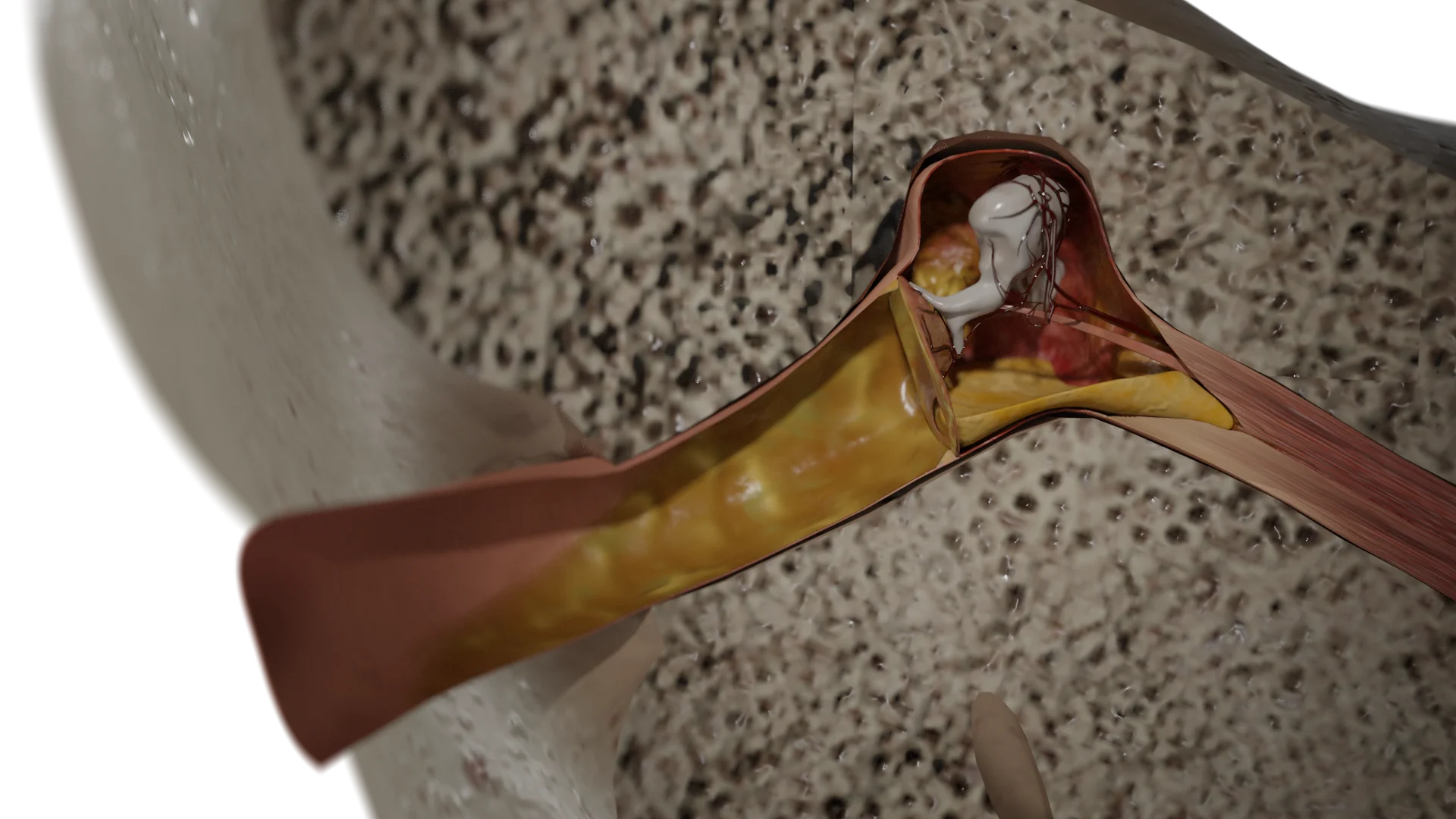
Acute otitis media is characterized by a staged process. Initially there is hyperemia of the middle ear mucosa (catarrhal stage), then a clear fluid component is detected.

With the progression of the process, the mucosa is infiltrated with leukocytes, the discharge is saturated with neutrophils, purulent exudate is formed (preperforative stage).
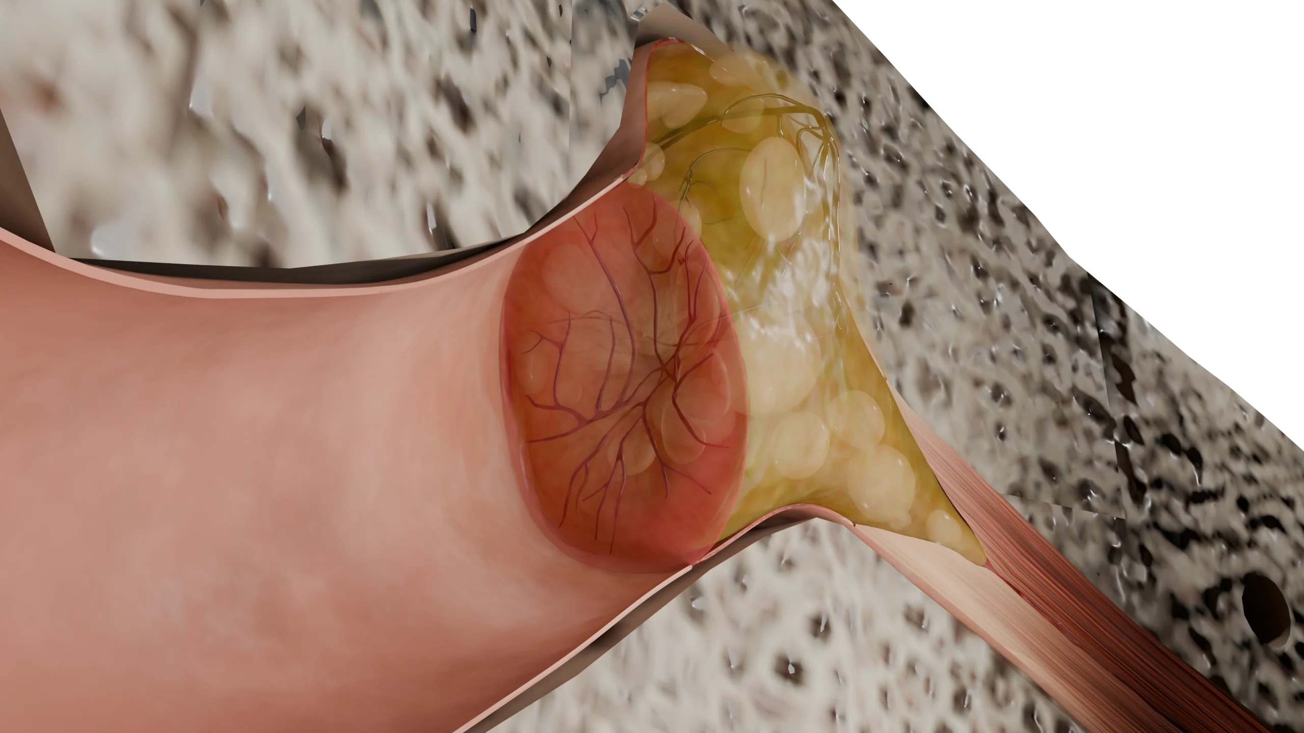
When the pressure of the purulent component is applied to the tympanic membrane, it ruptures (perforation stage), through which the pathologic content flows into the ear canal. After evacuation of the purulent content, the tympanic cavity is repaired and the perforation is scarred (reparative stage).
With an unfavorable course, purulent contents through the antrum can penetrate into the cells of the mastoid process, which leads to the spread of inflammation to the bone tissue and the development of a formidable complication – mastoiditis.
Acute otitis media are most characteristic of childhood due to anatomical features: wide and short auditory tube, more gentle angle between the bony and cartilaginous part of it, hypertrophied lymphoid tissue in the nasopharynx and the presence of myxoid tissue in the middle ear cavities.
For each stage of acute otitis media is characterized by its own clinical picture, which changes sequentially. Most often, the pathological process develops against the background of inflammatory changes in the nasopharynx, such as rhinitis, adenoiditis, sinusitis.
Stages of the disease:
In case of unfavorable outcome, the disease may become chronic or lead to complications such as chronic purulent otitis media, mastoiditis, labyrinthitis etc.
For the development of a pronounced clinical picture is enough 4-6 hours, with a favorable course of recovery comes on 5-7 days.
The gold standard for making this diagnosis is otoscopy.
Stages of acute otitis media, changes on otoscopy
| CCA stage | Tympanic membrane (TM) | External auditory canal (EAC) |
|---|---|---|
| Catarrhal stage | BP hyperemic; Full blood vessels; Light cone not defined; Malleus handle shortened; Short malleus protrudes sharply into the lumen of the NRS |
Unchanged |
| Purulent pre-perforative stage. | BP turbid, hyperemic, yellow, bulging into the lumen of the NSP; Purulent content is visible; Pulsation is possible; Identifying contours are not defined |
Unchanged |
| Purulent perforative stage | Perforation (more often slit-shaped, in the lower parts) in the PD; Purulent contents ooze through the perforation; Possible “pulsating reflex” – contents ooze in a jerky manner; Identifying contours are partially restored |
There’s a lot of purulent discharge in the NSP |
| Reparative stage | The tympanic membrane is gray, turbid, may be retracted; wind reflex is poorly defined; Perforation is slit-shaped, without pathological discharge, may be in the form of a scar |
Unchanged |
3D Models of the stages of acute otitis media:
 Acute purulent otitis media: preperforative stage (otoscopy)
 Acute purulent otitis media: perforation stage (otoscopy)
 Acute purulent otitis media: stage of repair (otoscopy)
Additionally performed:
In frequent, recurrent otitis media in children against the background of hypertrophied adenoids, adenotomy is indicated.
This type of otitis media is caused by the influenza virus and is more common during seasonal outbreaks. The virus enters the middle ear cavity through the auditory tube from the nasopharynx or hematogenously with the bloodstream.
The pathophysiology of this type of otitis media is generally no different from that of other etiologies.
Influenza otitis media is characterized by the following features:
The clinical picture is similar to that of common otitis media.
Main Symptoms:
In addition to localized complaints, the manifestation of other symptoms of influenza infection is noted:
If the outcome is unfavorable, acute purulent otitis media develops, and mastoiditis and meningitis may also occur.
To establish the diagnosis of acute otitis media, a complete otorhinolaryngologic examination (otorhinolaryngoscopy) is performed, if necessary – otomicroscopy.
From laboratory diagnostics, a blood test to determine inflammatory markers is mandatory. In the period of epidemic rise, the diagnosis of influenza is established clinically, but at low incidence of disease, PCR-study from the nasopharyngeal mucosa is performed.
A rapid test can be performed at home, but it has low sensitivity.
Specific antiviral therapy (neuraminidase inhibitors) is used. The rest of the otitis media treatment is carried out in accordance with the above recommendations. Antibacterial therapy is prescribed if indicated.
Measles infection is caused by the measles virus, which is transmitted by airborne droplets.
Measles virus causes upper respiratory tract catarrh and a specific skin rash. The general changes in the development of otitis media in measles are similar to those in other etiologies, with no significant differences.
Measles otitis media is characterized by the following differences:
In this type of otitis media, the following symptoms predominate:
Ear pain is not characteristic of this type of otitis media.
Diagnosis is based on otoscopy. Cameron tests are carried out, in which mixed hearing loss is determined, as well as vestibular tests with evaluation of nystagmus. In laboratory diagnosis, blood is tested for inflammatory markers, blood is tested for antibodies to measles virus.
The underlying disease is treated, but there is no specific treatment for measles at the moment, so symptomatic therapy is prescribed. Massive antibacterial therapy, sanitation of middle ear cavities, regular toileting are prescribed.
If complications occur, surgical treatment (antromastoidectomy) is performed. Vaccination is recommended for prevention.
The causative agent of this disease is beta-hemolytic group A streptococcus, which is transmitted by airborne droplets. The pathogen enters the middle ear cavity by hematogenous route or through the auditory tube.
Against the background of general changes in the body (rash, tonsillitis) there are specific changes in the middle ear cavity:
In this type of otitis media, the following symptoms predominate:
Diagnosis is based on otoscopy. Cameron tests are performed to determine conductive hearing loss in the affected ear, as well as vestibular tests to assess labyrinth function.
Obligatory laboratory diagnosis is carried out: the OAC is characterized by leukocytosis and an increase in the level of C-reactive protein. Microbiological examination determines the causative agent and its sensitivity to antibacterial drugs.
The underlying disease is treated, massive antibacterial therapy is prescribed. Penicillins or cephalosporins are preferred. Sanitation of the middle ear is performed, toilet with antiseptic solutions. If complications occur, surgical treatment (antromastoidectomy) is performed.
The underlying cause of this condition is obstruction of the auditory tube orifice in the nasopharynx, which can be either inflammatory or allergic in nature.
Children get sick more often due to a higher proportion of hyperplasia of the lymphoepithelial tissue of the nasopharynx. In adults, this pathology may indicate inflammatory processes in the nasopharynx. It is also necessary to take into account the possibility of neoplasms in the vault of the nasopharynx, obstructing the lumen of the aperture of the auditory tube.
The pathogenesis of the disease has a similar beginning with the development of otitis media, but the inflammation in this case is aseptic in nature.
Against the background of obstruction of the auditory tube in the middle ear cavity creates negative pressure, which contributes to the flow of transudate into its spaces.
The situation is aggravated by the fact that the squamous epithelium degenerates into secretory epithelium (the number of bocaloid cells and secretory glands increases). This process contributes to the transformation of transudate into exudate, due to the impregnation of the protein component and an increase in the viscosity of the contents.
Without adequate treatment, the process becomes chronic.
Clinically, the disease remains unrecognized for a long time, especially when the pathological process is present on both sides. The main symptoms are gradually progressive conductive hearing loss and ear stuffiness. Pain syndrome is usually not characteristic, but there may be tinnitus and autophony. The general condition is not impaired.
First of all, otoscopy is performed. On examination, a dense gray tympanic membrane with thickened vessels is visualized, which may be slightly retracted in the upper parts. The light cone is not defined. Transparent contents are visualized behind the tympanic membrane, sometimes with a level of exudate, and air bubbles may also be seen.
If exudate is suspected, tympanometry is performed, which is fundamental to the diagnosis. In the presence of contents, the tympanogram corresponds to type B. Cameron tests are performed to determine the nature of the hearing loss. Nasopharyngeal endoscopy is recommended to determine the cause of the disease. In doubtful cases, CT scan of temporal bones and nasopharynx is indicated.
First of all, it is necessary to eliminate the cause of the disease. Sanitation of the nasopharynx is performed. Systemic mucolytics are prescribed to improve secretion. It is also recommended that the patient perform a set of exercises to blow the auditory tube.
If it is impossible to master this technique, the doctor performs balloon blowing of the auditory tubes (according to Politzer) or their catheterization with the subsequent introduction of air.
Antibacterial drugs are prescribed very rarely, if indicated.
Find more scientifically accurate content on our social media


The cause of the disease is the impact of pathogenic microflora or activation of opportunistic microflora. Among the main causative agents distinguish:
Myringitis as an independent disease is rare and more often develops in conjunction with otitis media or otitis externa. It can also be the result of careless removal of foreign bodies from the ear canal, as well as exposure to thermal or chemical factors.
As a result of irritating factors (including hematogenous or contact route of infection), inflammatory changes occur on the tympanic membrane. As a result, there is full blood vessels and pronounced infiltration in the thickness of the membrane.
Serous or hemorrhagic blisters (bullae) may form on its surface. A serous discharge is secreted into the lumen of the ear canal.
Patients notice a sharp severe throbbing pain in the ear. There may be noise, crackling, itching in the affected ear, as well as scanty transparent or hemorrhagic discharge. There may be a decrease in hearing. In some cases, there is a pronounced general symptomatology, body temperature rises to 38-39 °C, accompanied by headache and weakness.
Diagnosis is based on otoscopy data. Hyperemic, infiltrated tympanic membrane with sharply thickened vessels is detected, bullae may be visualized on its surface. The identifying contours of the membrane are blurred.
Cameron tests and audiograms reveal conductive type hearing loss of varying degrees.
The general blood test reveals leukocytosis with a left shift in the formula and an increase in C-reactive protein (CRP). A swab of the tympanic membrane surface is also performed to determine the causative agent.
Anti-inflammatory drugs are prescribed to control pain and reduce inflammation, and antibacterial drugs are prescribed if indicated. Topical irrigation with antiseptic solutions is recommended to prevent the spread of infection and keep the ear canal clean.
1. What is acute otitis media and its main symptoms?
2. What causes can cause acute otitis media?
3. What are the clinical guidelines for the treatment of acute otitis media?
4. What are the dangers of acute otitis media?
5. How many days is acute otitis media treated?
List of Sources
1.
VOKA Catalog.
https://catalog.voka.io/
2.
Total Otolaryngology—Head and Neck Surgery, Anthony P. Sclafani, Robin A. Dyleski, Michael J. Pitman, Stimson P. Schantz. Thieme Medical Publishers, Inc., 2015. ISBN 978-1-60406-646-3.
3.
Бербом Х. Болезни уха, горла и носа / Ханс Бербом, Оливер Кашке, Тадеус Навка, Эндрю Свифт; пер. с англ. – 2-е изд. – М. : МЕДпреcс-информ, 2016. – 776 с. : ил. ISBN 978-5-00030- 322-1.
4.
Danishyar A, Ashurst JV. Acute Otitis Media. 2023 Apr 15. In: StatPearls [Internet]. Treasure Island (FL): StatPearls Publishing; 2025 Jan–. PMID: 29262176.
5.
Gaddey HL, Wright MT, Nelson TN. Otitis Media: Rapid Evidence Review. Am Fam Physician. 2019 Sep 15;100(6):350-356. PMID: 31524361.
6.
Jamal A, Alsabea A, Tarakmeh M, Safar A. Etiology, Diagnosis, Complications, and Management of Acute Otitis Media in Children. Cureus. 2022 Aug 15;14(8):e28019. doi: 10.7759/cureus.28019. PMID: 36134092; PMCID: PMC9471510.
7.
Pleșca VȘ, Marinescu AG, Voiosu C, Drăgănescu AC, Streinu-Cercel A, Vilaia A, Hainăroșie R, Pleșca DA, Săndulescu O. Occurrence of acute otitis and sinusitis in patients hospitalized for influenza. Germs. 2024 Mar 31;14(1):38-44. doi: 10.18683/germs.2024.1416. PMID: 39169978; PMCID: PMC11333841.
8.
Pardo S, Perera TB. Scarlet Fever. [Updated 2025 Feb 6]. In: StatPearls [Internet]. Treasure Island (FL): StatPearls Publishing; 2025 Jan-. Available from:
https://www.ncbi.nlm.nih.gov/books/NBK507889/
9.
Yano H, Suetake M, Endo H, Takayanagi R, Numata M, Ohyama K, Sagai S, Okitsu N, Okamoto M, Nishimura H, Kobayashi T. Isolation of measles virus from middle ear fluid of infants with acute otitis media. J Infect. 2005 Nov;51(4):e237-40. doi: 10.1016/j.jinf.2004.09.002. PMID: 16291278.
10.
Yano H, Okitsu N, Hori T, Watanabe O, Kisu T, Hatagishi E, Suzuki A, Okamoto M, Ohmi A, Suetake M, Sagai S, Kobayashi T, Nishimura H. Detection of respiratory viruses in nasopharyngeal secretions and middle ear fluid from children with acute otitis media. Acta Otolaryngol. 2009 Jan;129(1):19-24. doi: 10.1080/00016480802032777. PMID: 18607974.
11.
Searight FT, Singh R, Peterson DC. Otitis Media With Effusion. 2023 May 20. In: StatPearls [Internet]. Treasure Island (FL): StatPearls Publishing; 2025 Jan–. PMID: 30855877.
Loading test 6 questions
Table of Contents


Summarize article with AI
Choose your preferable AI assistant:
Link successfully copied to clipboard
Thank you!
Your message is sent!
Our experts will contact you shortly. If you have any additional questions, please contact us at info@voka.io


