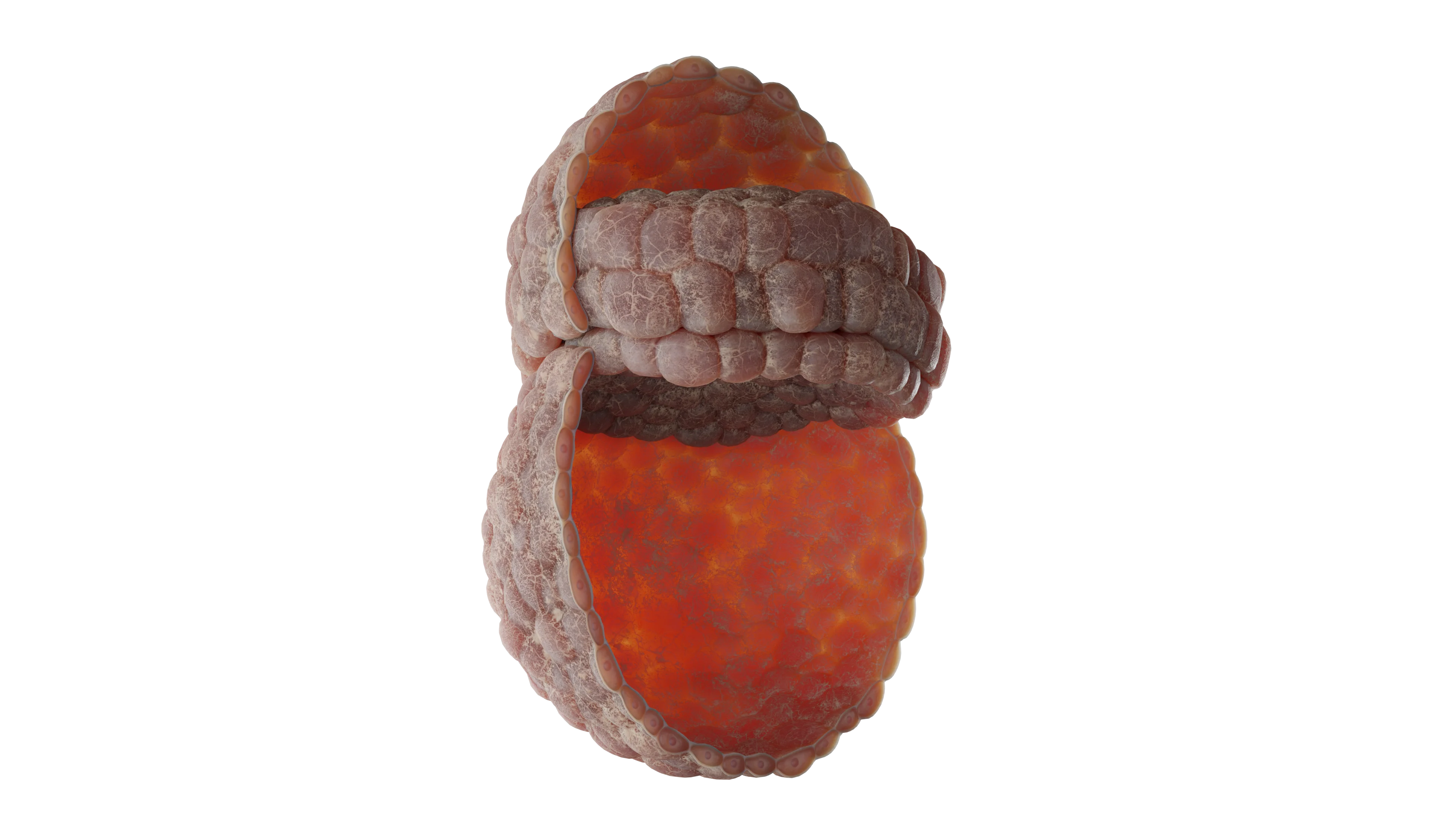
Week 2 of Embryonic Development: Structure, Morphogenetic Features, and Clinical and Anatomical Significance
Daria G.Gynecologist, fertility specialist, MD
20 min read·February 26, 2026
This article is for informational purposes only
The content on this website, including text, graphics, and other materials, is provided for informational purposes only. It is not intended as advice or guidance. Regarding your specific medical condition or treatment, please consult your healthcare provider.
The fourteenth day of postconceptional human development represents one of the most significant stages of early embryogenesis. During this period, gastrulation begins, transforming the bilaminar embryonic disc into three germ layers: the ectoderm, mesoderm, and endoderm.
This is a transitional stage between implantation and active gastrulation. At this point, interaction between the embryo and the maternal organism begins through the lacunae formed by the syncytiotrophoblast.
At the same time, the principal body axes that establish the organizational plan of the future organism are defined:
Cranial-caudal axis;
Dorsal-ventral axis;
Left-right axis.
While these processes are going on, the extraembryonic structures required for normal exchange between the embryo and the maternal organism continue to develop.
Embryo Morphology on Day 14
By this stage, the embryonic disc measures approximately 1.5–2 mm. Despite its small size, the disc retains its bilaminar structure but has already entered an active phase of remodeling.
Epiblast
The upper layer is represented by the epiblast, composed of high columnar cells with pronounced apical-basal polarity and high mitotic activity. The epiblast is the source of all embryonic tissues and also forms the wall of the amniotic cavity.
Its cells maintain strong intercellular adhesion through E-cadherin expression; however, these connections gradually weaken in the caudal region, preparing the epiblast for migration.
Hypoblast
The lower layer of the embryonic disc is represented by the hypoblast, which consists of flattened cells forming a thin sheet. Hypoblast cells do not directly contribute to the tissues of the embryo proper, but they participate in the formation of the extraembryonic endoderm and the yolk sac.
In addition, they secrete signaling molecules, including LEFTY1, Cer1, and DKK1, which inhibit WNT and NODAL expression in the cranial region. This prevents excessive mesoderm induction and promotes proper development of anterior structures.
Primitive Streak Formation
At the caudal end of the disc, the primitive streak forms as a thickening of the epiblast. It is the first morphological sign of the gastrulation onset which establishes the cranial-caudal axis of the embryo.
Cells within the primitive streak lose their apical-basal polarity, reorganize their cytoskeleton through the activity of Rho GTPases, and acquire migratory properties.
In the cranial portion of the streak, the primitive node (Hensen’s node) develops and serves as an organizing center. Its cells secrete NODAL, FGF8, and WNT3A, which activate epithelial-mesenchymal transition (EMT). As a result, epiblast cells begin to migrate actively into the interior of the embryonic disc.
Germ Layers Formation
Beginning on Day 14, migration of epiblast cells can be observed, following a highly ordered pattern.
The first wave of migrating cells displaces the hypoblast and forms the definitive endoderm, the germ layer that gives rise to the epithelium of the digestive tract and respiratory system.
Subsequent waves form the mesoderm, which develops between the epiblast and endoderm and later gives rise to muscles, blood vessels, bones, kidneys, and the heart.
Cells that remain on the surface form the ectoderm, from which the nervous system, sensory organs, and epidermis will develop.
Thus, it is at this stage that the three definitive germ layers, ectoderm, mesoderm, and endoderm, become fully established, providing the basis for subsequent organogenesis.
Body Axes
In the cranial region of the disc, the prechordal plate forms as a localized condensation of mesodermal cells. It serves as an early marker of the future oral membrane and an important landmark of the cranial end of the embryo.
Simultaneously with these morphogenetic events, the spatial axes of the embryo are established.
Cranial-caudal axis is determined by the formation of the primitive streak and the prechordal plate.
Dorsal-ventral axis is defined by the distinction between the dorsally positioned epiblast and the ventrally positioned hypoblast.
Left-right asymmetry is established through the activity of the Hensen’s node. The cilia of its cells generate a directional fluid flow, while localized expression of NODAL and LEFTY2 activates PITX2 exclusively on the left side, thereby initiating the left-sided program of organogenesis.
A detailed structural overview is given below:
Embryo on Day 14 of Development (3D-Diagram with Labeled Structures)Day 14 Human Embryo — 3D-Model
Amnion (blue sphere):
Derived from the epiblast;
Forms the amniotic cavity, which is filled with amniotic fluid;
Function: protects the embryo from mechanical injury and maintains a stable environment for development.
Establishes the head-to-tail (cranial-caudal) body axis;
Epiblast cells migrate through the primitive streak to form the mesoderm and endoderm.
Hensen’s Node (dark red focus at the cranial end of the streak):
Organizes the embryonic body axis;
Regulates cell migration, notochord formation, and establishment of body symmetry.
Cell Migration (arrows):
Green arrows → Endoderm Cells replace the hypoblast and form the innermost germ layer, which gives rise to the epithelial lining of the respiratory and digestive systems.
Blue arrows → Mesoderm Cells migrate between the ectoderm and endoderm to form skeletal muscle, bone, connective tissue, blood vessels, and kidneys.
Purple arrows → Ectoderm Cells that remain within the epiblast form the outer germ layer, from which the skin and nervous system develop.
Axes (labels surrounding the diagram):
Cranial-caudal axis (head ↔ tail);
Left-right axis (future body symmetry);
Dorsal-ventral axis (back ↔ ventral body wall).
Extraembryonic Structures
At this stage, embryonic development is closely linked to the formation of extraembryonic structures that provide nourishment, protection, and the foundation for future placental development.
Amnion
The amnion develops from amnioblasts derived from the epiblast. These cells form a thin membrane lining the amniotic cavity, which is located above the epiblast. The amniotic cavity gradually enlarges as it fills with amniotic fluid. Even at this early stage, the amnion begins to perform a protective function by creating an optimal environment for embryonic development.
Yolk sack
By Day 14, the yolk sac has acquired the form of the secondary (definitive) yolk sac. Its wall is lined by cells derived from the hypoblast and extraembryonic endoderm. Subsequently, the yolk sac becomes the site of primitive hematopoiesis and the origin of primordial germ cells.
Although the human yolk sac does not serve a nutritive function, it remains critically important as a source of cellular lineages and as a temporary hematopoietic organ.
Chorion
The chorion is formed from trophoblastic cells and the adjacent extraembryonic mesoderm. At this stage, critical changes occur within the chorion that determine subsequent placental development. Trophoblastic invasion of the endometrium is accompanied by development of the villous apparatus. Primary chorionic villi are formed, consisting of a central core of cytotrophoblast covered by syncytiotrophoblast.
Lacunae appear within the syncytiotrophoblast and gradually become filled with maternal blood. This process marks the beginning of a primitive maternal-embryonic exchange system, representing the earliest stage of placental circulation.
Allantois
Although the allantois begins to develop slightly later, approximately on Day 16, the region from which it will arise is already established within the caudal part of the embryonic disc. The allantois plays an important role in the development of placental blood vessels and in the formation of the urinary bladder.
Carnegie Stages of Embryonic Development
In embryology, human development is commonly described using the Carnegie staging system, a universal classification based on morphological characteristics rather than the embryo’s chronological age in days. A total of 23 Carnegie stages are recognized, covering the period from fertilization to the end of Week 8 of development (Day 56).
At this point, the embryo corresponds to Carnegie Stage 6. From a clinical perspective, this is the stage at which the embryo has not yet acquired clearly defined body axes (i.e. head-tail and dorsal-ventral), although the major extraembryonic structures required for continued growth and implantation are already being established.
This stage is marked by achieving the conditions necessary for gastrulation, which begins during the subsequent stage. Features Characteristic of Carnegie Stage 6:
Embryonic age: approximately 13–15 days after fertilization.
Embryonic size: approximately 0.1–0.2 mm.
Characteristic feature: the embryo is in the bilaminar embryonic disc stage.
Major developmental processes include the following:
The embryo is represented by a flattened disc composed of the epiblast and hypoblast.
An amniotic cavity, lined by amnioblasts, develops above the epiblast.
A primary yolk sac, subsequently replaced by the secondary yolk sac, develops beneath the hypoblast. Extraembryonic mesoderm appears between the cytotrophoblast and the exocoelomic membrane, within which the extraembryonic coelom, or chorionic cavity, soon forms.
The chorion starts to form along with its primary chorionic villi, composed of cytotrophoblast and syncytiotrophoblast.
Embryonic Disc with Amniotic Cavity and Yolk Sac — 3D-Model
Carnegie Progression of Embryogenesis (Stages 5–7)
Stage 5 (Days 11–13)
Stage 6 (Days 13–15)
Stage 7 (Days 16–19)
Completion of implantation
Secondary yolk sac
Primitive streak and Hensen’s node
Bilaminar embryonic disc
Amnion
Beginning of gastrulation
Amniotic cavity
Chorion with primary villi
Notochordal process
Primary yolk sac
Extraembryonic coelom
Formation of three germ layers
Syncytiotrophoblastic lacunae
Preparation for gastrulation
Active cell migration
Find more scientifically accurate content on our social media
Subscribe and don’t miss out the latest resources
Molecular Regulation of Gastrulation and Body Axis Formation
Formation of the primitive streak and segregation of the embryo into the three germ layers are controlled by a complex network of signaling cascades.
Central Role of NODAL (TGF-β Family)
The NODAL signaling pathway is the key regulator required for:
Initiation of epiblast cell migration;
Induction of endoderm formation;
Induction of mesoderm formation.
Its effect depends on the level of expression:
high expression → endoderm formation;
lower expression → mesoderm formation.
The NODAL spatial activity is tightly regulated by its antagonists, LEFTY1, LEFTY2, and CER1, which prevent excessive mesoderm formation.
WNT Signaling Pathway and BMP4 Interaction
WNT3 and WNT3A proteins:
Activate NODAL expression;
Promote mesodermal differentiation.
The interaction between WNT and BMP4:
Enhances formation of the primitive streak;
Promotes development of the lateral mesoderm.
However, strict regulatory balance is required:
BMP4 stimulates mesoderm formation;
Its inhibitors (CHORDIN, NOGGIN, FOLLISTATIN) restrict BMP4 activity within the dorsal region;
This restriction enables proper formation of the axial mesoderm and the future notochord.
Regulation of Cell Migration: FGF8 Role
FGF8 protein secreted by cells of the primitive streak:
Regulates epiblast cell migration;
Suppresses E-cadherin expression;
Reduces intercellular adhesion;
Initiates the epithelial-mesenchymal transition (EMT).
As a result, epiblast cells acquire the ability to migrate into the interior of the embryo.
At the same time, the principal embryonic body axes are established.
The cranial-caudal axis is formed through the expression of WNT and BMP inhibitors (CER1, DKK1, LEFTY1) in the cranial region. This prevents excessive mesoderm induction and allows proper development of cephalic structures.
The dorsal-ventral axis is established by gradients of BMP and its antagonists (CHORDIN, NOGGIN, FOLLISTATIN). High BMP levels promote ventral structure formation, whereas BMP inhibition in the dorsal region specifies dorsal structures;
Left-right asymmetry is initiated in the region of Hensen’s node. Here, NODAL and LEFTY2 are expressed, activating PITX2 transcription factor exclusively on the left side of the embryo. This is a key mechanism responsible for the correct positioning of the heart, stomach, and other asymmetric organs.
Several transcription factors also participate in these processes.
BRACHYURY (T) is essential for mesoderm formation, particularly the axial mesoderm (notochord);
GOOSECOID is expressed in Hensen’s node and regulates cell migration while supporting the development of cranial structures;
OTX2, LIM1, and HESX1 are key regulators of forebrain and head development;
HOX genes confer segmental identity to the mesoderm along the cranial-caudal axis, thereby determining the future arrangement of somites and internal organs.
Simultaneously, the development of extraembryonic structures continues. The syncytiotrophoblast secretes human chorionic gonadotropin (hCG), encoded by members of the CGB gene family, which maintains corpus luteum function and supports progesterone secretion.
Successful implantation and placental development require MMP2 and MMP9 matrix metalloproteinases, which degrade components of the endometrial extracellular matrix and facilitate trophoblast invasion. VEGF and PGF stimulate the growth of the chorionic vascular network, creating conditions necessary for maternal-embryonic exchange.
Finally, HLA-G expression promotes maternal immune tolerance by protecting trophoblastic cells from natural killer (NK) cell-mediated destruction and thereby preventing maternal-fetal immune conflict.
Thus, development at this stage can be viewed as the interaction of two coordinated systems:
Embryonic signals (NODAL, WNT, BMP, FGF, and their antagonists) establish the spatiotemporal organization of gastrulation and body-axis formation;
Extraembryonic signals (CGB, MMP, VEGF, and HLA-G) support embryonic nutrition, trophoblast invasion, vascular development, and immunologic protection.
FAQ
1. Can the embryo be visualized by ultrasound during Week 2?
At Week 2 of embryonic development (corresponding to 4 weeks of gestation), the embryo itself cannot yet be visualized by ultrasonography because it measures only about 1.5 mm in size. However, modern transvaginal ultrasound equipment is often capable of detecting the gestational sac, which has already implanted within the uterine cavity. On ultrasound imaging, it typically appears as a small anechoic (dark) structure measuring approximately 2–3 mm in diameter.
2. Where is the embryo located during Week 2 of development?
By the end of Day 14 of postconceptional development, the embryo is completely embedded within the endometrium, the functional mucosal lining of the uterus. At this stage, implantation is essentially complete. The outer cell layer, the trophoblast, actively invades the maternal tissues, forming the primary chorionic villi and syncytiotrophoblastic lacunae, which become filled with maternal blood and establish the basis for primitive maternal-fetal exchange.
3. What is the exact size of the embryo on Day 14?
On Day 14 of embryogenesis (corresponding to Carnegie Stage 6), the embryonic disc measures only approximately 1.5–2.0 mm in length, and the embryo appears as a minute, flattened bilaminar structure. Despite its microscopic size, highly complex morphogenetic processes are already underway within this cellular disc. The amniotic cavity and the secondary yolk sac are present and continue to develop, providing essential support for subsequent embryonic growth and differentiation.
4. What symptoms may a woman experience during Week 2 of embryogenesis?
During this period, which roughly corresponds to the beginning of a missed menstrual period, a woman may experience some of the earliest signs of pregnancy associated with rising levels of human chorionic gonadotropin (hCG). Common symptoms may include increased fatigue or drowsiness, breast tenderness or swelling, mild pulling or cramping sensations in the lower abdomen, light spotting or scant vaginal bleeding associated with trophoblastic invasion of the highly vascularized uterine wall.
References
1.
VOKA 3D Anatomy & Pathology – Complete Anatomy and Pathology 3D Atlas. VOKA 3D Anatomy & Pathology.
Deglincerti A, Croft GF, Pietila LN, et al. Self-organization of the in vitro attached human embryo. Nature. 2016;533(7602):251-254.
6.
Shahbazi MN, Jedrusik A, Vuoristo S, et al. Self-organization of the human embryo in the absence of maternal tissues. Nat Cell Biol. 2016;18(6):700–708.
7.
Xiang L, Yin Y, Zheng Y, et al. A developmental landscape of 3D-cultured human pre-gastrulation embryos. Nature. 2020;577(7791):537-542.
8.
Tyser RCV, Mahammadov E, Nakanoh S, et al. Single-cell transcriptomic characterization of a gastrulating human embryo. Nature. 2021;600(7888):285-289.
9.
Petropoulos S, Edsgärd D, Reinius B, et al. Single-cell RNA-seq reveals lineage and X chromosome dynamics in human preimplantation embryos. Cell. 2016;165(4):1012-1026.
10.
Blakeley P, Fogarty NM, del Valle I, et al. Defining the three cell lineages of the human blastocyst by single-cell RNA-seq. Development. 2015;142(18):3151-3165.
11.
Fogarty NME, McCarthy A, Snijders KE, et al. Genome editing reveals a role for OCT4 in human embryogenesis. Nature. 2017;550(7674):67-73.
12.
Rossant J. Genetic control of early cell lineages in the mammalian embryo. Annu Rev Genet. 2018;52:185-201.
13.
Arnold SJ, Robertson EJ. Making a commitment: cell lineage allocation and axis patterning in the early mouse embryo. Nat Rev Mol Cell Biol. 2009;10(2):91-103.
14.
Tam PPL, Behringer RR. Mouse gastrulation: the formation of a mammalian body plan. Mech Dev. 1997;68(1-2):3-25.
15.
Simunovic M, Brivanlou AH. Embryoids, organoids and gastruloids: new approaches to understanding embryogenesis. Development. 2017;144(6):976–985.
16.
Vento-Tormo R, Efremova M, Botting RA, et al. Single-cell reconstruction of the early maternal–fetal interface. Nature. 2018;563(7731):347-353.
17.
Turco MY, Moffett A. Development of the human placenta. Development. 2019;146(22):dev163428.
18.
Aplin JD, Ruane PT. Embryo–epithelium interactions during implantation. J Cell Sci. 2017;130(1):15-22.
19.
Norwitz ER, Schust DJ, Fisher SJ. Implantation and the survival of early pregnancy. N Engl J Med. 2001;345(19):1400-1408.
20.
Wang H, Dey SK. Roadmap to embryo implantation: clues from mouse models. Nat Rev Genet. 2006;7(3):185-199.
21.
Rossant J, Tam PPL. New insights into early human development: lessons from stem cell models. Nat Rev Genet. 2021;22(11):701-713.
50 Franklin Street, STE 203 Boston, MA 02110 United States
Thank you!
Your message is sent! Our experts will contact you shortly. If you have any additional questions, please contact us at info@voka.io
Cookie Consent
We use cookies to enhance your browsing experience, analyze site traffic, and deliver content. Please choose whether you accept all cookies or wish to reject non-essential tracking.
Cookie Preferences
Manage your cookie preferences below:
Essential cookies enable basic functions and are necessary for the proper function of the website.
Name
Description
Duration
Geolocation Config
This cookie is used to store the consent settings based on the visitor's location.
30 days
Cookie Preferences
This cookie is used to store the user's cookie consent preferences.
30 days
Google reCAPTCHA helps protect websites from spam and abuse by verifying user interactions through challenges.
Name
Description
Duration
_GRECAPTCHA
Google reCAPTCHA sets a necessary cookie (_GRECAPTCHA) when executed for the purpose of providing its risk analysis.
179 days
Statistics cookies collect information anonymously. This information helps us understand how visitors use our website.
Google Analytics is a powerful tool that tracks and analyzes website traffic for informed marketing decisions.
Used to monitor number of Google Analytics server requests when using Google Tag Manager
1 minute
_gac_
Contains information related to marketing campaigns of the user. These are shared with Google AdWords / Google Ads when the Google Ads and Google Analytics accounts are linked together.
90 days
__utma
ID used to identify users and sessions
2 years after last activity
__utmt
Used to monitor number of Google Analytics server requests
10 minutes
__utmb
Used to distinguish new sessions and visits. This cookie is set when the GA.js javascript library is loaded and there is no existing __utmb cookie. The cookie is updated every time data is sent to the Google Analytics server.
30 minutes after last activity
__utmc
Used only with old Urchin versions of Google Analytics and not with GA.js. Was used to distinguish between new sessions and visits at the end of a session.
End of session (browser)
__utmz
Contains information about the traffic source or campaign that directed user to the website. The cookie is set when the GA.js javascript is loaded and updated when data is sent to the Google Anaytics server
6 months after last activity
__utmv
Contains custom information set by the web developer via the _setCustomVar method in Google Analytics. This cookie is updated every time new data is sent to the Google Analytics server.
2 years after last activity
__utmx
Used to determine whether a user is included in an A / B or Multivariate test.
18 months
_ga
ID used to identify users
2 years
_gali
Used by Google Analytics to determine which links on a page are being clicked
30 seconds
_ga_
ID used to identify users
2 years
_gid
ID used to identify users for 24 hours after last activity
24 hours
Clarity is a web analytics service that tracks and reports website traffic.