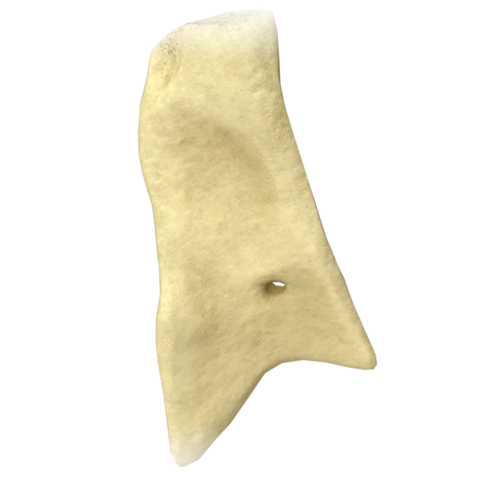
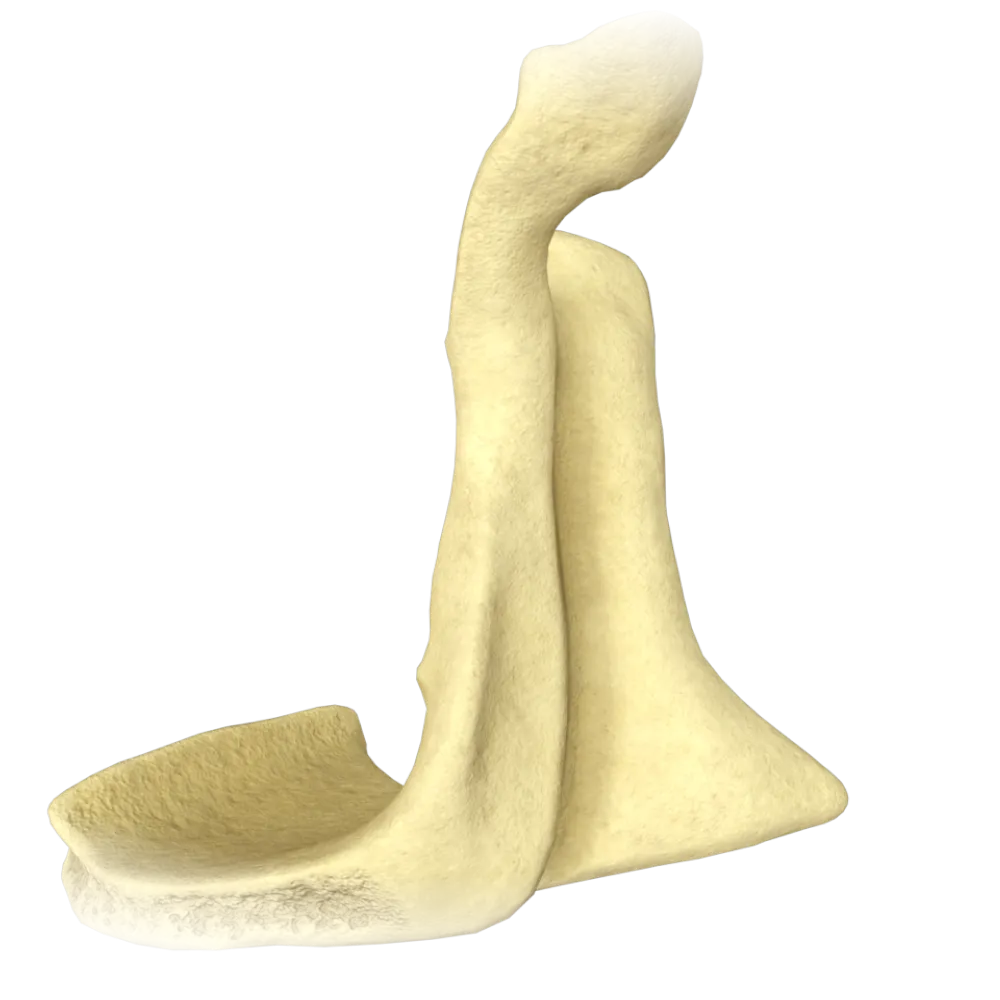
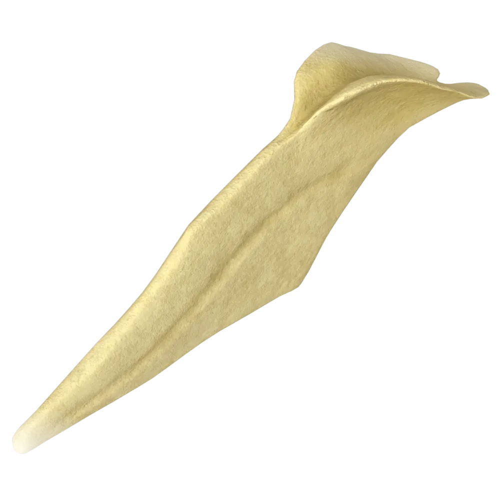
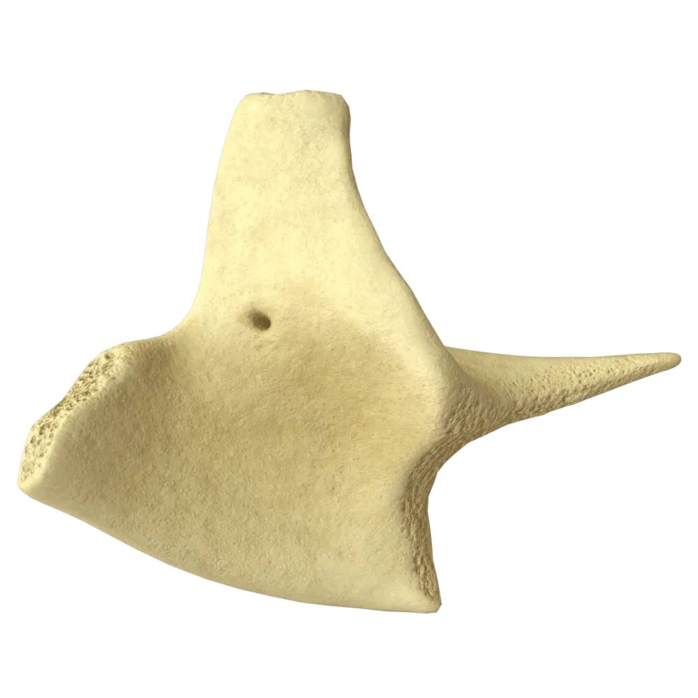
Zygomatic bone anatomy quiz
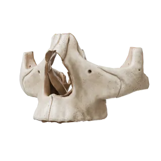
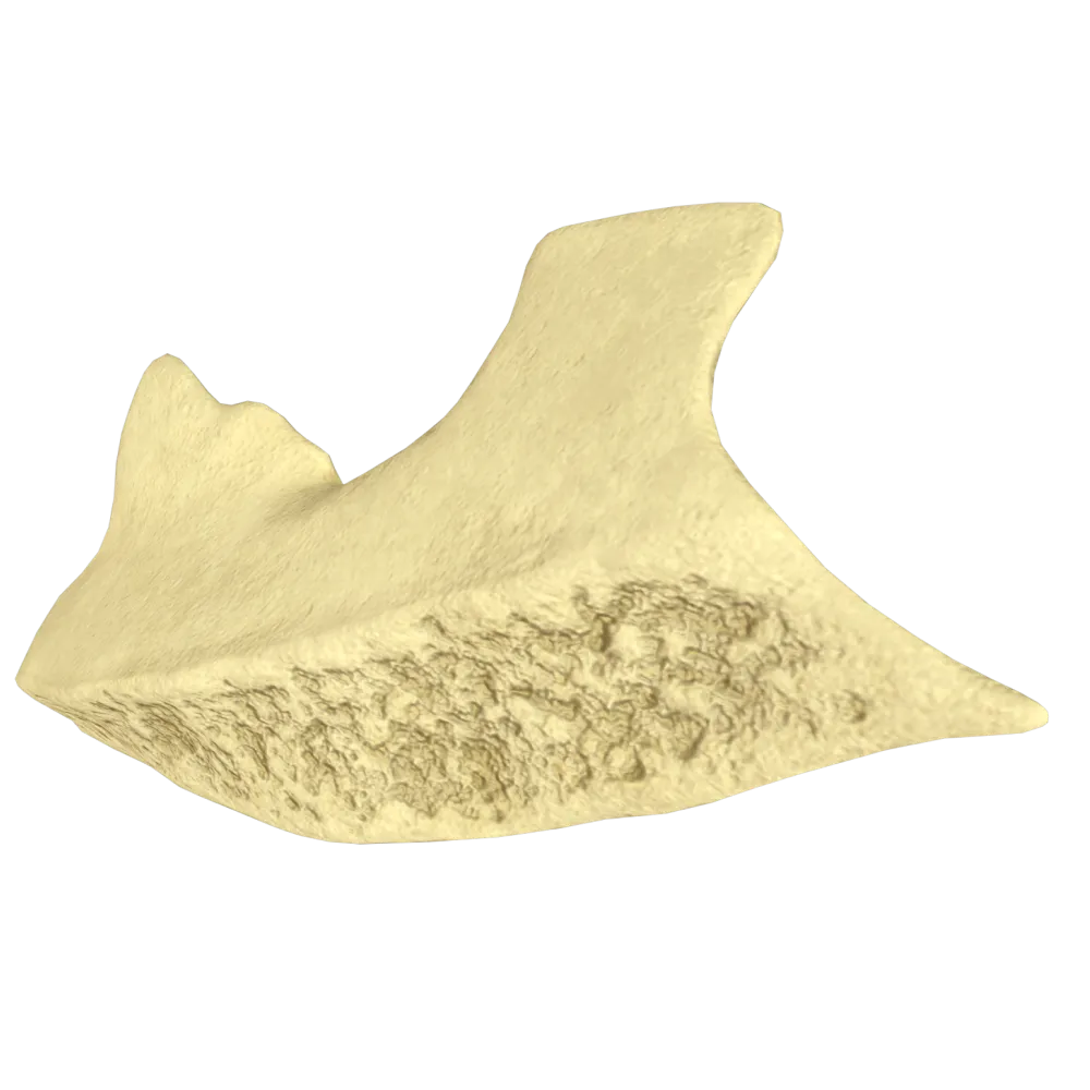
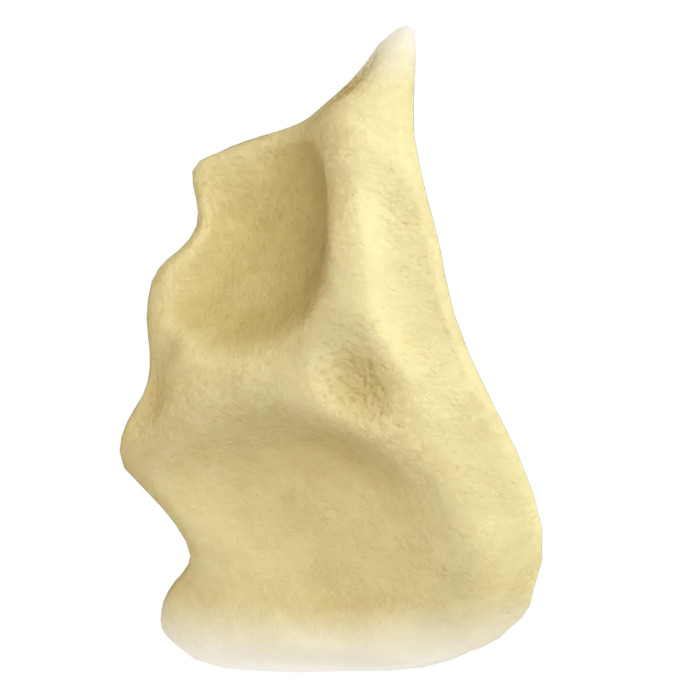
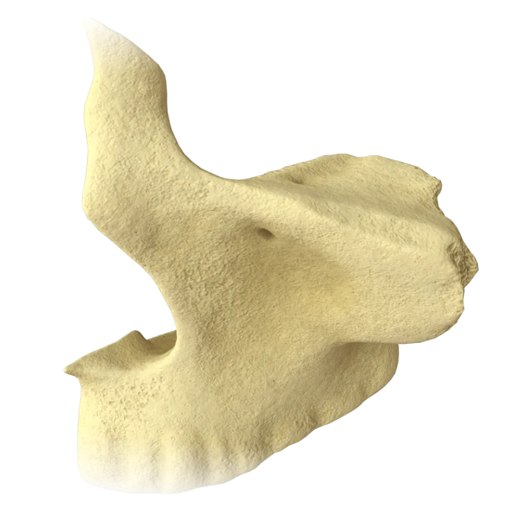
The quiz covers the structure, processes, surfaces, sutures, and anatomical relationships of the zygomatic bone.
1/20
bold
text
1. To which group of cranial bones does the zygomatic bone belong?
-
Neurocranium Bones
The zygomatic bone (os zygomaticum) is a bone of the Viscerocranium, forming the lateral wall of the face and the zygomatic arch.
-
Bones of the Viscerocranium
The zygomatic bone (os zygomaticum) is a bone of the Viscerocranium, forming the lateral wall of the face and the zygomatic arch.
-
Bones of the cranial base
The zygomatic bone (os zygomaticum) is a bone of the Viscerocranium, forming the lateral wall of the face and the zygomatic arch.
-
Bones of the Calvaria
The zygomatic bone (os zygomaticum) is a bone of the Viscerocranium, forming the lateral wall of the face and the zygomatic arch.
-
I find it difficult to answer
The zygomatic bone (os zygomaticum) is a bone of the Viscerocranium, forming the lateral wall of the face and the zygomatic arch.
2. How many processes does the zygomatic bone comprise?
-
Two
The zygomatic bone comprises three processes: the frontal process (processus frontalis), temporal process (processus temporalis), and maxillary process (processus maxillaris).
-
Four.
The zygomatic bone comprises three processes: the frontal process (processus frontalis), temporal process (processus temporalis), and maxillary process (processus maxillaris).
-
Three.
The zygomatic bone comprises three processes: the frontal process (processus frontalis), temporal process (processus temporalis), and maxillary process (processus maxillaris).
-
One
The zygomatic bone comprises three processes: the frontal process (processus frontalis), temporal process (processus temporalis), and maxillary process (processus maxillaris).
-
I find it difficult to answer
The zygomatic bone comprises three processes: the frontal process (processus frontalis), temporal process (processus temporalis), and maxillary process (processus maxillaris).
3. Which process of the zygomatic bone articulates with the Frontal bone?
-
Temporal process
The frontal process (processus frontalis) of the zygomatic bone articulates with the zygomatic process of the Frontal bone, forming the frontozygomatic suture (sutura frontozygomatica).
-
Maxillary process
The frontal process (processus frontalis) of the zygomatic bone articulates with the zygomatic process of the Frontal bone, forming the frontozygomatic suture (sutura frontozygomatica).
-
Orbital process
The frontal process (processus frontalis) of the zygomatic bone articulates with the zygomatic process of the Frontal bone, forming the frontozygomatic suture (sutura frontozygomatica).
-
Frontal process
The frontal process (processus frontalis) of the zygomatic bone articulates with the zygomatic process of the Frontal bone, forming the frontozygomatic suture (sutura frontozygomatica).
-
I find it difficult to answer
The frontal process (processus frontalis) of the zygomatic bone articulates with the zygomatic process of the Frontal bone, forming the frontozygomatic suture (sutura frontozygomatica).
4. Which suture is formed between the zygomatic bone and the maxilla?
-
Frontozygomatic suture
The zygomaticomaxillary suture (sutura zygomaticomaxillaris) is formed between the zygomatic bone and the maxilla, connecting the maxillary process of the zygomatic bone to the body of the maxilla.
-
Temporozygomatic suture
The zygomaticomaxillary suture (sutura zygomaticomaxillaris) is formed between the zygomatic bone and the maxilla, connecting the maxillary process of the zygomatic bone to the body of the maxilla.
-
Zygomaticomaxillary suture
The zygomaticomaxillary suture (sutura zygomaticomaxillaris) is formed between the zygomatic bone and the maxilla, connecting the maxillary process of the zygomatic bone to the body of the maxilla.
-
Sphenozygomatic suture
The zygomaticomaxillary suture (sutura zygomaticomaxillaris) is formed between the zygomatic bone and the maxilla, connecting the maxillary process of the zygomatic bone to the body of the maxilla.
-
I find it difficult to answer
The zygomaticomaxillary suture (sutura zygomaticomaxillaris) is formed between the zygomatic bone and the maxilla, connecting the maxillary process of the zygomatic bone to the body of the maxilla.
5. Which surface of the zygomatic bone is called facies lateralis?
-
The surface facing the orbit
Facies lateralis is the external (lateral) convex surface of the zygomatic bone, facing laterally and anteriorly; the zygomaticofacial foramen (foramen zygomaticofaciale) opens on it.
-
The surface facing the infratemporal fossa
Facies lateralis is the external (lateral) convex surface of the zygomatic bone, facing laterally and anteriorly; the zygomaticofacial foramen (foramen zygomaticofaciale) opens on it.
-
The external convex surface facing laterally and anteriorly
Facies lateralis is the external (lateral) convex surface of the zygomatic bone, facing laterally and anteriorly; the zygomaticofacial foramen (foramen zygomaticofaciale) opens on it.
-
The surface facing the palate
Facies lateralis is the external (lateral) convex surface of the zygomatic bone, facing laterally and anteriorly; the zygomaticofacial foramen (foramen zygomaticofaciale) opens on it.
-
I find it difficult to answer
Facies lateralis is the external (lateral) convex surface of the zygomatic bone, facing laterally and anteriorly; the zygomaticofacial foramen (foramen zygomaticofaciale) opens on it.
6. On which surface of the zygomatic bone is the zygomatico-orbital foramen (foramen zygomaticoorbitale) located?
-
Lateral surface
Foramen zygomaticoorbitale is located on the orbital surface (facies orbitalis) of the zygomatic bone and serves as the entrance to the zygomatic canal, which divides into two canaliculi.
-
Temporal surface
Foramen zygomaticoorbitale is located on the orbital surface (facies orbitalis) of the zygomatic bone and serves as the entrance to the zygomatic canal, which divides into two canaliculi.
-
Orbital surface
Foramen zygomaticoorbitale is located on the orbital surface (facies orbitalis) of the zygomatic bone and serves as the entrance to the zygomatic canal, which divides into two canaliculi.
-
Maxillary surface
Foramen zygomaticoorbitale is located on the orbital surface (facies orbitalis) of the zygomatic bone and serves as the entrance to the zygomatic canal, which divides into two canaliculi.
-
I find it difficult to answer
Foramen zygomaticoorbitale is located on the orbital surface (facies orbitalis) of the zygomatic bone and serves as the entrance to the zygomatic canal, which divides into two canaliculi.
7. Which nerve passes through foramen zygomaticofaciale?
-
Zygomaticotemporal nerve
The zygomaticofacial nerve (n. zygomaticofacialis), a branch of the Zygomatic nerve (n. zygomaticus from n. maxillaris), emerges through the zygomaticofacial foramen (foramen zygomaticofaciale).
-
infraorbital nerve
The zygomaticofacial nerve (n. zygomaticofacialis), a branch of the Zygomatic nerve (n. zygomaticus from n. maxillaris), emerges through the zygomaticofacial foramen (foramen zygomaticofaciale).
-
Zygomaticofacial nerve
The zygomaticofacial nerve (n. zygomaticofacialis), a branch of the Zygomatic nerve (n. zygomaticus from n. maxillaris), emerges through the zygomaticofacial foramen (foramen zygomaticofaciale).
-
Mental nerve
The zygomaticofacial nerve (n. zygomaticofacialis), a branch of the Zygomatic nerve (n. zygomaticus from n. maxillaris), emerges through the zygomaticofacial foramen (foramen zygomaticofaciale).
-
I find it difficult to answer
The zygomaticofacial nerve (n. zygomaticofacialis), a branch of the Zygomatic nerve (n. zygomaticus from n. maxillaris), emerges through the zygomaticofacial foramen (foramen zygomaticofaciale).
8. Which nerve passes through foramen zygomaticotemporale?
-
Zygomaticofacial nerve
The zygomaticotemporal branch (n. zygomaticotemporalis) of the Zygomatic nerve passes through the zygomaticotemporal foramen (foramen zygomaticotemporale) and innervates the skin of the temporal region.
-
Zygomaticotemporal nerve
The zygomaticotemporal branch (n. zygomaticotemporalis) of the Zygomatic nerve passes through the zygomaticotemporal foramen (foramen zygomaticotemporale) and innervates the skin of the temporal region.
-
N. auriculotemporalis
The zygomaticotemporal branch (n. zygomaticotemporalis) of the Zygomatic nerve passes through the zygomaticotemporal foramen (foramen zygomaticotemporale) and innervates the skin of the temporal region.
-
N. buccalis
The zygomaticotemporal branch (n. zygomaticotemporalis) of the Zygomatic nerve passes through the zygomaticotemporal foramen (foramen zygomaticotemporale) and innervates the skin of the temporal region.
-
I find it difficult to answer
The zygomaticotemporal branch (n. zygomaticotemporalis) of the Zygomatic nerve passes through the zygomaticotemporal foramen (foramen zygomaticotemporale) and innervates the skin of the temporal region.
9. Which suture is formed between the zygomatic bone and the sphenoid bone?
-
Frontozygomatic suture
Sutura sphenozygomatica (the sphenozygomatic suture) is formed on the medial wall of the orbit between the greater wing of the sphenoid bone and the orbital surface of the zygomatic bone.
-
Zygomaticomaxillary suture
Sutura sphenozygomatica (the sphenozygomatic suture) is formed on the medial wall of the orbit between the greater wing of the sphenoid bone and the orbital surface of the zygomatic bone.
-
Temporozygomatic suture
Sutura sphenozygomatica (the sphenozygomatic suture) is formed on the medial wall of the orbit between the greater wing of the sphenoid bone and the orbital surface of the zygomatic bone.
-
Sphenozygomatic suture
Sutura sphenozygomatica (the sphenozygomatic suture) is formed on the medial wall of the orbit between the greater wing of the sphenoid bone and the orbital surface of the zygomatic bone.
-
I find it difficult to answer
Sutura sphenozygomatica (the sphenozygomatic suture) is formed on the medial wall of the orbit between the greater wing of the sphenoid bone and the orbital surface of the zygomatic bone.
10. What role does the temporal process of the zygomatic bone (processus temporalis) play?
-
Contributes to the formation of the inferior orbital fissure
Processus temporalis of the zygomatic bone articulates with the zygomatic process of the temporal bone (processus zygomaticus ossis temporalis), forming the zygomatic arch (arcus zygomaticus).
-
Articulates with the zygomatic process of the temporal bone, forming the zygomatic arch
Processus temporalis of the zygomatic bone articulates with the zygomatic process of the temporal bone (processus zygomaticus ossis temporalis), forming the zygomatic arch (arcus zygomaticus).
-
Forms the inferior wall of the orbit
Processus temporalis of the zygomatic bone articulates with the zygomatic process of the temporal bone (processus zygomaticus ossis temporalis), forming the zygomatic arch (arcus zygomaticus).
-
Articulates with the zygomatic process of the Frontal bone
Processus temporalis of the zygomatic bone articulates with the zygomatic process of the temporal bone (processus zygomaticus ossis temporalis), forming the zygomatic arch (arcus zygomaticus).
-
I find it difficult to answer
Processus temporalis of the zygomatic bone articulates with the zygomatic process of the temporal bone (processus zygomaticus ossis temporalis), forming the zygomatic arch (arcus zygomaticus).
11. The orbital surface of the zygomatic bone contributes to the formation of which anatomical structure?
-
Medial wall of the orbit
Facies orbitalis of the zygomatic bone contributes to the formation of the lateral wall and the inferior portion of the lateral part of the floor (inferior wall) of the orbit (orbita).
-
Superior wall of the orbit
Facies orbitalis of the zygomatic bone contributes to the formation of the lateral wall and the inferior portion of the lateral part of the floor (inferior wall) of the orbit (orbita).
-
Lateral and inferior walls of the orbit
Facies orbitalis of the zygomatic bone contributes to the formation of the lateral wall and the inferior portion of the lateral part of the floor (inferior wall) of the orbit (orbita).
-
Posterior wall of the orbit
Facies orbitalis of the zygomatic bone contributes to the formation of the lateral wall and the inferior portion of the lateral part of the floor (inferior wall) of the orbit (orbita).
-
I find it difficult to answer
Facies orbitalis of the zygomatic bone contributes to the formation of the lateral wall and the inferior portion of the lateral part of the floor (inferior wall) of the orbit (orbita).
12. Which suture connects the zygomatic bone to the temporal bone?
-
Sphenozygomatic suture
The temporozygomatic suture (sutura temporozygomatica) connects the temporal process of the zygomatic bone to the zygomatic process of the temporal bone as part of the zygomatic arch.
-
Frontozygomatic suture
The temporozygomatic suture (sutura temporozygomatica) connects the temporal process of the zygomatic bone to the zygomatic process of the temporal bone as part of the zygomatic arch.
-
Temporozygomatic suture
The temporozygomatic suture (sutura temporozygomatica) connects the temporal process of the zygomatic bone to the zygomatic process of the temporal bone as part of the zygomatic arch.
-
Zygomaticomaxillary suture
The temporozygomatic suture (sutura temporozygomatica) connects the temporal process of the zygomatic bone to the zygomatic process of the temporal bone as part of the zygomatic arch.
-
I find it difficult to answer
The temporozygomatic suture (sutura temporozygomatica) connects the temporal process of the zygomatic bone to the zygomatic process of the temporal bone as part of the zygomatic arch.
13. How many constant foramina are anatomically identified in the zygomatic bone (os zygomaticum)?
-
4
The zygomatic bone has 3 constant foramina: the zygomatico-orbital foramen (foramen zygomaticoorbitale), the zygomaticofacial foramen (foramen zygomaticofaciale), and the zygomaticotemporal foramen (foramen zygomaticotemporale).
-
3
The zygomatic bone has 3 constant foramina: the zygomatico-orbital foramen (foramen zygomaticoorbitale), the zygomaticofacial foramen (foramen zygomaticofaciale), and the zygomaticotemporal foramen (foramen zygomaticotemporale).
-
2
The zygomatic bone has 3 constant foramina: the zygomatico-orbital foramen (foramen zygomaticoorbitale), the zygomaticofacial foramen (foramen zygomaticofaciale), and the zygomaticotemporal foramen (foramen zygomaticotemporale).
-
1
The zygomatic bone has 3 constant foramina: the zygomatico-orbital foramen (foramen zygomaticoorbitale), the zygomaticofacial foramen (foramen zygomaticofaciale), and the zygomaticotemporal foramen (foramen zygomaticotemporale).
-
I find it difficult to answer
The zygomatic bone has 3 constant foramina: the zygomatico-orbital foramen (foramen zygomaticoorbitale), the zygomaticofacial foramen (foramen zygomaticofaciale), and the zygomaticotemporal foramen (foramen zygomaticotemporale).
14. In terms of its development, the zygomatic bone is formed by:
-
Endochondral ossification
The zygomatic bone develops by desmal ossification—directly from mesenchyme, without passing through a cartilaginous stage.
-
Desmal (intramembranous) ossification
The zygomatic bone develops by desmal ossification—directly from mesenchyme, without passing through a cartilaginous stage.
-
Mixed ossification
The zygomatic bone develops by desmal ossification—directly from mesenchyme, without passing through a cartilaginous stage.
-
Perichondral ossification
The zygomatic bone develops by desmal ossification—directly from mesenchyme, without passing through a cartilaginous stage.
-
I find it difficult to answer
The zygomatic bone develops by desmal ossification—directly from mesenchyme, without passing through a cartilaginous stage.
15. How many surfaces are identified in the structure of the zygomatic bone (os zygomaticum)?
-
3
The zygomatic bone has 3 surfaces: the lateral surface (facies lateralis), the orbital surface (facies orbitalis), and the temporal surface (facies temporalis).
-
2
The zygomatic bone has 3 surfaces: the lateral surface (facies lateralis), the orbital surface (facies orbitalis), and the temporal surface (facies temporalis).
-
1
The zygomatic bone has 3 surfaces: the lateral surface (facies lateralis), the orbital surface (facies orbitalis), and the temporal surface (facies temporalis).
-
4
The zygomatic bone has 3 surfaces: the lateral surface (facies lateralis), the orbital surface (facies orbitalis), and the temporal surface (facies temporalis).
-
I find it difficult to answer
The zygomatic bone has 3 surfaces: the lateral surface (facies lateralis), the orbital surface (facies orbitalis), and the temporal surface (facies temporalis).
16. Is the zygomatic bone paired or unpaired?
-
Unpaired and located in the midline
The zygomatic bone (os zygomaticum) is a paired bone: the right and left bones are symmetrically located in the lateral regions of the Viscerocranium.
-
Paired and symmetrically located on both sides of the face
The zygomatic bone (os zygomaticum) is a paired bone: the right and left bones are symmetrically located in the lateral regions of the Viscerocranium.
-
Unpaired and asymmetrically located
The zygomatic bone (os zygomaticum) is a paired bone: the right and left bones are symmetrically located in the lateral regions of the Viscerocranium.
-
Represented by a single bone in the cranial base
The zygomatic bone (os zygomaticum) is a paired bone: the right and left bones are symmetrically located in the lateral regions of the Viscerocranium.
-
I find it difficult to answer
The zygomatic bone (os zygomaticum) is a paired bone: the right and left bones are symmetrically located in the lateral regions of the Viscerocranium.
17. Which of the following foramina does NOT belong to the zygomatic bone?
-
Zygomaticofacial foramen
The infraorbital foramen is located on the anterior surface of the body of the maxilla (maxilla), not on the zygomatic bone. The zygomatic bone has three foramina: zygomaticofaciale, zygomaticotemporale, and zygomaticoorbitale.
-
Zygomaticotemporal foramen
The infraorbital foramen is located on the anterior surface of the body of the maxilla (maxilla), not on the zygomatic bone. The zygomatic bone has three foramina: zygomaticofaciale, zygomaticotemporale, and zygomaticoorbitale.
-
Zygomatico-orbital foramen
The infraorbital foramen is located on the anterior surface of the body of the maxilla (maxilla), not on the zygomatic bone. The zygomatic bone has three foramina: zygomaticofaciale, zygomaticotemporale, and zygomaticoorbitale.
-
Infraorbital foramen
The infraorbital foramen is located on the anterior surface of the body of the maxilla (maxilla), not on the zygomatic bone. The zygomatic bone has three foramina: zygomaticofaciale, zygomaticotemporale, and zygomaticoorbitale.
-
I find it difficult to answer
The infraorbital foramen is located on the anterior surface of the body of the maxilla (maxilla), not on the zygomatic bone. The zygomatic bone has three foramina: zygomaticofaciale, zygomaticotemporale, and zygomaticoorbitale.
18. Which surface of the zygomatic bone faces the infratemporal fossa?
-
Orbital surface
Facies temporalis (the temporal surface) of the zygomatic bone faces posteriorly and medially, contributing to the boundaries of the infratemporal fossa and, in part, the temporal fossa.
-
Lateral surface
Facies temporalis (the temporal surface) of the zygomatic bone faces posteriorly and medially, contributing to the boundaries of the infratemporal fossa and, in part, the temporal fossa.
-
Temporal surface
Facies temporalis (the temporal surface) of the zygomatic bone faces posteriorly and medially, contributing to the boundaries of the infratemporal fossa and, in part, the temporal fossa.
-
Nasal surface
Facies temporalis (the temporal surface) of the zygomatic bone faces posteriorly and medially, contributing to the boundaries of the infratemporal fossa and, in part, the temporal fossa.
-
I find it difficult to answer
Facies temporalis (the temporal surface) of the zygomatic bone faces posteriorly and medially, contributing to the boundaries of the infratemporal fossa and, in part, the temporal fossa.
19. Which of the following sutures are located on the orbital surface of the zygomatic bone (in the orbital wall)?
-
Sutura temporozygomatica and sutura zygomaticomaxillaris
Two sutures are located on the orbital surface of the zygomatic bone (orbital wall): sutura sphenozygomatica (with the greater wing of the sphenoid bone) and sutura frontozygomatica (with the Frontal bone).
-
Sutura frontozygomatica and sutura sphenozygomatica
Two sutures are located on the orbital surface of the zygomatic bone (orbital wall): sutura sphenozygomatica (with the greater wing of the sphenoid bone) and sutura frontozygomatica (with the Frontal bone).
-
Sutura zygomaticomaxillaris and sutura temporozygomatica
Two sutures are located on the orbital surface of the zygomatic bone (orbital wall): sutura sphenozygomatica (with the greater wing of the sphenoid bone) and sutura frontozygomatica (with the Frontal bone).
-
Sutura sphenozygomatica and sutura frontozygomatica
Two sutures are located on the orbital surface of the zygomatic bone (orbital wall): sutura sphenozygomatica (with the greater wing of the sphenoid bone) and sutura frontozygomatica (with the Frontal bone).
-
I find it difficult to answer
Two sutures are located on the orbital surface of the zygomatic bone (orbital wall): sutura sphenozygomatica (with the greater wing of the sphenoid bone) and sutura frontozygomatica (with the Frontal bone).
20. What is the primary anatomical function of the orbital tubercle (tuberculum orbitale) of the zygomatic bone?
-
Serves as the exit site of the zygomatico-orbital nerve
The orbital tubercle (tuberculum orbitale) is located on the orbital surface of the zygomatic bone, closer to its lateral margin. The lateral palpebral ligament (ligamentum palpebrale laterale) attaches to it.
-
Serves as the attachment site of the lateral palpebral ligament
The orbital tubercle (tuberculum orbitale) is located on the orbital surface of the zygomatic bone, closer to its lateral margin. The lateral palpebral ligament (ligamentum palpebrale laterale) attaches to it.
-
Serves as the attachment site of the masseter muscle
The orbital tubercle (tuberculum orbitale) is located on the orbital surface of the zygomatic bone, closer to its lateral margin. The lateral palpebral ligament (ligamentum palpebrale laterale) attaches to it.
-
Forms an articulation with the wing of the Sphenoid bone
The orbital tubercle (tuberculum orbitale) is located on the orbital surface of the zygomatic bone, closer to its lateral margin. The lateral palpebral ligament (ligamentum palpebrale laterale) attaches to it.
-
I find it difficult to answer
The orbital tubercle (tuberculum orbitale) is located on the orbital surface of the zygomatic bone, closer to its lateral margin. The lateral palpebral ligament (ligamentum palpebrale laterale) attaches to it.
Retake this quiz?
Your current progress will be reset.
