Maxilla anatomy quiz
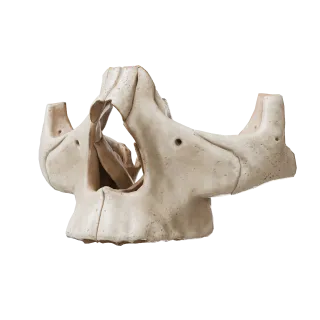
Normal anatomy quiz of the maxilla: structure, processes, canals, sinus, blood supply, and innervation.
1/20
bold
text
1. Which process of the maxilla participates in the formation of the hard palate?
-
Alveolar process
The palatine process of the maxilla (processus palatinus) is a horizontal plate that joins along the midline with the corresponding process on the opposite side. Together with the horizontal plates of the palatine bones, they form the bony framework of the hard palate (palatum durum), which separates the oral cavity from the nasal cavity.
-
Frontal process
The palatine process of the maxilla (processus palatinus) is a horizontal plate that joins along the midline with the corresponding process on the opposite side. Together with the horizontal plates of the palatine bones, they form the bony framework of the hard palate (palatum durum), which separates the oral cavity from the nasal cavity.
-
Zygomatic process
The palatine process of the maxilla (processus palatinus) is a horizontal plate that joins along the midline with the corresponding process on the opposite side. Together with the horizontal plates of the palatine bones, they form the bony framework of the hard palate (palatum durum), which separates the oral cavity from the nasal cavity.
-
Palatine process
The palatine process of the maxilla (processus palatinus) is a horizontal plate that joins along the midline with the corresponding process on the opposite side. Together with the horizontal plates of the palatine bones, they form the bony framework of the hard palate (palatum durum), which separates the oral cavity from the nasal cavity.
-
I find it difficult to answer
The palatine process of the maxilla (processus palatinus) is a horizontal plate that joins along the midline with the corresponding process on the opposite side. Together with the horizontal plates of the palatine bones, they form the bony framework of the hard palate (palatum durum), which separates the oral cavity from the nasal cavity.
2. Which process of the maxilla articulates with the frontal bone?
-
Zygomatic process
The frontal process (processus frontalis) of the maxilla extends superiorly and articulates with the nasal part of the frontal bone.
-
Frontal process
The frontal process (processus frontalis) of the maxilla extends superiorly and articulates with the nasal part of the frontal bone.
-
Alveolar process
The frontal process (processus frontalis) of the maxilla extends superiorly and articulates with the nasal part of the frontal bone.
-
Palatine process
The frontal process (processus frontalis) of the maxilla extends superiorly and articulates with the nasal part of the frontal bone.
-
I find it difficult to answer
The frontal process (processus frontalis) of the maxilla extends superiorly and articulates with the nasal part of the frontal bone.
3. Which air-filled cavity is located within the body of the maxilla?
-
Frontal sinus
The body of the maxilla contains the large air-filled maxillary sinus (sinus maxillaris, or antrum of Highmore), the largest of all the paranasal sinuses.
-
Sphenoidal sinus
The body of the maxilla contains the large air-filled maxillary sinus (sinus maxillaris, or antrum of Highmore), the largest of all the paranasal sinuses.
-
Maxillary (antral) sinus
The body of the maxilla contains the large air-filled maxillary sinus (sinus maxillaris, or antrum of Highmore), the largest of all the paranasal sinuses.
-
Ethmoidal labyrinth
The body of the maxilla contains the large air-filled maxillary sinus (sinus maxillaris, or antrum of Highmore), the largest of all the paranasal sinuses.
-
I find it difficult to answer
The body of the maxilla contains the large air-filled maxillary sinus (sinus maxillaris, or antrum of Highmore), the largest of all the paranasal sinuses.
4. Through which opening does the maxillary sinus communicate with the nasal cavity?
-
Through the round foramen
A large maxillary hiatus (hiatus maxillaris), leading into the sinus, is located on the nasal surface of the body of the maxilla.
-
Through the maxillary hiatus
A large maxillary hiatus (hiatus maxillaris), leading into the sinus, is located on the nasal surface of the body of the maxilla.
-
Through the inferior nasal meatus
A large maxillary hiatus (hiatus maxillaris), leading into the sinus, is located on the nasal surface of the body of the maxilla.
-
Through the choana
A large maxillary hiatus (hiatus maxillaris), leading into the sinus, is located on the nasal surface of the body of the maxilla.
-
I find it difficult to answer
A large maxillary hiatus (hiatus maxillaris), leading into the sinus, is located on the nasal surface of the body of the maxilla.
5. Which bone forms most of the floor of the orbit?
-
Palatine bone
The orbital surface (facies orbitalis) of the body of the maxilla forms most of the inferior wall (floor) of the orbit.
-
Lacrimal bone
The orbital surface (facies orbitalis) of the body of the maxilla forms most of the inferior wall (floor) of the orbit.
-
Upper jaw
The orbital surface (facies orbitalis) of the body of the maxilla forms most of the inferior wall (floor) of the orbit.
-
Ethmoid bone = Ethmoid = Ethmoidal bone
The orbital surface (facies orbitalis) of the body of the maxilla forms most of the inferior wall (floor) of the orbit.
-
I find it difficult to answer
The orbital surface (facies orbitalis) of the body of the maxilla forms most of the inferior wall (floor) of the orbit.
6. Through which foramen does the infraorbital nerve leave the maxilla and emerge onto the face?
-
Round foramen
The infraorbital nerve (n. infraorbitalis), as a continuation of the maxillary nerve, emerges through the foramen infraorbitale, located on the anterior (facial) surface of the body of the maxilla below the infraorbital margin, and innervates the skin of the face.
-
Oval foramen
The infraorbital nerve (n. infraorbitalis), as a continuation of the maxillary nerve, emerges through the foramen infraorbitale, located on the anterior (facial) surface of the body of the maxilla below the infraorbital margin, and innervates the skin of the face.
-
Infraorbital foramen
The infraorbital nerve (n. infraorbitalis), as a continuation of the maxillary nerve, emerges through the foramen infraorbitale, located on the anterior (facial) surface of the body of the maxilla below the infraorbital margin, and innervates the skin of the face.
-
Incisive foramen
The infraorbital nerve (n. infraorbitalis), as a continuation of the maxillary nerve, emerges through the foramen infraorbitale, located on the anterior (facial) surface of the body of the maxilla below the infraorbital margin, and innervates the skin of the face.
-
I find it difficult to answer
The infraorbital nerve (n. infraorbitalis), as a continuation of the maxillary nerve, emerges through the foramen infraorbitale, located on the anterior (facial) surface of the body of the maxilla below the infraorbital margin, and innervates the skin of the face.
7. The palatine process of the maxilla participates in the formation of which anatomical structure?
-
Lateral wall of the nasal cavity
The palatine process (processus palatinus) of the maxilla is a horizontal plate that, together with the corresponding process on the opposite side and the horizontal plates of the palatine bones, forms the bony hard palate.
-
Hard palate
The palatine process (processus palatinus) of the maxilla is a horizontal plate that, together with the corresponding process on the opposite side and the horizontal plates of the palatine bones, forms the bony hard palate.
-
Inferior wall of the orbit
The palatine process (processus palatinus) of the maxilla is a horizontal plate that, together with the corresponding process on the opposite side and the horizontal plates of the palatine bones, forms the bony hard palate.
-
Medial wall of the maxillary sinus
The palatine process (processus palatinus) of the maxilla is a horizontal plate that, together with the corresponding process on the opposite side and the horizontal plates of the palatine bones, forms the bony hard palate.
-
I find it difficult to answer
The palatine process (processus palatinus) of the maxilla is a horizontal plate that, together with the corresponding process on the opposite side and the horizontal plates of the palatine bones, forms the bony hard palate.
8. Which of the listed functions is NOT a function of the maxillary (antral) sinus?
-
Reduction of the weight of the facial skeleton
Saliva is produced by the salivary glands of the oral cavity, not by the paranasal sinuses. The maxillary sinus is filled with air and serves to reduce the weight of the skull, contribute to vocal resonance, and warm inspired air.
-
Resonance during voice production
Saliva is produced by the salivary glands of the oral cavity, not by the paranasal sinuses. The maxillary sinus is filled with air and serves to reduce the weight of the skull, contribute to vocal resonance, and warm inspired air.
-
Production and secretion of saliva
Saliva is produced by the salivary glands of the oral cavity, not by the paranasal sinuses. The maxillary sinus is filled with air and serves to reduce the weight of the skull, contribute to vocal resonance, and warm inspired air.
-
Warming and humidification of inspired air
Saliva is produced by the salivary glands of the oral cavity, not by the paranasal sinuses. The maxillary sinus is filled with air and serves to reduce the weight of the skull, contribute to vocal resonance, and warm inspired air.
-
I find it difficult to answer
Saliva is produced by the salivary glands of the oral cavity, not by the paranasal sinuses. The maxillary sinus is filled with air and serves to reduce the weight of the skull, contribute to vocal resonance, and warm inspired air.
9. How many processes does the maxilla comprise?
-
5
The maxilla comprises 4 processes: frontal, zygomatic, palatine, and alveolar.
-
4
The maxilla comprises 4 processes: frontal, zygomatic, palatine, and alveolar.
-
3
The maxilla comprises 4 processes: frontal, zygomatic, palatine, and alveolar.
-
2
The maxilla comprises 4 processes: frontal, zygomatic, palatine, and alveolar.
-
I find it difficult to answer
The maxilla comprises 4 processes: frontal, zygomatic, palatine, and alveolar.
10. With which anatomical structure does the zygomatic process of the maxilla articulate?
-
Zygomatic arch
The zygomatic process (processus zygomaticus) of the maxilla articulates directly with the zygomatic bone (os zygomaticum), forming the zygomaticomaxillary suture (sutura zygomaticomaxillaris).
-
Zygomatic process of the frontal bone
The zygomatic process (processus zygomaticus) of the maxilla articulates directly with the zygomatic bone (os zygomaticum), forming the zygomaticomaxillary suture (sutura zygomaticomaxillaris).
-
Zygomatic bone
The zygomatic process (processus zygomaticus) of the maxilla articulates directly with the zygomatic bone (os zygomaticum), forming the zygomaticomaxillary suture (sutura zygomaticomaxillaris).
-
Zygomatic process of the temporal bone
The zygomatic process (processus zygomaticus) of the maxilla articulates directly with the zygomatic bone (os zygomaticum), forming the zygomaticomaxillary suture (sutura zygomaticomaxillaris).
-
I find it difficult to answer
The zygomatic process (processus zygomaticus) of the maxilla articulates directly with the zygomatic bone (os zygomaticum), forming the zygomaticomaxillary suture (sutura zygomaticomaxillaris).
11. Which foramen is located in the anterior part of the hard palate and constitutes the outlet of the incisive canal?
-
Greater palatine foramen
The foramen incisivum (incisive foramen) is located in the anterior part of the hard palate, posterior to the maxillary incisors, along the median line. The nasopalatine nerve and vessels (branches of the a. sphenopalatina) pass through it.
-
Lesser palatine foramen
The foramen incisivum (incisive foramen) is located in the anterior part of the hard palate, posterior to the maxillary incisors, along the median line. The nasopalatine nerve and vessels (branches of the a. sphenopalatina) pass through it.
-
Incisive foramen
The foramen incisivum (incisive foramen) is located in the anterior part of the hard palate, posterior to the maxillary incisors, along the median line. The nasopalatine nerve and vessels (branches of the a. sphenopalatina) pass through it.
-
Pterygoid canal
The foramen incisivum (incisive foramen) is located in the anterior part of the hard palate, posterior to the maxillary incisors, along the median line. The nasopalatine nerve and vessels (branches of the a. sphenopalatina) pass through it.
-
I find it difficult to answer
The foramen incisivum (incisive foramen) is located in the anterior part of the hard palate, posterior to the maxillary incisors, along the median line. The nasopalatine nerve and vessels (branches of the a. sphenopalatina) pass through it.
12. On which process of the maxilla are the teeth located?
-
Frontal
The maxillary teeth are anchored in the alveolar process, which bears the dental alveoli (sockets) for the roots of the teeth. The other processes (frontal, zygomatic, and palatine) are not associated with the teeth.
-
Zygomatic
The maxillary teeth are anchored in the alveolar process, which bears the dental alveoli (sockets) for the roots of the teeth. The other processes (frontal, zygomatic, and palatine) are not associated with the teeth.
-
Alveolar
The maxillary teeth are anchored in the alveolar process, which bears the dental alveoli (sockets) for the roots of the teeth. The other processes (frontal, zygomatic, and palatine) are not associated with the teeth.
-
Palatine
The maxillary teeth are anchored in the alveolar process, which bears the dental alveoli (sockets) for the roots of the teeth. The other processes (frontal, zygomatic, and palatine) are not associated with the teeth.
-
I find it difficult to answer
The maxillary teeth are anchored in the alveolar process, which bears the dental alveoli (sockets) for the roots of the teeth. The other processes (frontal, zygomatic, and palatine) are not associated with the teeth.
13. On which surface of the maxilla is the canine fossa (fossa canina) located?
-
Nasal surface
The canine fossa (fossa canina) is a depression on the anterior (facial) surface of the body of the maxilla, located laterally to the canine eminence (jugum caninum).
-
Infratemporal surface
The canine fossa (fossa canina) is a depression on the anterior (facial) surface of the body of the maxilla, located laterally to the canine eminence (jugum caninum).
-
Anterior (facial) surface
The canine fossa (fossa canina) is a depression on the anterior (facial) surface of the body of the maxilla, located laterally to the canine eminence (jugum caninum).
-
Orbital surface
The canine fossa (fossa canina) is a depression on the anterior (facial) surface of the body of the maxilla, located laterally to the canine eminence (jugum caninum).
-
I find it difficult to answer
The canine fossa (fossa canina) is a depression on the anterior (facial) surface of the body of the maxilla, located laterally to the canine eminence (jugum caninum).
14. What is the infraorbital canal (canalis infraorbitalis), and what does it contain?
-
A canal in the zygomatic process containing the zygomatic nerve
Canalis infraorbitalis is a bony canal passing through the substance of the body of the maxilla. It contains the infraorbital nerve (n. infraorbitalis, a branch of V₂) and the infraorbital artery and vein, which emerge through the foramen infraorbitale.
-
A canal in the palatine process containing the nasopalatine nerve
Canalis infraorbitalis is a bony canal passing through the substance of the body of the maxilla. It contains the infraorbital nerve (n. infraorbitalis, a branch of V₂) and the infraorbital artery and vein, which emerge through the foramen infraorbitale.
-
A canal in the body of the maxilla containing the infraorbital nerve and vessels
Canalis infraorbitalis is a bony canal passing through the substance of the body of the maxilla. It contains the infraorbital nerve (n. infraorbitalis, a branch of V₂) and the infraorbital artery and vein, which emerge through the foramen infraorbitale.
-
A canal in the alveolar process containing the superior alveolar nerve
Canalis infraorbitalis is a bony canal passing through the substance of the body of the maxilla. It contains the infraorbital nerve (n. infraorbitalis, a branch of V₂) and the infraorbital artery and vein, which emerge through the foramen infraorbitale.
-
I find it difficult to answer
Canalis infraorbitalis is a bony canal passing through the substance of the body of the maxilla. It contains the infraorbital nerve (n. infraorbitalis, a branch of V₂) and the infraorbital artery and vein, which emerge through the foramen infraorbitale.
15. According to anatomical classification, to which division of the skull does the maxilla belong?
-
Neurocranium (neurocranium)
The maxilla (maxilla) is part of the Viscerocranium (viscerocranium).
-
Viscerocranium (viscerocranium)
The maxilla (maxilla) is part of the Viscerocranium (viscerocranium).
-
Cranial base (basis cranii)
The maxilla (maxilla) is part of the Viscerocranium (viscerocranium).
-
Calvaria (calvaria)
The maxilla (maxilla) is part of the Viscerocranium (viscerocranium).
-
I find it difficult to answer
The maxilla (maxilla) is part of the Viscerocranium (viscerocranium).
16. Through which foramen of the maxilla does the greater palatine nerve (n. palatinus major) emerge from the canal?
-
Incisive foramen
The greater palatine nerve (n. palatinus major) passes through the greater palatine canal (canalis palatinus major) and emerges through the foramen palatinum majus, located at the lateral margin of the hard palate at the level of the third molar.
-
Greater palatine foramen
The greater palatine nerve (n. palatinus major) passes through the greater palatine canal (canalis palatinus major) and emerges through the foramen palatinum majus, located at the lateral margin of the hard palate at the level of the third molar.
-
Lesser palatine foramen
The greater palatine nerve (n. palatinus major) passes through the greater palatine canal (canalis palatinus major) and emerges through the foramen palatinum majus, located at the lateral margin of the hard palate at the level of the third molar.
-
Round foramen
The greater palatine nerve (n. palatinus major) passes through the greater palatine canal (canalis palatinus major) and emerges through the foramen palatinum majus, located at the lateral margin of the hard palate at the level of the third molar.
-
I find it difficult to answer
The greater palatine nerve (n. palatinus major) passes through the greater palatine canal (canalis palatinus major) and emerges through the foramen palatinum majus, located at the lateral margin of the hard palate at the level of the third molar.
17. To which crest of the maxilla is the middle nasal concha of the ethmoid bone attached?
-
To the nasal crest (crista nasalis)
The ethmoidal crest (crista ethmoidalis) is located on the nasal surface of the frontal process of the maxilla; the middle nasal concha (which is part of the labyrinth of the ethmoid bone) is attached to it.
-
To the ethmoidal crest (crista ethmoidalis)
The ethmoidal crest (crista ethmoidalis) is located on the nasal surface of the frontal process of the maxilla; the middle nasal concha (which is part of the labyrinth of the ethmoid bone) is attached to it.
-
To the conchal crest (crista conchalis)
The ethmoidal crest (crista ethmoidalis) is located on the nasal surface of the frontal process of the maxilla; the middle nasal concha (which is part of the labyrinth of the ethmoid bone) is attached to it.
-
To the lacrimal crest (crista lacrimalis)
The ethmoidal crest (crista ethmoidalis) is located on the nasal surface of the frontal process of the maxilla; the middle nasal concha (which is part of the labyrinth of the ethmoid bone) is attached to it.
-
I find it difficult to answer
The ethmoidal crest (crista ethmoidalis) is located on the nasal surface of the frontal process of the maxilla; the middle nasal concha (which is part of the labyrinth of the ethmoid bone) is attached to it.
18. Which suture joins the palatine processes of the right and left maxillae?
-
Transverse palatine suture
The palatine processes of the two maxillae are joined along the median line by the median palatine suture, sutura palatina mediana.
-
Median palatine suture
The palatine processes of the two maxillae are joined along the median line by the median palatine suture, sutura palatina mediana.
-
Zygomaticomaxillary suture
The palatine processes of the two maxillae are joined along the median line by the median palatine suture, sutura palatina mediana.
-
Frontonasal suture
The palatine processes of the two maxillae are joined along the median line by the median palatine suture, sutura palatina mediana.
-
I find it difficult to answer
The palatine processes of the two maxillae are joined along the median line by the median palatine suture, sutura palatina mediana.
19. Which surface of the body of the maxilla forms the medial wall of the maxillary sinus and constitutes part of the lateral wall of the nasal cavity?
-
Anterior (facial) surface
The nasal surface (facies nasalis) of the body of the maxilla faces medially into the nasal cavity, forming the lateral wall of the nasal cavity while also constituting the medial wall of the maxillary sinus.
-
Orbital surface
The nasal surface (facies nasalis) of the body of the maxilla faces medially into the nasal cavity, forming the lateral wall of the nasal cavity while also constituting the medial wall of the maxillary sinus.
-
Nasal surface
The nasal surface (facies nasalis) of the body of the maxilla faces medially into the nasal cavity, forming the lateral wall of the nasal cavity while also constituting the medial wall of the maxillary sinus.
-
Infratemporal surface
The nasal surface (facies nasalis) of the body of the maxilla faces medially into the nasal cavity, forming the lateral wall of the nasal cavity while also constituting the medial wall of the maxillary sinus.
-
I find it difficult to answer
The nasal surface (facies nasalis) of the body of the maxilla faces medially into the nasal cavity, forming the lateral wall of the nasal cavity while also constituting the medial wall of the maxillary sinus.
20. Which anatomical space is bounded by the infratemporal surface of the body of the maxilla (facies infratemporalis)?
-
Pterygopalatine fossa anteriorly
The infratemporal surface (facies infratemporalis) of the maxilla forms the anterior wall of the infratemporal fossa (fossa infratemporalis). The alveolar foramina (foramina alveolaria), through which the posterior superior alveolar nerves and vessels pass, are located on this surface.
-
Pterygopalatine fossa posteriorly
The infratemporal surface (facies infratemporalis) of the maxilla forms the anterior wall of the infratemporal fossa (fossa infratemporalis). The alveolar foramina (foramina alveolaria), through which the posterior superior alveolar nerves and vessels pass, are located on this surface.
-
Infratemporal fossa anteriorly
The infratemporal surface (facies infratemporalis) of the maxilla forms the anterior wall of the infratemporal fossa (fossa infratemporalis). The alveolar foramina (foramina alveolaria), through which the posterior superior alveolar nerves and vessels pass, are located on this surface.
-
Temporal fossa inferiorly
The infratemporal surface (facies infratemporalis) of the maxilla forms the anterior wall of the infratemporal fossa (fossa infratemporalis). The alveolar foramina (foramina alveolaria), through which the posterior superior alveolar nerves and vessels pass, are located on this surface.
-
I find it difficult to answer
The infratemporal surface (facies infratemporalis) of the maxilla forms the anterior wall of the infratemporal fossa (fossa infratemporalis). The alveolar foramina (foramina alveolaria), through which the posterior superior alveolar nerves and vessels pass, are located on this surface.
Retake this quiz?
Your current progress will be reset.
