Lacrimal bone anatomy quiz
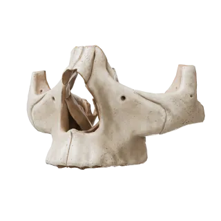
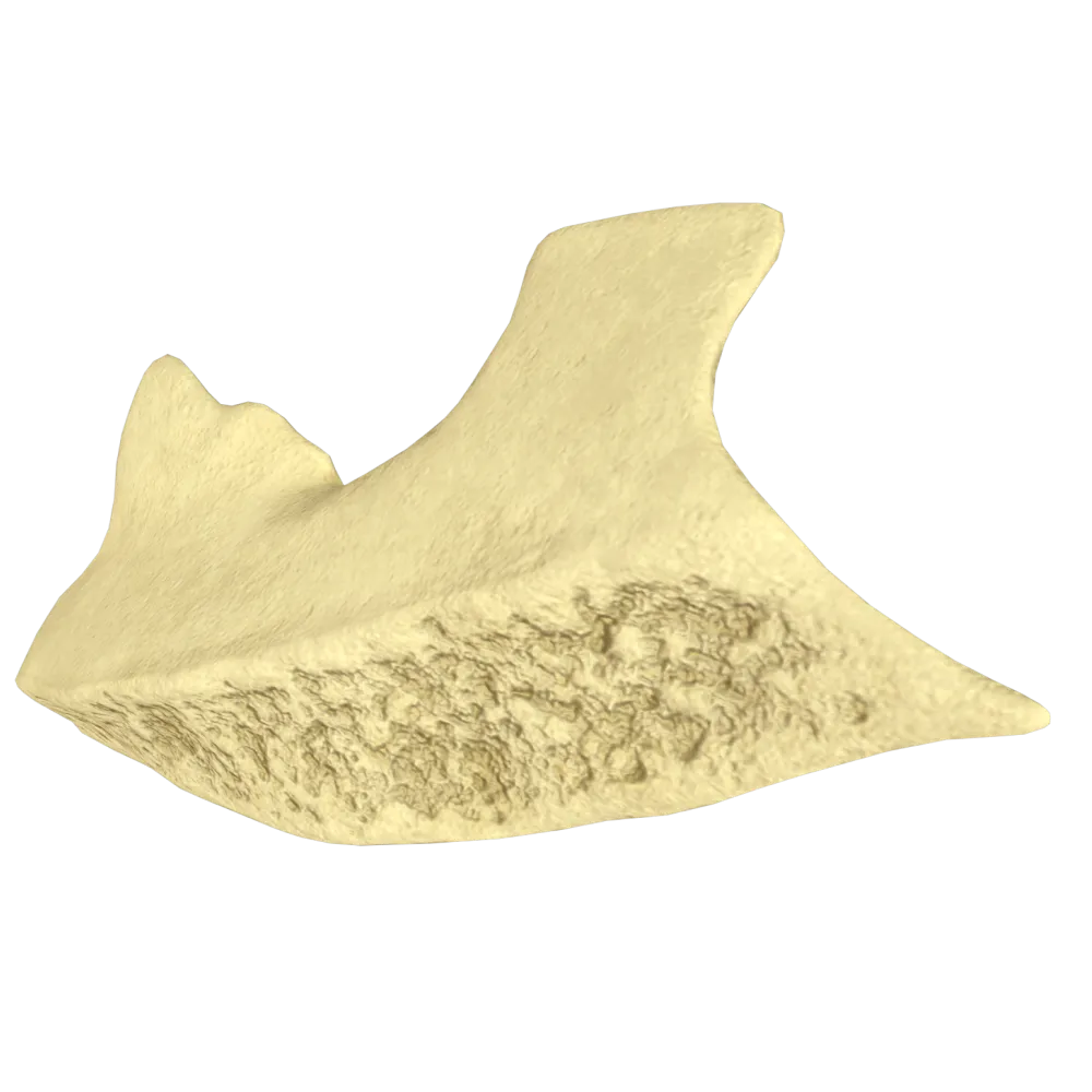
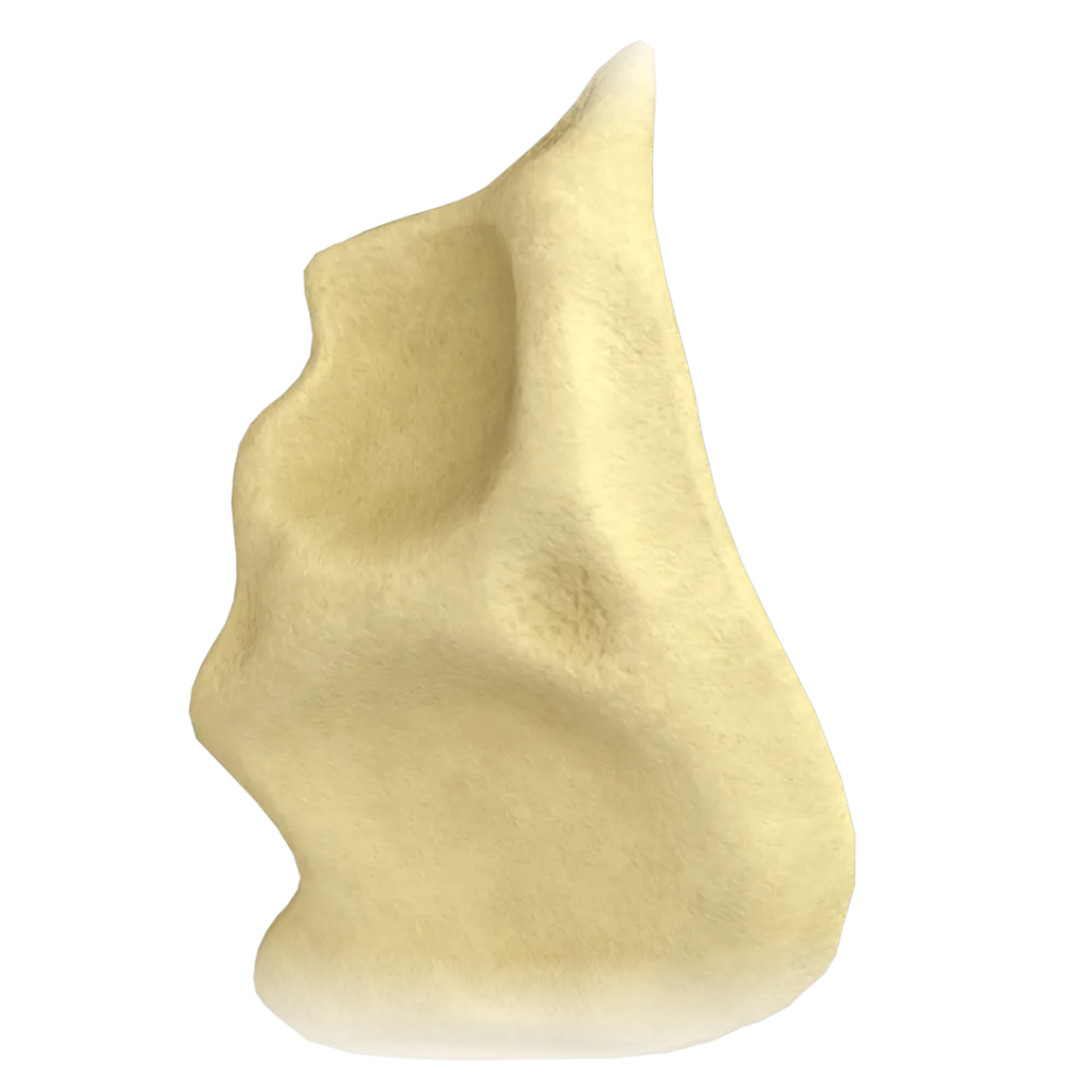
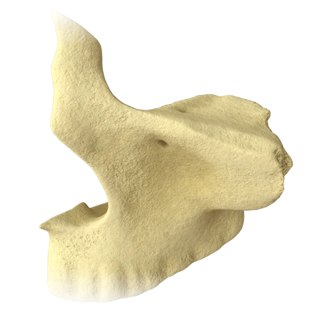
Lacrimal bone anatomy quiz: structure, topography, articular surfaces, canals, and anatomical relationships.
1/20
bold
text
1. To which anatomical group of bones does the lacrimal bone belong?
-
Neurocranium Bones
The lacrimal bone (os lacrimale) belongs to the bones of the viscerocranium.
-
Bones of the viscerocranium
The lacrimal bone (os lacrimale) belongs to the bones of the viscerocranium.
-
Bones of the cranial base
The lacrimal bone (os lacrimale) belongs to the bones of the viscerocranium.
-
Mixed bones of cranium
The lacrimal bone (os lacrimale) belongs to the bones of the viscerocranium.
-
I find it difficult to answer
The lacrimal bone (os lacrimale) belongs to the bones of the viscerocranium.
2. What is the typical shape of the lacrimal bone?
-
Rounded, resembling a pea
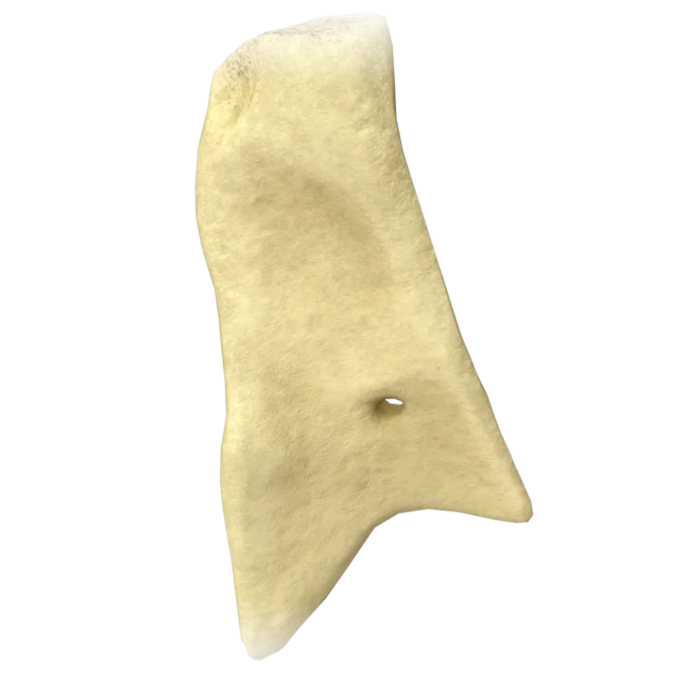
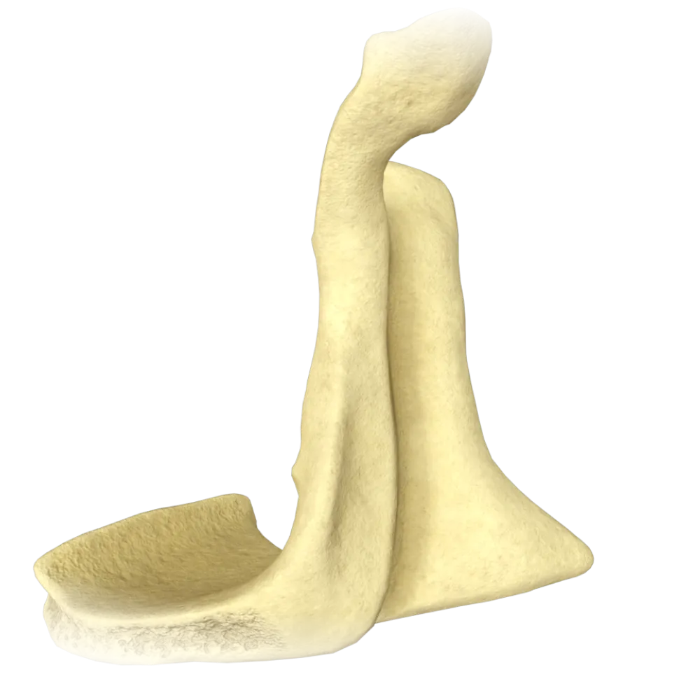
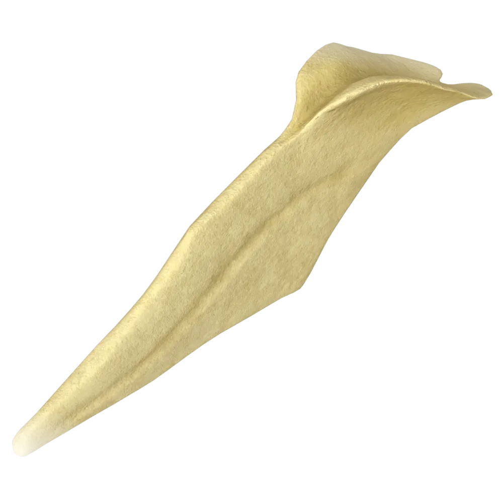
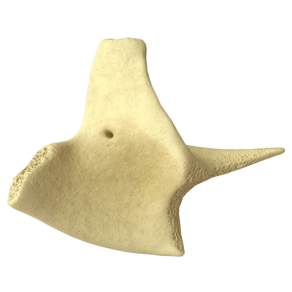
The lacrimal bone is a thin quadrangular osseous plate and is one of the smallest and most fragile bones of the viscerocranium.
-
Trapezoidal with prominent tubercles
The lacrimal bone is a thin quadrangular osseous plate and is one of the smallest and most fragile bones of the viscerocranium.
-
A thin quadrangular plate
The lacrimal bone is a thin quadrangular osseous plate and is one of the smallest and most fragile bones of the viscerocranium.
-
Triangular with a sharp inferior border
The lacrimal bone is a thin quadrangular osseous plate and is one of the smallest and most fragile bones of the viscerocranium.
-
I find it difficult to answer
The lacrimal bone is a thin quadrangular osseous plate and is one of the smallest and most fragile bones of the viscerocranium.
3. In which wall of the orbit is the lacrimal bone located?
-
Superior wall (roof) of the orbit
The lacrimal bone contributes to the formation of the medial wall of the orbit and is located between the frontal process of the maxilla anteriorly and the orbital plate of the ethmoid bone posteriorly.
-
Inferior wall (floor) of the orbit
The lacrimal bone contributes to the formation of the medial wall of the orbit and is located between the frontal process of the maxilla anteriorly and the orbital plate of the ethmoid bone posteriorly.
-
Lateral wall of the orbit
The lacrimal bone contributes to the formation of the medial wall of the orbit and is located between the frontal process of the maxilla anteriorly and the orbital plate of the ethmoid bone posteriorly.
-
Medial wall of the orbit
The lacrimal bone contributes to the formation of the medial wall of the orbit and is located between the frontal process of the maxilla anteriorly and the orbital plate of the ethmoid bone posteriorly.
-
I find it difficult to answer
The lacrimal bone contributes to the formation of the medial wall of the orbit and is located between the frontal process of the maxilla anteriorly and the orbital plate of the ethmoid bone posteriorly.
4. Which surface of the lacrimal bone faces the orbital cavity?
-
Nasal surface
The orbital surface (facies orbitalis) of the lacrimal bone faces the orbital cavity.
-
Orbital surface
The orbital surface (facies orbitalis) of the lacrimal bone faces the orbital cavity.
-
Facial surface
The orbital surface (facies orbitalis) of the lacrimal bone faces the orbital cavity.
-
Cerebral surface
The orbital surface (facies orbitalis) of the lacrimal bone faces the orbital cavity.
-
I find it difficult to answer
The orbital surface (facies orbitalis) of the lacrimal bone faces the orbital cavity.
5. What is the posteriorly located vertical crest on the orbital surface of the lacrimal bone called?
-
Anterior lacrimal crest
The orbital surface of the lacrimal bone bears the posterior lacrimal crest (crista lacrimalis posterior), anterior to which is the lacrimal groove, which contributes to the formation of the lacrimal sac fossa.
-
Ethmoidal crest
The orbital surface of the lacrimal bone bears the posterior lacrimal crest (crista lacrimalis posterior), anterior to which is the lacrimal groove, which contributes to the formation of the lacrimal sac fossa.
-
Posterior lacrimal crest
The orbital surface of the lacrimal bone bears the posterior lacrimal crest (crista lacrimalis posterior), anterior to which is the lacrimal groove, which contributes to the formation of the lacrimal sac fossa.
-
Nasal crest
The orbital surface of the lacrimal bone bears the posterior lacrimal crest (crista lacrimalis posterior), anterior to which is the lacrimal groove, which contributes to the formation of the lacrimal sac fossa.
-
I find it difficult to answer
The orbital surface of the lacrimal bone bears the posterior lacrimal crest (crista lacrimalis posterior), anterior to which is the lacrimal groove, which contributes to the formation of the lacrimal sac fossa.
6. The lacrimal sac fossa (fossa sacci lacrimalis) is formed by:
-
The lacrimal bone alone
The lacrimal sac fossa is formed by the posterior lacrimal crest of the lacrimal bone and the anterior lacrimal crest of the frontal process of the maxilla; the lacrimal groove lies between these crests.
-
The lacrimal bone and the frontal process of the maxilla
The lacrimal sac fossa is formed by the posterior lacrimal crest of the lacrimal bone and the anterior lacrimal crest of the frontal process of the maxilla; the lacrimal groove lies between these crests.
-
The lacrimal bone and the nasal bone
The lacrimal sac fossa is formed by the posterior lacrimal crest of the lacrimal bone and the anterior lacrimal crest of the frontal process of the maxilla; the lacrimal groove lies between these crests.
-
The lacrimal bone and the orbital plate of the ethmoid bone
The lacrimal sac fossa is formed by the posterior lacrimal crest of the lacrimal bone and the anterior lacrimal crest of the frontal process of the maxilla; the lacrimal groove lies between these crests.
-
I find it difficult to answer
The lacrimal sac fossa is formed by the posterior lacrimal crest of the lacrimal bone and the anterior lacrimal crest of the frontal process of the maxilla; the lacrimal groove lies between these crests.
7. Which anatomical feature makes the medial wall of the orbital cavity (orbit) the thinnest and most fragile?
-
It is formed exclusively by the thick squamous part of the frontal bone
Most of the medial (internal) wall of the orbit is formed by the lacrimal bone and the orbital (paper) plate of the ethmoid bone (lamina papyracea). It derives its designation “paper” specifically from its extremely small thickness—only 0.2 to 0.4 mm. Consequently, the medial wall is easily damaged by blunt trauma to the eye.
-
It is formed by the most fragile bones of cranium: the lacrimal bone and the orbital plate of the ethmoid bone
Most of the medial (internal) wall of the orbit is formed by the lacrimal bone and the orbital (paper) plate of the ethmoid bone (lamina papyracea). It derives its designation “paper” specifically from its extremely small thickness—only 0.2 to 0.4 mm. Consequently, the medial wall is easily damaged by blunt trauma to the eye.
-
It consists entirely of dense cartilaginous tissue without an osseous basis
Most of the medial (internal) wall of the orbit is formed by the lacrimal bone and the orbital (paper) plate of the ethmoid bone (lamina papyracea). It derives its designation “paper” specifically from its extremely small thickness—only 0.2 to 0.4 mm. Consequently, the medial wall is easily damaged by blunt trauma to the eye.
-
A large infraorbital canal passes through it, compromising the structural integrity of the bone
Most of the medial (internal) wall of the orbit is formed by the lacrimal bone and the orbital (paper) plate of the ethmoid bone (lamina papyracea). It derives its designation “paper” specifically from its extremely small thickness—only 0.2 to 0.4 mm. Consequently, the medial wall is easily damaged by blunt trauma to the eye.
-
I find it difficult to answer
Most of the medial (internal) wall of the orbit is formed by the lacrimal bone and the orbital (paper) plate of the ethmoid bone (lamina papyracea). It derives its designation “paper” specifically from its extremely small thickness—only 0.2 to 0.4 mm. Consequently, the medial wall is easily damaged by blunt trauma to the eye.
8. With which bone does the lacrimal bone articulate via the frontolacrimal suture?
-
With the nasal bone
The superior border of the lacrimal bone articulates with the nasal part of the frontal bone, forming the frontolacrimal suture (sutura frontolacrimalis).
-
With the maxilla
The superior border of the lacrimal bone articulates with the nasal part of the frontal bone, forming the frontolacrimal suture (sutura frontolacrimalis).
-
With the frontal bone
The superior border of the lacrimal bone articulates with the nasal part of the frontal bone, forming the frontolacrimal suture (sutura frontolacrimalis).
-
With the ethmoid bone
The superior border of the lacrimal bone articulates with the nasal part of the frontal bone, forming the frontolacrimal suture (sutura frontolacrimalis).
-
I find it difficult to answer
The superior border of the lacrimal bone articulates with the nasal part of the frontal bone, forming the frontolacrimal suture (sutura frontolacrimalis).
9. With which bone does the lacrimal bone articulate via the lacrimoethmoidal suture?
-
With the palatine bone
The posterior border of the lacrimal bone articulates with the orbital plate of the ethmoid bone via the lacrimoethmoidal suture (sutura lacrimoethmoidalis).
-
With the ethmoid bone
The posterior border of the lacrimal bone articulates with the orbital plate of the ethmoid bone via the lacrimoethmoidal suture (sutura lacrimoethmoidalis).
-
With the nasal bone
The posterior border of the lacrimal bone articulates with the orbital plate of the ethmoid bone via the lacrimoethmoidal suture (sutura lacrimoethmoidalis).
-
With the inferior turbinate
The posterior border of the lacrimal bone articulates with the orbital plate of the ethmoid bone via the lacrimoethmoidal suture (sutura lacrimoethmoidalis).
-
I find it difficult to answer
The posterior border of the lacrimal bone articulates with the orbital plate of the ethmoid bone via the lacrimoethmoidal suture (sutura lacrimoethmoidalis).
10. With which bone does the lacrimal bone articulate along its anterior border?
-
With the frontal bone
The anterior border of the lacrimal bone forms a suture with the frontal process of the maxilla (sutura lacrimomaxillaris).
-
With the sphenoid bone
The anterior border of the lacrimal bone forms a suture with the frontal process of the maxilla (sutura lacrimomaxillaris).
-
With the frontal process of the maxilla
The anterior border of the lacrimal bone forms a suture with the frontal process of the maxilla (sutura lacrimomaxillaris).
-
With the nasal bone
The anterior border of the lacrimal bone forms a suture with the frontal process of the maxilla (sutura lacrimomaxillaris).
-
I find it difficult to answer
The anterior border of the lacrimal bone forms a suture with the frontal process of the maxilla (sutura lacrimomaxillaris).
11. The lacrimal bone (os lacrimale) contributes to the formation of which wall of the nasal cavity?
-
It contributes to the formation of the nasal septum
The nasal surface of the lacrimal bone faces the nasal cavity and contributes to the formation of the lateral (side) wall of the nasal cavity in the region of the middle nasal meatus.
-
It contributes to the formation of the lateral wall of the nasal cavity
The nasal surface of the lacrimal bone faces the nasal cavity and contributes to the formation of the lateral (side) wall of the nasal cavity in the region of the middle nasal meatus.
-
It contributes to the formation of the floor of the nasal cavity
The nasal surface of the lacrimal bone faces the nasal cavity and contributes to the formation of the lateral (side) wall of the nasal cavity in the region of the middle nasal meatus.
-
It contributes to the formation of the roof of the nasal cavity
The nasal surface of the lacrimal bone faces the nasal cavity and contributes to the formation of the lateral (side) wall of the nasal cavity in the region of the middle nasal meatus.
-
I find it difficult to answer
The nasal surface of the lacrimal bone faces the nasal cavity and contributes to the formation of the lateral (side) wall of the nasal cavity in the region of the middle nasal meatus.
12. The nasal surface of the lacrimal bone bears a crest that articulates with a nasal concha. What is it called?
-
Ethmoidal crest
The conchal crest (crista conchalis) is located on the nasal surface of the lacrimal bone and articulates with the anterior end of the inferior turbinate.
-
Lacrimal crest
The conchal crest (crista conchalis) is located on the nasal surface of the lacrimal bone and articulates with the anterior end of the inferior turbinate.
-
Conchal crest
The conchal crest (crista conchalis) is located on the nasal surface of the lacrimal bone and articulates with the anterior end of the inferior turbinate.
-
Nasal crest
The conchal crest (crista conchalis) is located on the nasal surface of the lacrimal bone and articulates with the anterior end of the inferior turbinate.
-
I find it difficult to answer
The conchal crest (crista conchalis) is located on the nasal surface of the lacrimal bone and articulates with the anterior end of the inferior turbinate.
13. The lacrimomaxillary suture is located:
-
On the medial wall of the orbit between the lacrimal bone and the frontal process of the maxilla
The lacrimomaxillary suture (sutura lacrimomaxillaris) extends along the medial wall of the orbit between the anterior border of the lacrimal bone and the frontal process of the maxilla.
-
On the inferior wall of the orbit
The lacrimomaxillary suture (sutura lacrimomaxillaris) extends along the medial wall of the orbit between the anterior border of the lacrimal bone and the frontal process of the maxilla.
-
At the anterior border of the lacrimal bone, with the nasal bone
The lacrimomaxillary suture (sutura lacrimomaxillaris) extends along the medial wall of the orbit between the anterior border of the lacrimal bone and the frontal process of the maxilla.
-
Between the lacrimal bone and the palatine bone
The lacrimomaxillary suture (sutura lacrimomaxillaris) extends along the medial wall of the orbit between the anterior border of the lacrimal bone and the frontal process of the maxilla.
-
I find it difficult to answer
The lacrimomaxillary suture (sutura lacrimomaxillaris) extends along the medial wall of the orbit between the anterior border of the lacrimal bone and the frontal process of the maxilla.
14. Which opening is formed with the participation of the lacrimal hook and the maxilla?
-
Inferior orbital foramen
The lacrimal hook of the lacrimal bone, together with the lacrimal notch of the maxilla, forms the superior (orbital) opening of the nasolacrimal canal (canalis nasolacrimalis).
-
Cribriform foramen
The lacrimal hook of the lacrimal bone, together with the lacrimal notch of the maxilla, forms the superior (orbital) opening of the nasolacrimal canal (canalis nasolacrimalis).
-
Superior opening of the nasolacrimal canal
The lacrimal hook of the lacrimal bone, together with the lacrimal notch of the maxilla, forms the superior (orbital) opening of the nasolacrimal canal (canalis nasolacrimalis).
-
Lacrimal foramen of the nasal septum
The lacrimal hook of the lacrimal bone, together with the lacrimal notch of the maxilla, forms the superior (orbital) opening of the nasolacrimal canal (canalis nasolacrimalis).
-
I find it difficult to answer
The lacrimal hook of the lacrimal bone, together with the lacrimal notch of the maxilla, forms the superior (orbital) opening of the nasolacrimal canal (canalis nasolacrimalis).
15. The inferior part of the nasolacrimal canal opens into the:
-
Superior nasal meatus
The nasolacrimal canal opens through its inferior aperture into the inferior nasal meatus (meatus nasi inferior), beneath the anterior part of the inferior turbinate.
-
Middle nasal meatus
The nasolacrimal canal opens through its inferior aperture into the inferior nasal meatus (meatus nasi inferior), beneath the anterior part of the inferior turbinate.
-
Inferior nasal meatus
The nasolacrimal canal opens through its inferior aperture into the inferior nasal meatus (meatus nasi inferior), beneath the anterior part of the inferior turbinate.
-
Common nasal meatus
The nasolacrimal canal opens through its inferior aperture into the inferior nasal meatus (meatus nasi inferior), beneath the anterior part of the inferior turbinate.
-
I find it difficult to answer
The nasolacrimal canal opens through its inferior aperture into the inferior nasal meatus (meatus nasi inferior), beneath the anterior part of the inferior turbinate.
16. Which anatomical structure is located in the depression between the anterior and posterior lacrimal crests?
-
Infraorbital foramen (foramen infraorbitale)
The concave osseous depression formed between these crests is called the lacrimal sac fossa (fossa sacci lacrimalis). It contains the lacrimal sac.
-
Cribriform plate (lamina cribrosa)
The concave osseous depression formed between these crests is called the lacrimal sac fossa (fossa sacci lacrimalis). It contains the lacrimal sac.
-
Lacrimal sac fossa (fossa sacci lacrimalis)
The concave osseous depression formed between these crests is called the lacrimal sac fossa (fossa sacci lacrimalis). It contains the lacrimal sac.
-
Pterygopalatine fossa (fossa pterygopalatina)
The concave osseous depression formed between these crests is called the lacrimal sac fossa (fossa sacci lacrimalis). It contains the lacrimal sac.
-
I find it difficult to answer
The concave osseous depression formed between these crests is called the lacrimal sac fossa (fossa sacci lacrimalis). It contains the lacrimal sac.
17. Into which anatomical structure does the lacrimal sac fossa (fossa sacci lacrimalis) continue directly inferiorly?
-
Nasolacrimal canal (canalis nasolacrimalis)
The lacrimal sac fossa (fossa sacci lacrimalis), located in the anterior part of the medial wall of the orbit, narrows inferiorly and continues directly into the nasolacrimal canal (canalis nasolacrimalis).
-
Infraorbital groove (sulcus infraorbitalis)
The lacrimal sac fossa (fossa sacci lacrimalis), located in the anterior part of the medial wall of the orbit, narrows inferiorly and continues directly into the nasolacrimal canal (canalis nasolacrimalis).
-
Superior orbital fissure (fissura orbitalis superior)
The lacrimal sac fossa (fossa sacci lacrimalis), located in the anterior part of the medial wall of the orbit, narrows inferiorly and continues directly into the nasolacrimal canal (canalis nasolacrimalis).
-
Pterygopalatine fossa (fossa pterygopalatina)
The lacrimal sac fossa (fossa sacci lacrimalis), located in the anterior part of the medial wall of the orbit, narrows inferiorly and continues directly into the nasolacrimal canal (canalis nasolacrimalis).
-
I find it difficult to answer
The lacrimal sac fossa (fossa sacci lacrimalis), located in the anterior part of the medial wall of the orbit, narrows inferiorly and continues directly into the nasolacrimal canal (canalis nasolacrimalis).
18. To which structural type does the lacrimal bone belong?
-
Cancellous bone
The lacrimal bone is classified as a flat bone. It is a thin osseous plate consisting of two layers of compact bone with a minimal amount of cancellous bone between them.
-
Flat bone
The lacrimal bone is classified as a flat bone. It is a thin osseous plate consisting of two layers of compact bone with a minimal amount of cancellous bone between them.
-
Tubular bone
The lacrimal bone is classified as a flat bone. It is a thin osseous plate consisting of two layers of compact bone with a minimal amount of cancellous bone between them.
-
Mixed bone
The lacrimal bone is classified as a flat bone. It is a thin osseous plate consisting of two layers of compact bone with a minimal amount of cancellous bone between them.
-
I find it difficult to answer
The lacrimal bone is classified as a flat bone. It is a thin osseous plate consisting of two layers of compact bone with a minimal amount of cancellous bone between them.
19. Which of the following sutures is NOT a suture of the lacrimal bone?
-
Sutura frontolacrimalis
Sutura temporozygomatica (temporozygomatic suture) connects the zygomatic process of the temporal bone with the zygomatic bone and is unrelated to the lacrimal bone. The lacrimal bone forms sutures with the frontal, ethmoid, and maxillary bones.
-
Sutura lacrimoethmoidalis
Sutura temporozygomatica (temporozygomatic suture) connects the zygomatic process of the temporal bone with the zygomatic bone and is unrelated to the lacrimal bone. The lacrimal bone forms sutures with the frontal, ethmoid, and maxillary bones.
-
Sutura lacrimomaxillaris
Sutura temporozygomatica (temporozygomatic suture) connects the zygomatic process of the temporal bone with the zygomatic bone and is unrelated to the lacrimal bone. The lacrimal bone forms sutures with the frontal, ethmoid, and maxillary bones.
-
Sutura temporozygomatica
Sutura temporozygomatica (temporozygomatic suture) connects the zygomatic process of the temporal bone with the zygomatic bone and is unrelated to the lacrimal bone. The lacrimal bone forms sutures with the frontal, ethmoid, and maxillary bones.
-
I find it difficult to answer
Sutura temporozygomatica (temporozygomatic suture) connects the zygomatic process of the temporal bone with the zygomatic bone and is unrelated to the lacrimal bone. The lacrimal bone forms sutures with the frontal, ethmoid, and maxillary bones.
20. The lacrimal groove (sulcus lacrimalis) of the lacrimal bone is located:
-
Posterior to the posterior lacrimal crest
The lacrimal groove is located anterior to the posterior lacrimal crest on the orbital surface of the lacrimal bone. Together with the corresponding groove of the frontal process of the maxilla, it forms the lacrimal sac fossa.
-
Anterior to the posterior lacrimal crest
The lacrimal groove is located anterior to the posterior lacrimal crest on the orbital surface of the lacrimal bone. Together with the corresponding groove of the frontal process of the maxilla, it forms the lacrimal sac fossa.
-
On the nasal surface of the bone
The lacrimal groove is located anterior to the posterior lacrimal crest on the orbital surface of the lacrimal bone. Together with the corresponding groove of the frontal process of the maxilla, it forms the lacrimal sac fossa.
-
On the inferior border of the bone in the region of the lacrimal hook
The lacrimal groove is located anterior to the posterior lacrimal crest on the orbital surface of the lacrimal bone. Together with the corresponding groove of the frontal process of the maxilla, it forms the lacrimal sac fossa.
-
I find it difficult to answer
The lacrimal groove is located anterior to the posterior lacrimal crest on the orbital surface of the lacrimal bone. Together with the corresponding groove of the frontal process of the maxilla, it forms the lacrimal sac fossa.
Retake this quiz?
Your current progress will be reset.
