
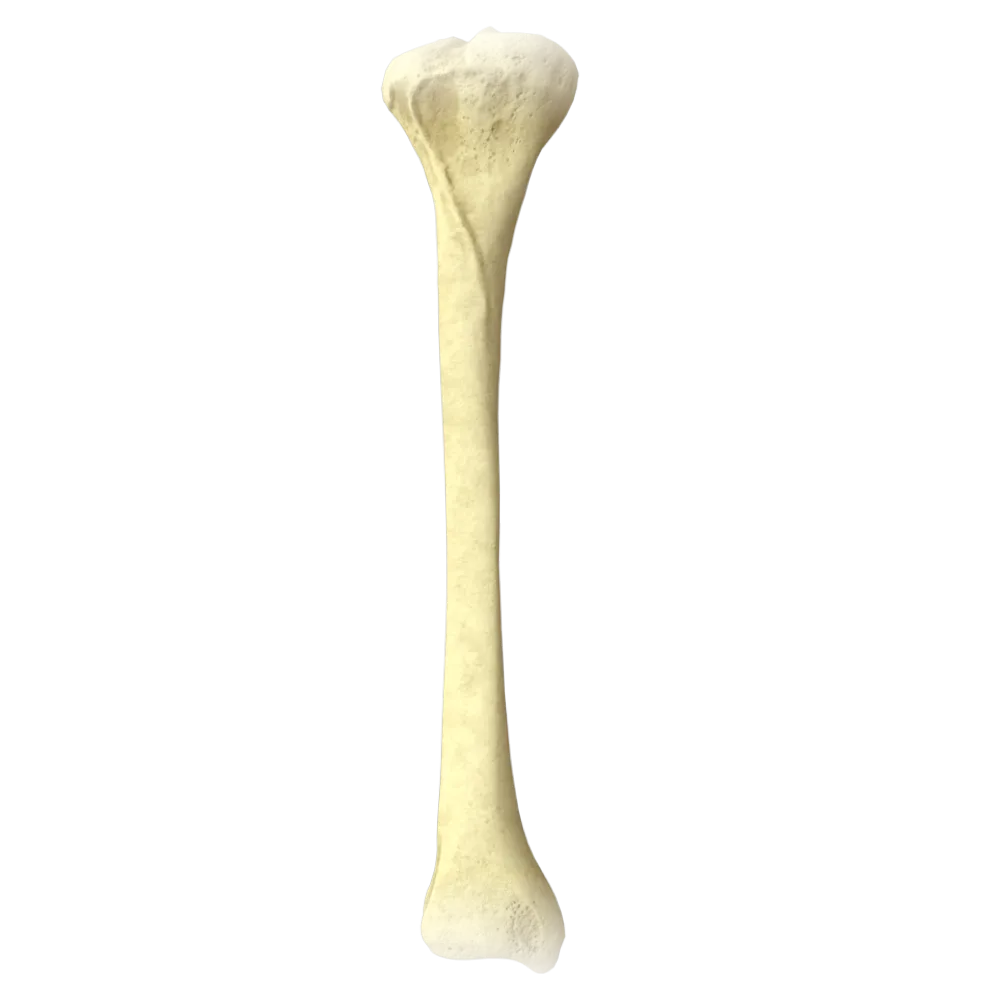
Tibia anatomy quiz
Quiz on the normal anatomy of the tibia: structure, articular surfaces, muscle attachments, vessels, and nerves.
1/20
bold
text
1. Which surface of the tibia is readily palpable beneath the skin along the entire length of the bone?
-
Anterior surface
The medial surface of the tibia has no muscular covering and is covered only by skin and subcutaneous tissue; therefore, it is readily palpable along the entire length of the leg. The anterior and lateral surfaces are covered by muscles, and the posterior surface by the deep muscles of the posterior compartment of the leg.
-
Lateral surface
The medial surface of the tibia has no muscular covering and is covered only by skin and subcutaneous tissue; therefore, it is readily palpable along the entire length of the leg. The anterior and lateral surfaces are covered by muscles, and the posterior surface by the deep muscles of the posterior compartment of the leg.
-
Medial surface
The medial surface of the tibia has no muscular covering and is covered only by skin and subcutaneous tissue; therefore, it is readily palpable along the entire length of the leg. The anterior and lateral surfaces are covered by muscles, and the posterior surface by the deep muscles of the posterior compartment of the leg.
-
Posterior surface
The medial surface of the tibia has no muscular covering and is covered only by skin and subcutaneous tissue; therefore, it is readily palpable along the entire length of the leg. The anterior and lateral surfaces are covered by muscles, and the posterior surface by the deep muscles of the posterior compartment of the leg.
-
I find it difficult to answer
The medial surface of the tibia has no muscular covering and is covered only by skin and subcutaneous tissue; therefore, it is readily palpable along the entire length of the leg. The anterior and lateral surfaces are covered by muscles, and the posterior surface by the deep muscles of the posterior compartment of the leg.
2. Which bony landmark of the tibia is the attachment site of the patellar ligament (lig. patellae)?
-
Medial condyle of the tibia
The patellar ligament (lig. patellae), which is a continuation of the quadriceps femoris tendon, attaches to the tibial tuberosity (tuberositas tibiae), located on the anterior surface of the proximal epiphysis of the bone.
-
Tibial tuberosity
The patellar ligament (lig. patellae), which is a continuation of the quadriceps femoris tendon, attaches to the tibial tuberosity (tuberositas tibiae), located on the anterior surface of the proximal epiphysis of the bone.
-
Intercondylar eminence
The patellar ligament (lig. patellae), which is a continuation of the quadriceps femoris tendon, attaches to the tibial tuberosity (tuberositas tibiae), located on the anterior surface of the proximal epiphysis of the bone.
-
Lateral condyle of tibia
The patellar ligament (lig. patellae), which is a continuation of the quadriceps femoris tendon, attaches to the tibial tuberosity (tuberositas tibiae), located on the anterior surface of the proximal epiphysis of the bone.
-
I find it difficult to answer
The patellar ligament (lig. patellae), which is a continuation of the quadriceps femoris tendon, attaches to the tibial tuberosity (tuberositas tibiae), located on the anterior surface of the proximal epiphysis of the bone.
3. What is the shape of the articular surface of the medial condyle of the tibia?
-
Convex (convex)
The medial articular surface of the tibia (facies articularis superior medialis) is concave. This corresponds to the convex medial condyle of the femur and, together with the medial meniscus, ensures congruence of the knee joint.
-
Flat
The medial articular surface of the tibia (facies articularis superior medialis) is concave. This corresponds to the convex medial condyle of the femur and, together with the medial meniscus, ensures congruence of the knee joint.
-
Concave (concave)
The medial articular surface of the tibia (facies articularis superior medialis) is concave. This corresponds to the convex medial condyle of the femur and, together with the medial meniscus, ensures congruence of the knee joint.
-
Saddle-shaped
The medial articular surface of the tibia (facies articularis superior medialis) is concave. This corresponds to the convex medial condyle of the femur and, together with the medial meniscus, ensures congruence of the knee joint.
-
I find it difficult to answer
The medial articular surface of the tibia (facies articularis superior medialis) is concave. This corresponds to the convex medial condyle of the femur and, together with the medial meniscus, ensures congruence of the knee joint.
4. Where on the tibia is the sulcus malleolaris (groove of the medial malleolus) located?
-
On the anterior surface of the medial malleolus
The sulcus malleolaris is located on the posterior surface of the medial malleolus.
-
On the posterior surface of the medial malleolus
The sulcus malleolaris is located on the posterior surface of the medial malleolus.
-
On the lateral surface of the distal epiphysis
The sulcus malleolaris is located on the posterior surface of the medial malleolus.
-
On the medial surface of the shaft
The sulcus malleolaris is located on the posterior surface of the medial malleolus.
-
I find it difficult to answer
The sulcus malleolaris is located on the posterior surface of the medial malleolus.
5. To which feature of the tibia does the iliotibial tract (iliotibial tract) attach?
-
Tibial tuberosity
The iliotibial tract (tractus iliotibialis) attaches to the lateral condyle of the tibia in the region of the so-called Gerdy’s tubercle (tuberculum tractus iliotibialis). This is an important anatomical landmark on the proximal epiphysis.
-
Intercondylar eminence
The iliotibial tract (tractus iliotibialis) attaches to the lateral condyle of the tibia in the region of the so-called Gerdy’s tubercle (tuberculum tractus iliotibialis). This is an important anatomical landmark on the proximal epiphysis.
-
Lateral condyle of the tibia (Gerdy’s tubercle)
The iliotibial tract (tractus iliotibialis) attaches to the lateral condyle of the tibia in the region of the so-called Gerdy’s tubercle (tuberculum tractus iliotibialis). This is an important anatomical landmark on the proximal epiphysis.
-
Medial condyle of the tibia
The iliotibial tract (tractus iliotibialis) attaches to the lateral condyle of the tibia in the region of the so-called Gerdy’s tubercle (tuberculum tractus iliotibialis). This is an important anatomical landmark on the proximal epiphysis.
-
I find it difficult to answer
The iliotibial tract (tractus iliotibialis) attaches to the lateral condyle of the tibia in the region of the so-called Gerdy’s tubercle (tuberculum tractus iliotibialis). This is an important anatomical landmark on the proximal epiphysis.
6. Which of the following structures is located between the two articular surfaces of the proximal epiphysis of the tibia?
-
Tibial tuberosity
The intercondylar eminence (eminentia intercondylaris) is located between the medial and lateral articular surfaces of the proximal epiphysis of the tibia and comprises medial and lateral intercondylar tubercles, to which the cruciate ligaments and horns of the menisci attach.
-
Intercondylar eminence (eminentia intercondylaris)
The intercondylar eminence (eminentia intercondylaris) is located between the medial and lateral articular surfaces of the proximal epiphysis of the tibia and comprises medial and lateral intercondylar tubercles, to which the cruciate ligaments and horns of the menisci attach.
-
Fibular notch
The intercondylar eminence (eminentia intercondylaris) is located between the medial and lateral articular surfaces of the proximal epiphysis of the tibia and comprises medial and lateral intercondylar tubercles, to which the cruciate ligaments and horns of the menisci attach.
-
Medial malleolus
The intercondylar eminence (eminentia intercondylaris) is located between the medial and lateral articular surfaces of the proximal epiphysis of the tibia and comprises medial and lateral intercondylar tubercles, to which the cruciate ligaments and horns of the menisci attach.
-
I find it difficult to answer
The intercondylar eminence (eminentia intercondylaris) is located between the medial and lateral articular surfaces of the proximal epiphysis of the tibia and comprises medial and lateral intercondylar tubercles, to which the cruciate ligaments and horns of the menisci attach.
7. Which muscle attaches to the medial surface of the tibia as part of the “goose’s foot” (pes anserinus)?
-
Sartorius muscle (m. sartorius)
The “goose’s foot” (pes anserinus) comprises three muscles: the sartorius (m. sartorius), gracilis (m. gracilis), and semitendinosus (m. semitendinosus). They attach to the medial surface of the proximal metaphysis of the tibia inferior to the medial condyle. The semimembranosus muscle attaches separately.
-
Rectus femoris muscle (m. rectus femoris)
The “goose’s foot” (pes anserinus) comprises three muscles: the sartorius (m. sartorius), gracilis (m. gracilis), and semitendinosus (m. semitendinosus). They attach to the medial surface of the proximal metaphysis of the tibia inferior to the medial condyle. The semimembranosus muscle attaches separately.
-
Biceps femoris muscle (m. biceps femoris)
The “goose’s foot” (pes anserinus) comprises three muscles: the sartorius (m. sartorius), gracilis (m. gracilis), and semitendinosus (m. semitendinosus). They attach to the medial surface of the proximal metaphysis of the tibia inferior to the medial condyle. The semimembranosus muscle attaches separately.
-
Semimembranosus muscle (m. semimembranosus)
The “goose’s foot” (pes anserinus) comprises three muscles: the sartorius (m. sartorius), gracilis (m. gracilis), and semitendinosus (m. semitendinosus). They attach to the medial surface of the proximal metaphysis of the tibia inferior to the medial condyle. The semimembranosus muscle attaches separately.
-
I find it difficult to answer
The “goose’s foot” (pes anserinus) comprises three muscles: the sartorius (m. sartorius), gracilis (m. gracilis), and semitendinosus (m. semitendinosus). They attach to the medial surface of the proximal metaphysis of the tibia inferior to the medial condyle. The semimembranosus muscle attaches separately.
8. Which artery is the main source of blood supply to the tibial diaphysis through the nutrient (nutritive) foramen?
-
Anterior tibial artery (a. tibialis anterior)
The nutrient artery of the tibia (a. nutricia tibiae) is a branch of the posterior tibial artery (a. tibialis posterior). It enters the bone through the nutrient foramen on the posterior surface of the diaphysis in its proximal third and supplies the bone marrow and endosteum.
-
Fibular artery (a. peronea)
The nutrient artery of the tibia (a. nutricia tibiae) is a branch of the posterior tibial artery (a. tibialis posterior). It enters the bone through the nutrient foramen on the posterior surface of the diaphysis in its proximal third and supplies the bone marrow and endosteum.
-
Posterior tibial artery (a. tibialis posterior)
The nutrient artery of the tibia (a. nutricia tibiae) is a branch of the posterior tibial artery (a. tibialis posterior). It enters the bone through the nutrient foramen on the posterior surface of the diaphysis in its proximal third and supplies the bone marrow and endosteum.
-
Popliteal artery (a. poplitea)
The nutrient artery of the tibia (a. nutricia tibiae) is a branch of the posterior tibial artery (a. tibialis posterior). It enters the bone through the nutrient foramen on the posterior surface of the diaphysis in its proximal third and supplies the bone marrow and endosteum.
-
I find it difficult to answer
The nutrient artery of the tibia (a. nutricia tibiae) is a branch of the posterior tibial artery (a. tibialis posterior). It enters the bone through the nutrient foramen on the posterior surface of the diaphysis in its proximal third and supplies the bone marrow and endosteum.
9. Which nerve passes through the interosseous space of the leg, lying directly on the lateral surface of the tibia?
-
Tibial nerve (n. tibialis)
The deep fibular nerve (n. peroneus profundus) passes through the anterior fascial compartment of the leg together with the anterior tibial artery, lying on the interosseous membrane directly adjacent to the lateral surface of the tibia. It innervates the muscles of the anterior compartment of the leg.
-
Deep fibular nerve (n. peroneus profundus)
The deep fibular nerve (n. peroneus profundus) passes through the anterior fascial compartment of the leg together with the anterior tibial artery, lying on the interosseous membrane directly adjacent to the lateral surface of the tibia. It innervates the muscles of the anterior compartment of the leg.
-
Superficial fibular nerve (n. peroneus superficialis)
The deep fibular nerve (n. peroneus profundus) passes through the anterior fascial compartment of the leg together with the anterior tibial artery, lying on the interosseous membrane directly adjacent to the lateral surface of the tibia. It innervates the muscles of the anterior compartment of the leg.
-
Saphenous nerve (n. saphenus)
The deep fibular nerve (n. peroneus profundus) passes through the anterior fascial compartment of the leg together with the anterior tibial artery, lying on the interosseous membrane directly adjacent to the lateral surface of the tibia. It innervates the muscles of the anterior compartment of the leg.
-
I find it difficult to answer
The deep fibular nerve (n. peroneus profundus) passes through the anterior fascial compartment of the leg together with the anterior tibial artery, lying on the interosseous membrane directly adjacent to the lateral surface of the tibia. It innervates the muscles of the anterior compartment of the leg.
10. Which fibrous joint (syndesmosis) connects the tibia and fibula along the entire length of their diaphyses?
-
Syndesmosis tibiofibularis
The diaphyses of the tibia and fibula are connected by the interosseous membrane of the leg (membrana interossea cruris), which extends between the interosseous borders of the two bones. Syndesmosis tibiofibularis is the distal articulation, whereas lig. tibiofibulare anterius is part of it.
-
Membrana interossea cruris
The diaphyses of the tibia and fibula are connected by the interosseous membrane of the leg (membrana interossea cruris), which extends between the interosseous borders of the two bones. Syndesmosis tibiofibularis is the distal articulation, whereas lig. tibiofibulare anterius is part of it.
-
Ligamentum tibiofibulare anterius
The diaphyses of the tibia and fibula are connected by the interosseous membrane of the leg (membrana interossea cruris), which extends between the interosseous borders of the two bones. Syndesmosis tibiofibularis is the distal articulation, whereas lig. tibiofibulare anterius is part of it.
-
Ligamentum collaterale fibulare
The diaphyses of the tibia and fibula are connected by the interosseous membrane of the leg (membrana interossea cruris), which extends between the interosseous borders of the two bones. Syndesmosis tibiofibularis is the distal articulation, whereas lig. tibiofibulare anterius is part of it.
-
I find it difficult to answer
The diaphyses of the tibia and fibula are connected by the interosseous membrane of the leg (membrana interossea cruris), which extends between the interosseous borders of the two bones. Syndesmosis tibiofibularis is the distal articulation, whereas lig. tibiofibulare anterius is part of it.
11. Which process forms the medial malleolus and contributes to the ankle joint?
-
Lateral condyle of tibia
The medial malleolus (malleolus medialis) is the distal medial process of the tibia.
-
Posterior process of talus
The medial malleolus (malleolus medialis) is the distal medial process of the tibia.
-
Medial malleolus (malleolus medialis) of the tibia
The medial malleolus (malleolus medialis) is the distal medial process of the tibia.
-
Fibular notch
The medial malleolus (malleolus medialis) is the distal medial process of the tibia.
-
I find it difficult to answer
The medial malleolus (malleolus medialis) is the distal medial process of the tibia.
12. Which muscle originates from the posterior surface of the tibia inferior to the linea musculi solei?
-
Soleus muscle (m. soleus)
The flexor digitorum longus muscle (m. flexor digitorum longus) originates from the posterior surface of the tibia inferior to the soleal line (linea musculi solei). The soleus muscle originates superiorly from the same line and from the posterior surface of the head of the fibula.
-
Gastrocnemius muscle (m. gastrocnemius)
The flexor digitorum longus muscle (m. flexor digitorum longus) originates from the posterior surface of the tibia inferior to the soleal line (linea musculi solei). The soleus muscle originates superiorly from the same line and from the posterior surface of the head of the fibula.
-
Flexor digitorum longus muscle (m. flexor digitorum longus)
The flexor digitorum longus muscle (m. flexor digitorum longus) originates from the posterior surface of the tibia inferior to the soleal line (linea musculi solei). The soleus muscle originates superiorly from the same line and from the posterior surface of the head of the fibula.
-
Plantaris muscle (m. plantaris)
The flexor digitorum longus muscle (m. flexor digitorum longus) originates from the posterior surface of the tibia inferior to the soleal line (linea musculi solei). The soleus muscle originates superiorly from the same line and from the posterior surface of the head of the fibula.
-
I find it difficult to answer
The flexor digitorum longus muscle (m. flexor digitorum longus) originates from the posterior surface of the tibia inferior to the soleal line (linea musculi solei). The soleus muscle originates superiorly from the same line and from the posterior surface of the head of the fibula.
13. Which ligament attaches to the anterior intercondylar area (area intercondylaris anterior) of the tibia?
-
Posterior cruciate ligament
The anterior cruciate ligament (lig. cruciatum anterius) attaches to the anterior intercondylar area of the tibia (area intercondylaris anterior). The posterior cruciate ligament attaches to the posterior intercondylar area (area intercondylaris posterior).
-
Anterior cruciate ligament
The anterior cruciate ligament (lig. cruciatum anterius) attaches to the anterior intercondylar area of the tibia (area intercondylaris anterior). The posterior cruciate ligament attaches to the posterior intercondylar area (area intercondylaris posterior).
-
Fibular collateral ligament
The anterior cruciate ligament (lig. cruciatum anterius) attaches to the anterior intercondylar area of the tibia (area intercondylaris anterior). The posterior cruciate ligament attaches to the posterior intercondylar area (area intercondylaris posterior).
-
Transverse ligament of the knee (lig. transversum genus)
The anterior cruciate ligament (lig. cruciatum anterius) attaches to the anterior intercondylar area of the tibia (area intercondylaris anterior). The posterior cruciate ligament attaches to the posterior intercondylar area (area intercondylaris posterior).
-
I find it difficult to answer
The anterior cruciate ligament (lig. cruciatum anterius) attaches to the anterior intercondylar area of the tibia (area intercondylaris anterior). The posterior cruciate ligament attaches to the posterior intercondylar area (area intercondylaris posterior).
14. Which border of the tibia serves as an origin site for the tibialis anterior muscle (m. tibialis anterior)?
-
Medial border (margo medialis)
The tibialis anterior muscle (m. tibialis anterior) originates from the lateral surface of the tibia and the interosseous membrane. Its origin is bounded superiorly by the interosseous border (margo interosseus). The interosseous border is the attachment site of the interosseous membrane and separates the anterior and posterior muscle compartments of the leg.
-
Posterior border (margo posterior)
The tibialis anterior muscle (m. tibialis anterior) originates from the lateral surface of the tibia and the interosseous membrane. Its origin is bounded superiorly by the interosseous border (margo interosseus). The interosseous border is the attachment site of the interosseous membrane and separates the anterior and posterior muscle compartments of the leg.
-
Interosseous border (margo interosseus)
The tibialis anterior muscle (m. tibialis anterior) originates from the lateral surface of the tibia and the interosseous membrane. Its origin is bounded superiorly by the interosseous border (margo interosseus). The interosseous border is the attachment site of the interosseous membrane and separates the anterior and posterior muscle compartments of the leg.
-
Linea musculi solei
The tibialis anterior muscle (m. tibialis anterior) originates from the lateral surface of the tibia and the interosseous membrane. Its origin is bounded superiorly by the interosseous border (margo interosseus). The interosseous border is the attachment site of the interosseous membrane and separates the anterior and posterior muscle compartments of the leg.
-
I find it difficult to answer
The tibialis anterior muscle (m. tibialis anterior) originates from the lateral surface of the tibia and the interosseous membrane. Its origin is bounded superiorly by the interosseous border (margo interosseus). The interosseous border is the attachment site of the interosseous membrane and separates the anterior and posterior muscle compartments of the leg.
15. Which structure forms the distal articulation between the tibia and fibula?
-
Interosseous membrane of leg (membrana interossea cruris)
The distal ends of the tibia and fibula are connected by the distal tibiofibular syndesmosis (syndesmosis tibiofibularis distalis), reinforced by the anterior and posterior tibiofibular ligaments. This structure forms the ankle mortise.
-
Distal tibiofibular syndesmosis (syndesmosis tibiofibularis distalis)
The distal ends of the tibia and fibula are connected by the distal tibiofibular syndesmosis (syndesmosis tibiofibularis distalis), reinforced by the anterior and posterior tibiofibular ligaments. This structure forms the ankle mortise.
-
Head of fibula
The distal ends of the tibia and fibula are connected by the distal tibiofibular syndesmosis (syndesmosis tibiofibularis distalis), reinforced by the anterior and posterior tibiofibular ligaments. This structure forms the ankle mortise.
-
Proximal tibiofibular joint (articulatio tibiofibularis proximalis)
The distal ends of the tibia and fibula are connected by the distal tibiofibular syndesmosis (syndesmosis tibiofibularis distalis), reinforced by the anterior and posterior tibiofibular ligaments. This structure forms the ankle mortise.
-
I find it difficult to answer
The distal ends of the tibia and fibula are connected by the distal tibiofibular syndesmosis (syndesmosis tibiofibularis distalis), reinforced by the anterior and posterior tibiofibular ligaments. This structure forms the ankle mortise.
16. On which surface of the tibia is the linea musculi solei (soleal line) located?
-
Anterior surface
The linea musculi solei (soleal line) is located on the posterior surface of the tibia. It runs obliquely inferiorly and medially from the lateral condyle to the medial border of the bone. The m. soleus originates from this line and the area inferior to it, whereas part of the popliteus muscle originates superior to it.
-
Medial surface
The linea musculi solei (soleal line) is located on the posterior surface of the tibia. It runs obliquely inferiorly and medially from the lateral condyle to the medial border of the bone. The m. soleus originates from this line and the area inferior to it, whereas part of the popliteus muscle originates superior to it.
-
Lateral surface
The linea musculi solei (soleal line) is located on the posterior surface of the tibia. It runs obliquely inferiorly and medially from the lateral condyle to the medial border of the bone. The m. soleus originates from this line and the area inferior to it, whereas part of the popliteus muscle originates superior to it.
-
Posterior surface
The linea musculi solei (soleal line) is located on the posterior surface of the tibia. It runs obliquely inferiorly and medially from the lateral condyle to the medial border of the bone. The m. soleus originates from this line and the area inferior to it, whereas part of the popliteus muscle originates superior to it.
-
I find it difficult to answer
The linea musculi solei (soleal line) is located on the posterior surface of the tibia. It runs obliquely inferiorly and medially from the lateral condyle to the medial border of the bone. The m. soleus originates from this line and the area inferior to it, whereas part of the popliteus muscle originates superior to it.
17. Which inferior articular surface of the tibia articulates with the talus?
-
Facies articularis fibularis
The inferior articular surface of the tibia (facies articularis inferior tibiae) articulates with the trochlea of the talus (trochlea tali). It is rectangular, slightly concave in the sagittal direction, and forms the “roof” of the ankle joint.
-
Facies malleolaris medialis
The inferior articular surface of the tibia (facies articularis inferior tibiae) articulates with the trochlea of the talus (trochlea tali). It is rectangular, slightly concave in the sagittal direction, and forms the “roof” of the ankle joint.
-
Facies articularis inferior tibiae
The inferior articular surface of the tibia (facies articularis inferior tibiae) articulates with the trochlea of the talus (trochlea tali). It is rectangular, slightly concave in the sagittal direction, and forms the “roof” of the ankle joint.
-
Facies articularis superior tibiae
The inferior articular surface of the tibia (facies articularis inferior tibiae) articulates with the trochlea of the talus (trochlea tali). It is rectangular, slightly concave in the sagittal direction, and forms the “roof” of the ankle joint.
-
I find it difficult to answer
The inferior articular surface of the tibia (facies articularis inferior tibiae) articulates with the trochlea of the talus (trochlea tali). It is rectangular, slightly concave in the sagittal direction, and forms the “roof” of the ankle joint.
18. To which feature of the tibia does the posterior cruciate ligament attach?
-
Anterior intercondylar area (area intercondylaris anterior)
The posterior cruciate ligament (lig. cruciatum posterius) originates from the inner surface of the medial condyle of the femur and attaches to the posterior intercondylar area of the tibia (area intercondylaris posterior), partially extending onto the posterior surface of the bone.
-
Intercondylar eminence (eminentia intercondylaris)
The posterior cruciate ligament (lig. cruciatum posterius) originates from the inner surface of the medial condyle of the femur and attaches to the posterior intercondylar area of the tibia (area intercondylaris posterior), partially extending onto the posterior surface of the bone.
-
Posterior intercondylar area (area intercondylaris posterior)
The posterior cruciate ligament (lig. cruciatum posterius) originates from the inner surface of the medial condyle of the femur and attaches to the posterior intercondylar area of the tibia (area intercondylaris posterior), partially extending onto the posterior surface of the bone.
-
Posterior surface of the medial condyle
The posterior cruciate ligament (lig. cruciatum posterius) originates from the inner surface of the medial condyle of the femur and attaches to the posterior intercondylar area of the tibia (area intercondylaris posterior), partially extending onto the posterior surface of the bone.
-
I find it difficult to answer
The posterior cruciate ligament (lig. cruciatum posterius) originates from the inner surface of the medial condyle of the femur and attaches to the posterior intercondylar area of the tibia (area intercondylaris posterior), partially extending onto the posterior surface of the bone.
19. Which muscle attaches to the posterior surface of the medial condyle of the tibia?
-
Gracilis muscle (m. gracilis)
The semimembranosus muscle (m. semimembranosus) attaches via three expansions: the direct expansion to the posterior surface of the medial condyle of the tibia, the descending expansion to the fascia of the leg, and the ascending tendon to the lateral condyle of the femur as part of the oblique popliteal ligament.
-
Semitendinosus muscle (m. semitendinosus)
The semimembranosus muscle (m. semimembranosus) attaches via three expansions: the direct expansion to the posterior surface of the medial condyle of the tibia, the descending expansion to the fascia of the leg, and the ascending tendon to the lateral condyle of the femur as part of the oblique popliteal ligament.
-
Semimembranosus muscle (m. semimembranosus)
The semimembranosus muscle (m. semimembranosus) attaches via three expansions: the direct expansion to the posterior surface of the medial condyle of the tibia, the descending expansion to the fascia of the leg, and the ascending tendon to the lateral condyle of the femur as part of the oblique popliteal ligament.
-
Popliteus muscle (m. popliteus)
The semimembranosus muscle (m. semimembranosus) attaches via three expansions: the direct expansion to the posterior surface of the medial condyle of the tibia, the descending expansion to the fascia of the leg, and the ascending tendon to the lateral condyle of the femur as part of the oblique popliteal ligament.
-
I find it difficult to answer
The semimembranosus muscle (m. semimembranosus) attaches via three expansions: the direct expansion to the posterior surface of the medial condyle of the tibia, the descending expansion to the fascia of the leg, and the ascending tendon to the lateral condyle of the femur as part of the oblique popliteal ligament.
20. Which nutrient (nutritive) foramen of the tibia is located on the posterior surface of the diaphysis, and in which direction is it oriented?
-
Inferiorly (distally)
The nutrient foramen of the tibia (foramen nutricium tibiae) is located on the posterior surface of the diaphysis in the proximal third and is directed inferiorly (distally). This follows the general rule: the nutrient canals of the long bones of the lower limb are directed away from the knee joint—that is, distally in the tibia.
-
Superiorly (proximally)
The nutrient foramen of the tibia (foramen nutricium tibiae) is located on the posterior surface of the diaphysis in the proximal third and is directed inferiorly (distally). This follows the general rule: the nutrient canals of the long bones of the lower limb are directed away from the knee joint—that is, distally in the tibia.
-
Medially
The nutrient foramen of the tibia (foramen nutricium tibiae) is located on the posterior surface of the diaphysis in the proximal third and is directed inferiorly (distally). This follows the general rule: the nutrient canals of the long bones of the lower limb are directed away from the knee joint—that is, distally in the tibia.
-
Laterally
The nutrient foramen of the tibia (foramen nutricium tibiae) is located on the posterior surface of the diaphysis in the proximal third and is directed inferiorly (distally). This follows the general rule: the nutrient canals of the long bones of the lower limb are directed away from the knee joint—that is, distally in the tibia.
-
I find it difficult to answer
The nutrient foramen of the tibia (foramen nutricium tibiae) is located on the posterior surface of the diaphysis in the proximal third and is directed inferiorly (distally). This follows the general rule: the nutrient canals of the long bones of the lower limb are directed away from the knee joint—that is, distally in the tibia.
Retake this quiz?
Your current progress will be reset.
