
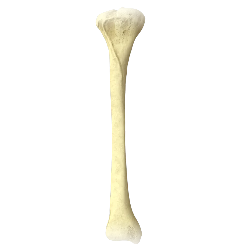
Femur anatomy quiz
The quiz covers the structure, parts, anatomical structures, and functional relationships of the femur.
1/20
bold
text
1. Which part of the femur belongs to its proximal epiphysis?
-
Medial condyle
The proximal epiphysis of the femur includes the head (caput ossis femoris), neck, greater trochanter, and lesser trochanter. The condyles and epicondyles belong to the distal epiphysis.
-
Patellar surface
The proximal epiphysis of the femur includes the head (caput ossis femoris), neck, greater trochanter, and lesser trochanter. The condyles and epicondyles belong to the distal epiphysis.
-
Head of femur
The proximal epiphysis of the femur includes the head (caput ossis femoris), neck, greater trochanter, and lesser trochanter. The condyles and epicondyles belong to the distal epiphysis.
-
Lateral epicondyle
The proximal epiphysis of the femur includes the head (caput ossis femoris), neck, greater trochanter, and lesser trochanter. The condyles and epicondyles belong to the distal epiphysis.
-
I find it difficult to answer
The proximal epiphysis of the femur includes the head (caput ossis femoris), neck, greater trochanter, and lesser trochanter. The condyles and epicondyles belong to the distal epiphysis.
2. What is the normal angle between the axis of the femoral neck and the femoral shaft in an adult called?
-
Angle of anteversion
The angle between the axis of the femoral neck and the axis of the femoral shaft is called the neck–shaft (cervicodiaphyseal) angle. In adults, it is normally approximately 126–128°.
-
Neck–shaft angle
The angle between the axis of the femoral neck and the axis of the femoral shaft is called the neck–shaft (cervicodiaphyseal) angle. In adults, it is normally approximately 126–128°.
-
Beimer angle
The angle between the axis of the femoral neck and the axis of the femoral shaft is called the neck–shaft (cervicodiaphyseal) angle. In adults, it is normally approximately 126–128°.
-
Epiphyseal angle
The angle between the axis of the femoral neck and the axis of the femoral shaft is called the neck–shaft (cervicodiaphyseal) angle. In adults, it is normally approximately 126–128°.
-
I find it difficult to answer
The angle between the axis of the femoral neck and the axis of the femoral shaft is called the neck–shaft (cervicodiaphyseal) angle. In adults, it is normally approximately 126–128°.
3. Which anatomical structure connects the greater and lesser trochanters on the posterior surface of the femur?
-
Intertrochanteric line
On the posterior surface of the femur, the greater and lesser trochanters are connected by the intertrochanteric crest (crista intertrochanterica). The intertrochanteric line is located on the anterior surface.
-
Pectineal line
On the posterior surface of the femur, the greater and lesser trochanters are connected by the intertrochanteric crest (crista intertrochanterica). The intertrochanteric line is located on the anterior surface.
-
Intertrochanteric crest
On the posterior surface of the femur, the greater and lesser trochanters are connected by the intertrochanteric crest (crista intertrochanterica). The intertrochanteric line is located on the anterior surface.
-
Gluteal tuberosity
On the posterior surface of the femur, the greater and lesser trochanters are connected by the intertrochanteric crest (crista intertrochanterica). The intertrochanteric line is located on the anterior surface.
-
I find it difficult to answer
On the posterior surface of the femur, the greater and lesser trochanters are connected by the intertrochanteric crest (crista intertrochanterica). The intertrochanteric line is located on the anterior surface.
4. To which structure of the femur does the iliopsoas muscle attach?
-
Greater trochanter
The iliopsoas muscle (m. iliopsoas) attaches to the lesser trochanter (trochanter minor) of the femur.
-
Gluteal tuberosity
The iliopsoas muscle (m. iliopsoas) attaches to the lesser trochanter (trochanter minor) of the femur.
-
Lesser trochanter
The iliopsoas muscle (m. iliopsoas) attaches to the lesser trochanter (trochanter minor) of the femur.
-
Pectineal line
The iliopsoas muscle (m. iliopsoas) attaches to the lesser trochanter (trochanter minor) of the femur.
-
I find it difficult to answer
The iliopsoas muscle (m. iliopsoas) attaches to the lesser trochanter (trochanter minor) of the femur.
5. Where is the fovea of the femoral head (fovea capitis) located?
-
At the center of the articular surface of the femoral head
The fovea of the femoral head (fovea capitis femoris) is located at the center of the articular surface of the femoral head; the ligament of the head of the femur (lig. capitis femoris) attaches to it.
-
On the femoral neck, medially
The fovea of the femoral head (fovea capitis femoris) is located at the center of the articular surface of the femoral head; the ligament of the head of the femur (lig. capitis femoris) attaches to it.
-
At the base of the trochanteric fossa
The fovea of the femoral head (fovea capitis femoris) is located at the center of the articular surface of the femoral head; the ligament of the head of the femur (lig. capitis femoris) attaches to it.
-
On the posterior surface of the greater trochanter
The fovea of the femoral head (fovea capitis femoris) is located at the center of the articular surface of the femoral head; the ligament of the head of the femur (lig. capitis femoris) attaches to it.
-
I find it difficult to answer
The fovea of the femoral head (fovea capitis femoris) is located at the center of the articular surface of the femoral head; the ligament of the head of the femur (lig. capitis femoris) attaches to it.
6. Which muscle attaches to the gluteal tuberosity of the femur?
-
Gluteus medius muscle
The gluteus maximus muscle (m. gluteus maximus) attaches to the gluteal tuberosity (tuberositas glutea) on the posterior surface of the femoral shaft.
-
Gluteus minimus muscle
The gluteus maximus muscle (m. gluteus maximus) attaches to the gluteal tuberosity (tuberositas glutea) on the posterior surface of the femoral shaft.
-
Gluteus maximus muscle
The gluteus maximus muscle (m. gluteus maximus) attaches to the gluteal tuberosity (tuberositas glutea) on the posterior surface of the femoral shaft.
-
Piriformis muscle
The gluteus maximus muscle (m. gluteus maximus) attaches to the gluteal tuberosity (tuberositas glutea) on the posterior surface of the femoral shaft.
-
I find it difficult to answer
The gluteus maximus muscle (m. gluteus maximus) attaches to the gluteal tuberosity (tuberositas glutea) on the posterior surface of the femoral shaft.
7. What is the articular surface of the distal epiphysis of the femur that articulates with the patella called?
-
Medial condyle
The patellar surface (facies patellaris) is the articular surface on the anterior aspect of the distal epiphysis of the femur that articulates with the patella.
-
Patellar surface
The patellar surface (facies patellaris) is the articular surface on the anterior aspect of the distal epiphysis of the femur that articulates with the patella.
-
Intercondylar fossa
The patellar surface (facies patellaris) is the articular surface on the anterior aspect of the distal epiphysis of the femur that articulates with the patella.
-
Lateral condyle
The patellar surface (facies patellaris) is the articular surface on the anterior aspect of the distal epiphysis of the femur that articulates with the patella.
-
I find it difficult to answer
The patellar surface (facies patellaris) is the articular surface on the anterior aspect of the distal epiphysis of the femur that articulates with the patella.
8. On which surface of the shaft is the linea aspera (linea aspera) of the femur located?
-
Anterior
The linea aspera (linea aspera) is located on the posterior surface of the femoral shaft and serves as an attachment site for numerous muscles of the thigh.
-
Lateral
The linea aspera (linea aspera) is located on the posterior surface of the femoral shaft and serves as an attachment site for numerous muscles of the thigh.
-
Medial
The linea aspera (linea aspera) is located on the posterior surface of the femoral shaft and serves as an attachment site for numerous muscles of the thigh.
-
Posterior
The linea aspera (linea aspera) is located on the posterior surface of the femoral shaft and serves as an attachment site for numerous muscles of the thigh.
-
I find it difficult to answer
The linea aspera (linea aspera) is located on the posterior surface of the femoral shaft and serves as an attachment site for numerous muscles of the thigh.
9. Which of the following muscles attaches to the pectineal line (linea pectinea) of the femur?
-
Sartorius muscle
The pectineus muscle (m. pectineus) attaches to the pectineal line (linea pectinea), which is located on the posterior surface of the proximal femur below the lesser trochanter.
-
Pectineus muscle
The pectineus muscle (m. pectineus) attaches to the pectineal line (linea pectinea), which is located on the posterior surface of the proximal femur below the lesser trochanter.
-
Rectus femoris
The pectineus muscle (m. pectineus) attaches to the pectineal line (linea pectinea), which is located on the posterior surface of the proximal femur below the lesser trochanter.
-
Biceps femoris muscle
The pectineus muscle (m. pectineus) attaches to the pectineal line (linea pectinea), which is located on the posterior surface of the proximal femur below the lesser trochanter.
-
I find it difficult to answer
The pectineus muscle (m. pectineus) attaches to the pectineal line (linea pectinea), which is located on the posterior surface of the proximal femur below the lesser trochanter.
10. Which structure is located posteriorly between the medial and lateral condyles of the femur?
-
Patellar surface
The intercondylar fossa (fossa intercondylaris) is located posteriorly between the femoral condyles, and the cruciate ligaments of the knee joint originate within it.
-
Popliteal surface
The intercondylar fossa (fossa intercondylaris) is located posteriorly between the femoral condyles, and the cruciate ligaments of the knee joint originate within it.
-
Intercondylar fossa
The intercondylar fossa (fossa intercondylaris) is located posteriorly between the femoral condyles, and the cruciate ligaments of the knee joint originate within it.
-
Intercondylar eminence
The intercondylar fossa (fossa intercondylaris) is located posteriorly between the femoral condyles, and the cruciate ligaments of the knee joint originate within it.
-
I find it difficult to answer
The intercondylar fossa (fossa intercondylaris) is located posteriorly between the femoral condyles, and the cruciate ligaments of the knee joint originate within it.
11. The angle of anteversion of the femoral neck is the angle between the axis of the neck and:
-
The frontal plane of the pelvis
The angle of anteversion (angle of torsion) is the angle between the axis of the femoral neck and the transverse axis passing through the femoral condyles. In adults, it is normally approximately 10–15°.
-
The transverse axis of the femoral condyles
The angle of anteversion (angle of torsion) is the angle between the axis of the femoral neck and the transverse axis passing through the femoral condyles. In adults, it is normally approximately 10–15°.
-
The sagittal plane of the body
The angle of anteversion (angle of torsion) is the angle between the axis of the femoral neck and the transverse axis passing through the femoral condyles. In adults, it is normally approximately 10–15°.
-
The vertical axis of the shaft
The angle of anteversion (angle of torsion) is the angle between the axis of the femoral neck and the transverse axis passing through the femoral condyles. In adults, it is normally approximately 10–15°.
-
I find it difficult to answer
The angle of anteversion (angle of torsion) is the angle between the axis of the femoral neck and the transverse axis passing through the femoral condyles. In adults, it is normally approximately 10–15°.
12. To which structure of the femur does the gluteus medius muscle attach?
-
Lesser trochanter
The gluteus medius muscle (m. gluteus medius) attaches to the external surface of the greater trochanter (trochanter major) of the femur.
-
Intertrochanteric crest
The gluteus medius muscle (m. gluteus medius) attaches to the external surface of the greater trochanter (trochanter major) of the femur.
-
Greater trochanter
The gluteus medius muscle (m. gluteus medius) attaches to the external surface of the greater trochanter (trochanter major) of the femur.
-
Gluteal tuberosity
The gluteus medius muscle (m. gluteus medius) attaches to the external surface of the greater trochanter (trochanter major) of the femur.
-
I find it difficult to answer
The gluteus medius muscle (m. gluteus medius) attaches to the external surface of the greater trochanter (trochanter major) of the femur.
13. Which of the following structures of the femur is an attachment site for the rectus femoris muscle (m. rectus femoris)?
-
Intertrochanteric line
The rectus femoris muscle (m. rectus femoris) is part of the quadriceps femoris muscle and attaches via the common tendon to the base of the patella (and then via the patellar ligament to the tibial tuberosity).
-
Base of the patella via the tendon
The rectus femoris muscle (m. rectus femoris) is part of the quadriceps femoris muscle and attaches via the common tendon to the base of the patella (and then via the patellar ligament to the tibial tuberosity).
-
Pectineal line
The rectus femoris muscle (m. rectus femoris) is part of the quadriceps femoris muscle and attaches via the common tendon to the base of the patella (and then via the patellar ligament to the tibial tuberosity).
-
Medial epicondyle
The rectus femoris muscle (m. rectus femoris) is part of the quadriceps femoris muscle and attaches via the common tendon to the base of the patella (and then via the patellar ligament to the tibial tuberosity).
-
I find it difficult to answer
The rectus femoris muscle (m. rectus femoris) is part of the quadriceps femoris muscle and attaches via the common tendon to the base of the patella (and then via the patellar ligament to the tibial tuberosity).
14. What is the surface of the femoral neck that faces the cavity of the hip joint and is covered with articular cartilage called?
-
Head of femur
The femoral neck is not covered with articular cartilage. Only the femoral head (caput ossis femoris) is covered with cartilage, except for the fovea of the femoral head (fovea capitis).
-
Trochanteric fossa
The femoral neck is not covered with articular cartilage. Only the femoral head (caput ossis femoris) is covered with cartilage, except for the fovea of the femoral head (fovea capitis).
-
The articular surface of the neck is not covered with cartilage
The femoral neck is not covered with articular cartilage. Only the femoral head (caput ossis femoris) is covered with cartilage, except for the fovea of the femoral head (fovea capitis).
-
Supra-alar surface
The femoral neck is not covered with articular cartilage. Only the femoral head (caput ossis femoris) is covered with cartilage, except for the fovea of the femoral head (fovea capitis).
-
I find it difficult to answer
The femoral neck is not covered with articular cartilage. Only the femoral head (caput ossis femoris) is covered with cartilage, except for the fovea of the femoral head (fovea capitis).
15. The following collateral ligament of the knee joint attaches to the medial epicondyle of the femur:
-
Anterior cruciate ligament
The tibial (medial) collateral ligament of the knee joint (lig. collaterale tibiale) attaches to the medial epicondyle of the femur. The fibular collateral ligament attaches to the lateral epicondyle.
-
Fibular collateral ligament
The tibial (medial) collateral ligament of the knee joint (lig. collaterale tibiale) attaches to the medial epicondyle of the femur. The fibular collateral ligament attaches to the lateral epicondyle.
-
Tibial collateral ligament
The tibial (medial) collateral ligament of the knee joint (lig. collaterale tibiale) attaches to the medial epicondyle of the femur. The fibular collateral ligament attaches to the lateral epicondyle.
-
Posterior cruciate ligament
The tibial (medial) collateral ligament of the knee joint (lig. collaterale tibiale) attaches to the medial epicondyle of the femur. The fibular collateral ligament attaches to the lateral epicondyle.
-
I find it difficult to answer
The tibial (medial) collateral ligament of the knee joint (lig. collaterale tibiale) attaches to the medial epicondyle of the femur. The fibular collateral ligament attaches to the lateral epicondyle.
16. Distally, the linea aspera of the femur divides into two lips that bound the:
-
Popliteal surface
In the distal third of the thigh, the medial and lateral lips of the linea aspera diverge, bounding the triangular popliteal surface (facies poplitea) on the posterior aspect of the distal femur.
-
Intercondylar fossa
In the distal third of the thigh, the medial and lateral lips of the linea aspera diverge, bounding the triangular popliteal surface (facies poplitea) on the posterior aspect of the distal femur.
-
Patellar surface
In the distal third of the thigh, the medial and lateral lips of the linea aspera diverge, bounding the triangular popliteal surface (facies poplitea) on the posterior aspect of the distal femur.
-
Trochanteric fossa
In the distal third of the thigh, the medial and lateral lips of the linea aspera diverge, bounding the triangular popliteal surface (facies poplitea) on the posterior aspect of the distal femur.
-
I find it difficult to answer
In the distal third of the thigh, the medial and lateral lips of the linea aspera diverge, bounding the triangular popliteal surface (facies poplitea) on the posterior aspect of the distal femur.
17. What type of bone is the femoral shaft structurally?
-
Cancellous bone
The femoral shaft is a cylindrical rod formed by compact bone substance (substantia compacta), within which the medullary cavity is located. The femur as a whole is classified as a long tubular bone.
-
Flat bone
The femoral shaft is a cylindrical rod formed by compact bone substance (substantia compacta), within which the medullary cavity is located. The femur as a whole is classified as a long tubular bone.
-
Compact tubular bone
The femoral shaft is a cylindrical rod formed by compact bone substance (substantia compacta), within which the medullary cavity is located. The femur as a whole is classified as a long tubular bone.
-
Mixed bone
The femoral shaft is a cylindrical rod formed by compact bone substance (substantia compacta), within which the medullary cavity is located. The femur as a whole is classified as a long tubular bone.
-
I find it difficult to answer
The femoral shaft is a cylindrical rod formed by compact bone substance (substantia compacta), within which the medullary cavity is located. The femur as a whole is classified as a long tubular bone.
18. The trochanteric fossa (fossa trochanterica) is located on the:
-
Anterior surface of the lesser trochanter
The trochanteric fossa (fossa trochanterica) is a depression on the medial surface of the greater trochanter to which the external rotators of the thigh attach: the piriformis, obturator internus, and gemelli muscles.
-
Medial surface of the greater trochanter
The trochanteric fossa (fossa trochanterica) is a depression on the medial surface of the greater trochanter to which the external rotators of the thigh attach: the piriformis, obturator internus, and gemelli muscles.
-
Posterior surface of the neck between the trochanters
The trochanteric fossa (fossa trochanterica) is a depression on the medial surface of the greater trochanter to which the external rotators of the thigh attach: the piriformis, obturator internus, and gemelli muscles.
-
Lateral surface of the greater trochanter
The trochanteric fossa (fossa trochanterica) is a depression on the medial surface of the greater trochanter to which the external rotators of the thigh attach: the piriformis, obturator internus, and gemelli muscles.
-
I find it difficult to answer
The trochanteric fossa (fossa trochanterica) is a depression on the medial surface of the greater trochanter to which the external rotators of the thigh attach: the piriformis, obturator internus, and gemelli muscles.
19. Which femoral condyle projects farther inferiorly when the limb is in the neutral position?
-
Both condyles project equally
The medial condyle of the femur is longer and projects more distally than the lateral condyle. This compensates for the oblique orientation of the femur (physiological valgus), maintaining a horizontal knee joint space.
-
Lateral condyle
The medial condyle of the femur is longer and projects more distally than the lateral condyle. This compensates for the oblique orientation of the femur (physiological valgus), maintaining a horizontal knee joint space.
-
Medial condyle
The medial condyle of the femur is longer and projects more distally than the lateral condyle. This compensates for the oblique orientation of the femur (physiological valgus), maintaining a horizontal knee joint space.
-
Posterior condyle
The medial condyle of the femur is longer and projects more distally than the lateral condyle. This compensates for the oblique orientation of the femur (physiological valgus), maintaining a horizontal knee joint space.
-
I find it difficult to answer
The medial condyle of the femur is longer and projects more distally than the lateral condyle. This compensates for the oblique orientation of the femur (physiological valgus), maintaining a horizontal knee joint space.
20. Which structure of the femur corresponds to the anterior attachment site of the hip joint capsule?
-
Intertrochanteric crest
On the anterior surface of the femur, the hip joint capsule attaches along the intertrochanteric line (linea intertrochanterica). On the posterior surface, the capsule attaches approximately midway along the neck without reaching the intertrochanteric crest.
-
Intertrochanteric line
On the anterior surface of the femur, the hip joint capsule attaches along the intertrochanteric line (linea intertrochanterica). On the posterior surface, the capsule attaches approximately midway along the neck without reaching the intertrochanteric crest.
-
Base of the neck on the posterior surface
On the anterior surface of the femur, the hip joint capsule attaches along the intertrochanteric line (linea intertrochanterica). On the posterior surface, the capsule attaches approximately midway along the neck without reaching the intertrochanteric crest.
-
Lesser trochanter
On the anterior surface of the femur, the hip joint capsule attaches along the intertrochanteric line (linea intertrochanterica). On the posterior surface, the capsule attaches approximately midway along the neck without reaching the intertrochanteric crest.
-
I find it difficult to answer
On the anterior surface of the femur, the hip joint capsule attaches along the intertrochanteric line (linea intertrochanterica). On the posterior surface, the capsule attaches approximately midway along the neck without reaching the intertrochanteric crest.
Retake this quiz?
Your current progress will be reset.
