
Bones of Foot Quiz in the Skeletal System
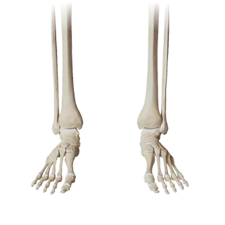
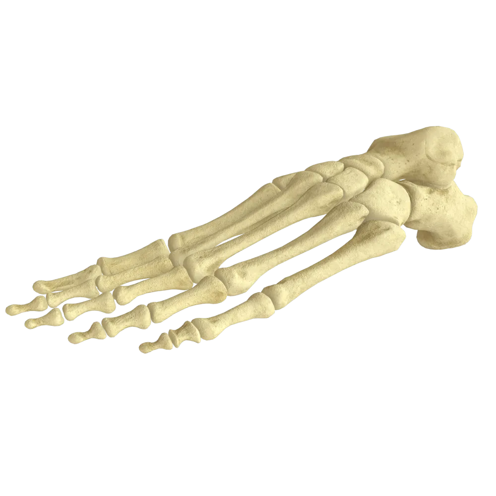
Bones of foot quiz: structure, classification, articular surfaces, and topography of bones in the skeletal system.
1/20
bold
text
1. Which bone of the foot is the largest?
-
Talus
The calcaneus (calcaneus) is the largest bone of the foot. It forms the posterior part of the tarsus and bears the primary load during weight-bearing.
-
Scaphoid
The calcaneus (calcaneus) is the largest bone of the foot. It forms the posterior part of the tarsus and bears the primary load during weight-bearing.
-
Calcaneus (left)
The calcaneus (calcaneus) is the largest bone of the foot. It forms the posterior part of the tarsus and bears the primary load during weight-bearing.
-
Cuboid bone
The calcaneus (calcaneus) is the largest bone of the foot. It forms the posterior part of the tarsus and bears the primary load during weight-bearing.
-
I find it difficult to answer
The calcaneus (calcaneus) is the largest bone of the foot. It forms the posterior part of the tarsus and bears the primary load during weight-bearing.
2. How many bones comprise the tarsus (tarsus)?
-
5
The tarsus consists of 7 bones: the talus, calcaneus, navicular, cuboid, and three cuneiform bones (medial, intermediate, and lateral).
-
8
The tarsus consists of 7 bones: the talus, calcaneus, navicular, cuboid, and three cuneiform bones (medial, intermediate, and lateral).
-
7
The tarsus consists of 7 bones: the talus, calcaneus, navicular, cuboid, and three cuneiform bones (medial, intermediate, and lateral).
-
6
The tarsus consists of 7 bones: the talus, calcaneus, navicular, cuboid, and three cuneiform bones (medial, intermediate, and lateral).
-
I find it difficult to answer
The tarsus consists of 7 bones: the talus, calcaneus, navicular, cuboid, and three cuneiform bones (medial, intermediate, and lateral).
3. Which surface of the talus articulates with the tibia?
-
Inferior articular surface
The trochlea of the talus (trochlea tali) is the superior articular surface that articulates with the inferior articular surface of the tibia, forming the ankle joint.
-
Trochlea of the talus (trochlea tali)
The trochlea of the talus (trochlea tali) is the superior articular surface that articulates with the inferior articular surface of the tibia, forming the ankle joint.
-
Head of talus
The trochlea of the talus (trochlea tali) is the superior articular surface that articulates with the inferior articular surface of the tibia, forming the ankle joint.
-
Posterior process of talus
The trochlea of the talus (trochlea tali) is the superior articular surface that articulates with the inferior articular surface of the tibia, forming the ankle joint.
-
I find it difficult to answer
The trochlea of the talus (trochlea tali) is the superior articular surface that articulates with the inferior articular surface of the tibia, forming the ankle joint.
4. What is the process of the calcaneus that supports the talus called?
-
Processus anterior calcanei
Sustentaculum tali is the medial process of the calcaneus, forming a shelf on which the head of the talus rests. The tendon of the flexor hallucis longus passes beneath it.
-
Tuber calcanei
Sustentaculum tali is the medial process of the calcaneus, forming a shelf on which the head of the talus rests. The tendon of the flexor hallucis longus passes beneath it.
-
Sustentaculum tali
Sustentaculum tali is the medial process of the calcaneus, forming a shelf on which the head of the talus rests. The tendon of the flexor hallucis longus passes beneath it.
-
Trochlea peronealis
Sustentaculum tali is the medial process of the calcaneus, forming a shelf on which the head of the talus rests. The tendon of the flexor hallucis longus passes beneath it.
-
I find it difficult to answer
Sustentaculum tali is the medial process of the calcaneus, forming a shelf on which the head of the talus rests. The tendon of the flexor hallucis longus passes beneath it.
5. Which tarsal bone has no distinct body and consists predominantly of a head and neck?
-
Cuboid bone
The talus (talus) is divided into a body, neck, and head. It has no muscular attachments and approximately 60–70% of its surface is covered by articular surfaces.
-
Scaphoid
The talus (talus) is divided into a body, neck, and head. It has no muscular attachments and approximately 60–70% of its surface is covered by articular surfaces.
-
Talus
The talus (talus) is divided into a body, neck, and head. It has no muscular attachments and approximately 60–70% of its surface is covered by articular surfaces.
-
Calcaneus (left)
The talus (talus) is divided into a body, neck, and head. It has no muscular attachments and approximately 60–70% of its surface is covered by articular surfaces.
-
I find it difficult to answer
The talus (talus) is divided into a body, neck, and head. It has no muscular attachments and approximately 60–70% of its surface is covered by articular surfaces.
6. How many metatarsal bones (ossa metatarsi) are present in the foot?
-
4
The metatarsus consists of 5 metatarsal bones (os metatarsi I–V). Each comprises a base, body, and head.
-
6
The metatarsus consists of 5 metatarsal bones (os metatarsi I–V). Each comprises a base, body, and head.
-
5
The metatarsus consists of 5 metatarsal bones (os metatarsi I–V). Each comprises a base, body, and head.
-
7
The metatarsus consists of 5 metatarsal bones (os metatarsi I–V). Each comprises a base, body, and head.
-
I find it difficult to answer
The metatarsus consists of 5 metatarsal bones (os metatarsi I–V). Each comprises a base, body, and head.
7. Which bone of the foot comprises a groove (sulcus tendinis musculi fibularis longi) on its plantar surface?
-
Scaphoid
The calcaneus comprises a groove for the tendon of the fibularis longus muscle (sulcus tendinis musculi fibularis longi).
-
Medial cuneiform bone
The calcaneus comprises a groove for the tendon of the fibularis longus muscle (sulcus tendinis musculi fibularis longi).
-
Talus
The calcaneus comprises a groove for the tendon of the fibularis longus muscle (sulcus tendinis musculi fibularis longi).
-
Calcaneus (left)
The calcaneus comprises a groove for the tendon of the fibularis longus muscle (sulcus tendinis musculi fibularis longi).
-
I find it difficult to answer
The calcaneus comprises a groove for the tendon of the fibularis longus muscle (sulcus tendinis musculi fibularis longi).
8. How many sesamoid bones are normally found beneath the head of the first metatarsal bone?
-
3
Normally, 2 sesamoid bones are located beneath the head of the first metatarsal bone: medial and lateral. They lie within the tendon of the flexor hallucis brevis muscle.
-
1
Normally, 2 sesamoid bones are located beneath the head of the first metatarsal bone: medial and lateral. They lie within the tendon of the flexor hallucis brevis muscle.
-
2
Normally, 2 sesamoid bones are located beneath the head of the first metatarsal bone: medial and lateral. They lie within the tendon of the flexor hallucis brevis muscle.
-
4
Normally, 2 sesamoid bones are located beneath the head of the first metatarsal bone: medial and lateral. They lie within the tendon of the flexor hallucis brevis muscle.
-
I find it difficult to answer
Normally, 2 sesamoid bones are located beneath the head of the first metatarsal bone: medial and lateral. They lie within the tendon of the flexor hallucis brevis muscle.
9. Tuber calcanei forms:
-
The medial border of the calcaneus
Tuber calcanei is the calcaneal tuberosity, which forms the posterior part of the calcaneus and, consequently, the anatomical basis of the heel. The calcaneal (Achilles) tendon attaches to it.
-
The posterior prominence of the calcaneus forming the heel
Tuber calcanei is the calcaneal tuberosity, which forms the posterior part of the calcaneus and, consequently, the anatomical basis of the heel. The calcaneal (Achilles) tendon attaches to it.
-
The superior articular surface of the calcaneus
Tuber calcanei is the calcaneal tuberosity, which forms the posterior part of the calcaneus and, consequently, the anatomical basis of the heel. The calcaneal (Achilles) tendon attaches to it.
-
The anterior process of the calcaneus
Tuber calcanei is the calcaneal tuberosity, which forms the posterior part of the calcaneus and, consequently, the anatomical basis of the heel. The calcaneal (Achilles) tendon attaches to it.
-
I find it difficult to answer
Tuber calcanei is the calcaneal tuberosity, which forms the posterior part of the calcaneus and, consequently, the anatomical basis of the heel. The calcaneal (Achilles) tendon attaches to it.
10. Which tarsal bone does not articulate directly with the metatarsal bones?
-
Cuboid bone
The talus (talus) does not articulate directly with the metatarsal bones. It articulates with the calcaneus and navicular bone, whereas the metatarsal bones articulate with the cuneiform bones and cuboid bone.
-
Medial cuneiform bone
The talus (talus) does not articulate directly with the metatarsal bones. It articulates with the calcaneus and navicular bone, whereas the metatarsal bones articulate with the cuneiform bones and cuboid bone.
-
Talus
The talus (talus) does not articulate directly with the metatarsal bones. It articulates with the calcaneus and navicular bone, whereas the metatarsal bones articulate with the cuneiform bones and cuboid bone.
-
Lateral cuneiform bone
The talus (talus) does not articulate directly with the metatarsal bones. It articulates with the calcaneus and navicular bone, whereas the metatarsal bones articulate with the cuneiform bones and cuboid bone.
-
I find it difficult to answer
The talus (talus) does not articulate directly with the metatarsal bones. It articulates with the calcaneus and navicular bone, whereas the metatarsal bones articulate with the cuneiform bones and cuboid bone.
11. Which part of the foot is formed by the cuneiform bones and cuboid bone?
-
Distal part of the tarsus
The cuneiform bones (medial, intermediate, and lateral) and the cuboid bone form the distal row of the tarsus. The proximal row is represented by the talus and calcaneus.
-
Metatarsus
The cuneiform bones (medial, intermediate, and lateral) and the cuboid bone form the distal row of the tarsus. The proximal row is represented by the talus and calcaneus.
-
Proximal part of the tarsus
The cuneiform bones (medial, intermediate, and lateral) and the cuboid bone form the distal row of the tarsus. The proximal row is represented by the talus and calcaneus.
-
Toes
The cuneiform bones (medial, intermediate, and lateral) and the cuboid bone form the distal row of the tarsus. The proximal row is represented by the talus and calcaneus.
-
I find it difficult to answer
The cuneiform bones (medial, intermediate, and lateral) and the cuboid bone form the distal row of the tarsus. The proximal row is represented by the talus and calcaneus.
12. Which metatarsal bone comprises the most prominent tuberosity (tuberositas ossis metatarsi) at its base?
-
Second metatarsal bone
The fifth metatarsal bone comprises a well-developed tuberosity (tuberositas ossis metatarsi V) at its base, to which the tendon of the fibularis brevis muscle attaches. It is readily palpable on the lateral surface of the foot.
-
Fourth metatarsal bone
The fifth metatarsal bone comprises a well-developed tuberosity (tuberositas ossis metatarsi V) at its base, to which the tendon of the fibularis brevis muscle attaches. It is readily palpable on the lateral surface of the foot.
-
Third metatarsal bone
The fifth metatarsal bone comprises a well-developed tuberosity (tuberositas ossis metatarsi V) at its base, to which the tendon of the fibularis brevis muscle attaches. It is readily palpable on the lateral surface of the foot.
-
Fifth metatarsal bone
The fifth metatarsal bone comprises a well-developed tuberosity (tuberositas ossis metatarsi V) at its base, to which the tendon of the fibularis brevis muscle attaches. It is readily palpable on the lateral surface of the foot.
-
I find it difficult to answer
The fifth metatarsal bone comprises a well-developed tuberosity (tuberositas ossis metatarsi V) at its base, to which the tendon of the fibularis brevis muscle attaches. It is readily palpable on the lateral surface of the foot.
13. How many phalanges does the great toe (hallux) have?
-
3
The great toe (hallux, digitus I) comprises 2 phalanges: proximal and distal. The remaining toes (II–V) each comprise 3 phalanges.
-
4
The great toe (hallux, digitus I) comprises 2 phalanges: proximal and distal. The remaining toes (II–V) each comprise 3 phalanges.
-
1
The great toe (hallux, digitus I) comprises 2 phalanges: proximal and distal. The remaining toes (II–V) each comprise 3 phalanges.
-
2
The great toe (hallux, digitus I) comprises 2 phalanges: proximal and distal. The remaining toes (II–V) each comprise 3 phalanges.
-
I find it difficult to answer
The great toe (hallux, digitus I) comprises 2 phalanges: proximal and distal. The remaining toes (II–V) each comprise 3 phalanges.
14. Where is the navicular bone (os naviculare) located in the tarsus?
-
Between the calcaneus and cuboid bone
The scaphoid is located between the head of the talus proximally and the three cuneiform bones distally. It occupies the medial part of the distal tarsus.
-
Between the talus and cuneiform bones
The scaphoid is located between the head of the talus proximally and the three cuneiform bones distally. It occupies the medial part of the distal tarsus.
-
Between the talus and cuboid bone
The scaphoid is located between the head of the talus proximally and the three cuneiform bones distally. It occupies the medial part of the distal tarsus.
-
Between the calcaneus and cuneiform bones
The scaphoid is located between the head of the talus proximally and the three cuneiform bones distally. It occupies the medial part of the distal tarsus.
-
I find it difficult to answer
The scaphoid is located between the head of the talus proximally and the three cuneiform bones distally. It occupies the medial part of the distal tarsus.
15. Which surface of the navicular bone comprises the tuberosity (tuberositas ossis navicularis)?
-
Lateral
The tuberosity of the navicular bone (tuberositas ossis navicularis) is located on its medial surface. The tendon of the tibialis posterior muscle attaches to it. The tuberosity is readily palpable through the skin.
-
Superior
The tuberosity of the navicular bone (tuberositas ossis navicularis) is located on its medial surface. The tendon of the tibialis posterior muscle attaches to it. The tuberosity is readily palpable through the skin.
-
Medial
The tuberosity of the navicular bone (tuberositas ossis navicularis) is located on its medial surface. The tendon of the tibialis posterior muscle attaches to it. The tuberosity is readily palpable through the skin.
-
Plantar
The tuberosity of the navicular bone (tuberositas ossis navicularis) is located on its medial surface. The tendon of the tibialis posterior muscle attaches to it. The tuberosity is readily palpable through the skin.
-
I find it difficult to answer
The tuberosity of the navicular bone (tuberositas ossis navicularis) is located on its medial surface. The tendon of the tibialis posterior muscle attaches to it. The tuberosity is readily palpable through the skin.
16. Which bone forms the posterolateral angle of the tarsus and articulates with the fourth and fifth metatarsal bones?
-
Lateral cuneiform bone
The Cuboid bone (left) (os cuboideum) occupies the lateral part of the tarsus and articulates distally with the bases of metatarsal bones IV and V, proximally—with the calcaneus, medially—with the navicular and Lateral cuneiform bone.
-
Intermediate cuneiform bone
The Cuboid bone (left) (os cuboideum) occupies the lateral part of the tarsus and articulates distally with the bases of metatarsal bones IV and V, proximally—with the calcaneus, medially—with the navicular and Lateral cuneiform bone.
-
Calcaneus (left)
The Cuboid bone (left) (os cuboideum) occupies the lateral part of the tarsus and articulates distally with the bases of metatarsal bones IV and V, proximally—with the calcaneus, medially—with the navicular and Lateral cuneiform bone.
-
Cuboid bone
The Cuboid bone (left) (os cuboideum) occupies the lateral part of the tarsus and articulates distally with the bases of metatarsal bones IV and V, proximally—with the calcaneus, medially—with the navicular and Lateral cuneiform bone.
-
I find it difficult to answer
The Cuboid bone (left) (os cuboideum) occupies the lateral part of the tarsus and articulates distally with the bases of metatarsal bones IV and V, proximally—with the calcaneus, medially—with the navicular and Lateral cuneiform bone.
17. Which of the cuneiform bones is the largest?
-
Intermediate cuneiform (os cuneiforme intermedium)
The medial cuneiform bone (os cuneiforme mediale) is the largest of the three cuneiform bones. The intermediate cuneiform is the smallest. The medial cuneiform articulates with the navicular bone and the first metatarsal bone.
-
All three are equal in size
The medial cuneiform bone (os cuneiforme mediale) is the largest of the three cuneiform bones. The intermediate cuneiform is the smallest. The medial cuneiform articulates with the navicular bone and the first metatarsal bone.
-
Lateral cuneiform (os cuneiforme laterale)
The medial cuneiform bone (os cuneiforme mediale) is the largest of the three cuneiform bones. The intermediate cuneiform is the smallest. The medial cuneiform articulates with the navicular bone and the first metatarsal bone.
-
Medial cuneiform (os cuneiforme mediale)
The medial cuneiform bone (os cuneiforme mediale) is the largest of the three cuneiform bones. The intermediate cuneiform is the smallest. The medial cuneiform articulates with the navicular bone and the first metatarsal bone.
-
I find it difficult to answer
The medial cuneiform bone (os cuneiforme mediale) is the largest of the three cuneiform bones. The intermediate cuneiform is the smallest. The medial cuneiform articulates with the navicular bone and the first metatarsal bone.
18. What is the articular surface of the calcaneus that articulates anteriorly with the talus called?
-
Facies articularis talaris anterior
The calcaneus comprises three articular surfaces for articulation with the talus: anterior (facies articularis talaris anterior), middle (on the sustentaculum tali), and posterior (facies articularis talaris posterior). The anterior surface is located most distally.
-
Sustentaculum tali
The calcaneus comprises three articular surfaces for articulation with the talus: anterior (facies articularis talaris anterior), middle (on the sustentaculum tali), and posterior (facies articularis talaris posterior). The anterior surface is located most distally.
-
Facies articularis talaris posterior
The calcaneus comprises three articular surfaces for articulation with the talus: anterior (facies articularis talaris anterior), middle (on the sustentaculum tali), and posterior (facies articularis talaris posterior). The anterior surface is located most distally.
-
Sulcus calcanei
The calcaneus comprises three articular surfaces for articulation with the talus: anterior (facies articularis talaris anterior), middle (on the sustentaculum tali), and posterior (facies articularis talaris posterior). The anterior surface is located most distally.
-
I find it difficult to answer
The calcaneus comprises three articular surfaces for articulation with the talus: anterior (facies articularis talaris anterior), middle (on the sustentaculum tali), and posterior (facies articularis talaris posterior). The anterior surface is located most distally.
19. What is the total number of bones in the adult foot skeleton (tarsus + metatarsus + phalanges, excluding variable sesamoid bones)?
-
30
The adult foot skeleton contains 26 constant bones: 7 tarsal bones + 5 metatarsal bones + 14 phalanges (2 phalanges in the first toe and 3 phalanges in each of the second through fifth toes). Sesamoid bones are variable and are traditionally not included in this number.
-
28
The adult foot skeleton contains 26 constant bones: 7 tarsal bones + 5 metatarsal bones + 14 phalanges (2 phalanges in the first toe and 3 phalanges in each of the second through fifth toes). Sesamoid bones are variable and are traditionally not included in this number.
-
26
The adult foot skeleton contains 26 constant bones: 7 tarsal bones + 5 metatarsal bones + 14 phalanges (2 phalanges in the first toe and 3 phalanges in each of the second through fifth toes). Sesamoid bones are variable and are traditionally not included in this number.
-
24
The adult foot skeleton contains 26 constant bones: 7 tarsal bones + 5 metatarsal bones + 14 phalanges (2 phalanges in the first toe and 3 phalanges in each of the second through fifth toes). Sesamoid bones are variable and are traditionally not included in this number.
-
I find it difficult to answer
The adult foot skeleton contains 26 constant bones: 7 tarsal bones + 5 metatarsal bones + 14 phalanges (2 phalanges in the first toe and 3 phalanges in each of the second through fifth toes). Sesamoid bones are variable and are traditionally not included in this number.
20. Which process of the talus bounds the groove (sulcus tali) posteriorly and serves as the attachment site for the posterior talofibular ligament?
-
Processus lateralis tali
Processus posterior tali is the posterior process of the talus, located posterior to the trochlea. It is divided by the groove for the tendon of the flexor hallucis longus into medial and lateral tubercles. The posterior talofibular ligament attaches to the lateral tubercle.
-
Caput tali
Processus posterior tali is the posterior process of the talus, located posterior to the trochlea. It is divided by the groove for the tendon of the flexor hallucis longus into medial and lateral tubercles. The posterior talofibular ligament attaches to the lateral tubercle.
-
Collum tali
Processus posterior tali is the posterior process of the talus, located posterior to the trochlea. It is divided by the groove for the tendon of the flexor hallucis longus into medial and lateral tubercles. The posterior talofibular ligament attaches to the lateral tubercle.
-
Processus posterior tali
Processus posterior tali is the posterior process of the talus, located posterior to the trochlea. It is divided by the groove for the tendon of the flexor hallucis longus into medial and lateral tubercles. The posterior talofibular ligament attaches to the lateral tubercle.
-
I find it difficult to answer
Processus posterior tali is the posterior process of the talus, located posterior to the trochlea. It is divided by the groove for the tendon of the flexor hallucis longus into medial and lateral tubercles. The posterior talofibular ligament attaches to the lateral tubercle.
Retake this quiz?
Your current progress will be reset.
