1/20

Quiz on the Anatomy of the Free Part of the Lower Limb
The quiz covers bones, joints, muscles, vessels, and nerves of the free part of the lower limb.
1. Which bone articulates with the condyles of the femur?
-
Fibula
The tibia, with its medial and lateral condyles, articulates with the femoral condyles, forming the knee joint. The fibula does not play a role in the knee joint.
-
Tibia
The tibia, with its medial and lateral condyles, articulates with the femoral condyles, forming the knee joint. The fibula does not play a role in the knee joint.
-
Patella
The tibia, with its medial and lateral condyles, articulates with the femoral condyles, forming the knee joint. The fibula does not play a role in the knee joint.
-
Talus
The tibia, with its medial and lateral condyles, articulates with the femoral condyles, forming the knee joint. The fibula does not play a role in the knee joint.
-
I find it difficult to answer
The tibia, with its medial and lateral condyles, articulates with the femoral condyles, forming the knee joint. The fibula does not play a role in the knee joint.
2. Which knee joint ligament attaches to the medial femoral condyle and the superior surface of the medial tibial condyle?
-
Lateral collateral ligament
The medial (tibial) collateral ligament extends from the medial epicondyle of the femur to the medial tibial condyle and is fused with the medial meniscus. The lateral collateral ligament attaches to the head of the fibula.
-
Anterior cruciate ligament
The medial (tibial) collateral ligament extends from the medial epicondyle of the femur to the medial tibial condyle and is fused with the medial meniscus. The lateral collateral ligament attaches to the head of the fibula.
-
Posterior cruciate ligament
The medial (tibial) collateral ligament extends from the medial epicondyle of the femur to the medial tibial condyle and is fused with the medial meniscus. The lateral collateral ligament attaches to the head of the fibula.
-
Medial collateral ligament
The medial (tibial) collateral ligament extends from the medial epicondyle of the femur to the medial tibial condyle and is fused with the medial meniscus. The lateral collateral ligament attaches to the head of the fibula.
-
I find it difficult to answer
The medial (tibial) collateral ligament extends from the medial epicondyle of the femur to the medial tibial condyle and is fused with the medial meniscus. The lateral collateral ligament attaches to the head of the fibula.
3. Which thigh muscle is part of the quadriceps and originates from the intertrochanteric line of the femur?
-
Rectus femoris.
The vastus intermedius muscle originates from the anterior and lateral surfaces of the femur, including the intertrochanteric line. The rectus femoris muscle originates from the anterior inferior iliac spine.
-
Vastus lateralis muscle
The vastus intermedius muscle originates from the anterior and lateral surfaces of the femur, including the intertrochanteric line. The rectus femoris muscle originates from the anterior inferior iliac spine.
-
Vastus intermedius muscle
The vastus intermedius muscle originates from the anterior and lateral surfaces of the femur, including the intertrochanteric line. The rectus femoris muscle originates from the anterior inferior iliac spine.
-
Vastus medialis
The vastus intermedius muscle originates from the anterior and lateral surfaces of the femur, including the intertrochanteric line. The rectus femoris muscle originates from the anterior inferior iliac spine.
-
I find it difficult to answer
The vastus intermedius muscle originates from the anterior and lateral surfaces of the femur, including the intertrochanteric line. The rectus femoris muscle originates from the anterior inferior iliac spine.
4. The obturator nerve innervates the muscles of the adductor group of the thigh. Which of the following muscles is NOT innervated by the obturator nerve?
-
Adductor longus
The adductor magnus is innervated by two nerves: the anterior (adductor) part by the obturator nerve, and the posterior (hamstring) part by the tibial part of the sciatic nerve. It is the posterior part that does not receive innervation from the obturator nerve.
-
Gracilis muscle.
The adductor magnus is innervated by two nerves: the anterior (adductor) part by the obturator nerve, and the posterior (hamstring) part by the tibial part of the sciatic nerve. It is the posterior part that does not receive innervation from the obturator nerve.
-
Adductor brevis
The adductor magnus is innervated by two nerves: the anterior (adductor) part by the obturator nerve, and the posterior (hamstring) part by the tibial part of the sciatic nerve. It is the posterior part that does not receive innervation from the obturator nerve.
-
Adductor magnus (posterior part)
The adductor magnus is innervated by two nerves: the anterior (adductor) part by the obturator nerve, and the posterior (hamstring) part by the tibial part of the sciatic nerve. It is the posterior part that does not receive innervation from the obturator nerve.
-
I find it difficult to answer
The adductor magnus is innervated by two nerves: the anterior (adductor) part by the obturator nerve, and the posterior (hamstring) part by the tibial part of the sciatic nerve. It is the posterior part that does not receive innervation from the obturator nerve.
5. Which nerve passes through the adductor canal (Hunter's canal) together with the femoral artery and vein?
-
Obturator nerve
The saphenous nerve — the medial branch of the femoral nerve — enters the adductor canal with the femoral artery and vein, then exits through the anterior wall and accompanies the great saphenous vein of the leg.
-
Saphenous nerve (n. saphenus)
The saphenous nerve — the medial branch of the femoral nerve — enters the adductor canal with the femoral artery and vein, then exits through the anterior wall and accompanies the great saphenous vein of the leg.
-
Common fibular nerve
The saphenous nerve — the medial branch of the femoral nerve — enters the adductor canal with the femoral artery and vein, then exits through the anterior wall and accompanies the great saphenous vein of the leg.
-
Posterior femoral cutaneous nerve
The saphenous nerve — the medial branch of the femoral nerve — enters the adductor canal with the femoral artery and vein, then exits through the anterior wall and accompanies the great saphenous vein of the leg.
-
I find it difficult to answer
The saphenous nerve — the medial branch of the femoral nerve — enters the adductor canal with the femoral artery and vein, then exits through the anterior wall and accompanies the great saphenous vein of the leg.
6. Which artery is the main source of blood supply to the femoral head in adults?
-
Artery of the ligament of the head of the femur
In adults, the main source of blood supply to the head of the femur is the medial circumflex femoral artery, giving a branch to the neck and head.
-
Medial circumflex femoral artery
In adults, the main source of blood supply to the head of the femur is the medial circumflex femoral artery, giving a branch to the neck and head.
-
Lateral circumflex femoral artery
In adults, the main source of blood supply to the head of the femur is the medial circumflex femoral artery, giving a branch to the neck and head.
-
First perforating artery
In adults, the main source of blood supply to the head of the femur is the medial circumflex femoral artery, giving a branch to the neck and head.
-
I find it difficult to answer
In adults, the main source of blood supply to the head of the femur is the medial circumflex femoral artery, giving a branch to the neck and head.
7. Which posterior leg muscle belongs to the superficial layer and has two heads originating from the femoral condyles?
-
Soleus muscle.
The gastrocnemius muscle has medial and lateral heads, originating respectively from the medial and lateral femoral condyles. The soleus muscle originates from the bones of the leg.
-
Plantar muscle
The gastrocnemius muscle has medial and lateral heads, originating respectively from the medial and lateral femoral condyles. The soleus muscle originates from the bones of the leg.
-
Gastrocnemius muscle.
The gastrocnemius muscle has medial and lateral heads, originating respectively from the medial and lateral femoral condyles. The soleus muscle originates from the bones of the leg.
-
Flexor digitorum longus
The gastrocnemius muscle has medial and lateral heads, originating respectively from the medial and lateral femoral condyles. The soleus muscle originates from the bones of the leg.
-
I find it difficult to answer
The gastrocnemius muscle has medial and lateral heads, originating respectively from the medial and lateral femoral condyles. The soleus muscle originates from the bones of the leg.
8. Which bone of the foot articulates with the tibia and fibula to form the ankle joint?
-
Calcaneus
The talus via its trochlea fits into the 'mortise' of the ankle joint, formed by the distal epiphyses of the tibia (medial malleolus and articular surface) and fibula (lateral malleolus).
-
Navicular bone
The talus via its trochlea fits into the 'mortise' of the ankle joint, formed by the distal epiphyses of the tibia (medial malleolus and articular surface) and fibula (lateral malleolus).
-
Cuboid bone
The talus via its trochlea fits into the 'mortise' of the ankle joint, formed by the distal epiphyses of the tibia (medial malleolus and articular surface) and fibula (lateral malleolus).
-
Talus
The talus via its trochlea fits into the 'mortise' of the ankle joint, formed by the distal epiphyses of the tibia (medial malleolus and articular surface) and fibula (lateral malleolus).
-
I find it difficult to answer
The talus via its trochlea fits into the 'mortise' of the ankle joint, formed by the distal epiphyses of the tibia (medial malleolus and articular surface) and fibula (lateral malleolus).
9. What is the primary movement in the ankle joint due to its hinge-like shape?
-
Pronation and supination
The ankle joint is a hinge-like joint with a single axis of movement — the frontal one. In it, plantar flexion and dorsiflexion of the foot occur. Pronation and supination are carried out in the subtalar and transverse joints of the tarsal bones.
-
Abduction and adduction
The ankle joint is a hinge-like joint with a single axis of movement — the frontal one. In it, plantar flexion and dorsiflexion of the foot occur. Pronation and supination are carried out in the subtalar and transverse joints of the tarsal bones.
-
Plantar flexion and dorsiflexion
The ankle joint is a hinge-like joint with a single axis of movement — the frontal one. In it, plantar flexion and dorsiflexion of the foot occur. Pronation and supination are carried out in the subtalar and transverse joints of the tarsal bones.
-
Rotation around the vertical axis
The ankle joint is a hinge-like joint with a single axis of movement — the frontal one. In it, plantar flexion and dorsiflexion of the foot occur. Pronation and supination are carried out in the subtalar and transverse joints of the tarsal bones.
-
I find it difficult to answer
The ankle joint is a hinge-like joint with a single axis of movement — the frontal one. In it, plantar flexion and dorsiflexion of the foot occur. Pronation and supination are carried out in the subtalar and transverse joints of the tarsal bones.
10. The great saphenous vein drains into:
-
The popliteal vein
The great saphenous vein drains into the femoral vein through the hiatus saphenus (saphenous opening of the fascia lata), located below the inguinal ligament. This is an important anatomical point for the transition of the superficial and deep venous systems.
-
The femoral vein at the hiatus saphenus
The great saphenous vein drains into the femoral vein through the hiatus saphenus (saphenous opening of the fascia lata), located below the inguinal ligament. This is an important anatomical point for the transition of the superficial and deep venous systems.
-
The external iliac vein
The great saphenous vein drains into the femoral vein through the hiatus saphenus (saphenous opening of the fascia lata), located below the inguinal ligament. This is an important anatomical point for the transition of the superficial and deep venous systems.
-
Deep femoral vein
The great saphenous vein drains into the femoral vein through the hiatus saphenus (saphenous opening of the fascia lata), located below the inguinal ligament. This is an important anatomical point for the transition of the superficial and deep venous systems.
-
I find it difficult to answer
The great saphenous vein drains into the femoral vein through the hiatus saphenus (saphenous opening of the fascia lata), located below the inguinal ligament. This is an important anatomical point for the transition of the superficial and deep venous systems.
11. Which thigh muscle extends at the hip joint and flexes at the knee joint?
-
Rectus femoris.
The biceps femoris belongs to the posterior group of the thigh muscles. It extends the thigh at the hip joint and flexes the leg at the knee joint. The rectus femoris muscle, on the other hand, flexes the thigh and extends the leg.
-
Biceps femoris muscle.
The biceps femoris belongs to the posterior group of the thigh muscles. It extends the thigh at the hip joint and flexes the leg at the knee joint. The rectus femoris muscle, on the other hand, flexes the thigh and extends the leg.
-
Sartorius muscle.
The biceps femoris belongs to the posterior group of the thigh muscles. It extends the thigh at the hip joint and flexes the leg at the knee joint. The rectus femoris muscle, on the other hand, flexes the thigh and extends the leg.
-
Tensor fasciae latae
The biceps femoris belongs to the posterior group of the thigh muscles. It extends the thigh at the hip joint and flexes the leg at the knee joint. The rectus femoris muscle, on the other hand, flexes the thigh and extends the leg.
-
I find it difficult to answer
The biceps femoris belongs to the posterior group of the thigh muscles. It extends the thigh at the hip joint and flexes the leg at the knee joint. The rectus femoris muscle, on the other hand, flexes the thigh and extends the leg.
12. The following structures pass through the infrapiriform foramen from the pelvic cavity to the buttock:
-
Superior gluteal nerve
The following structures exit trough the infrapiriform foramen: the inferior gluteal nerve and vessels, sciatic nerve, pudendal nerve, posterior femoral cutaneous nerve. The superior gluteal nerve exits through the suprapiriform foramen.
-
Femoral nerve
The following structures exit trough the infrapiriform foramen: the inferior gluteal nerve and vessels, sciatic nerve, pudendal nerve, posterior femoral cutaneous nerve. The superior gluteal nerve exits through the suprapiriform foramen.
-
Inferior gluteal nerve.
The following structures exit trough the infrapiriform foramen: the inferior gluteal nerve and vessels, sciatic nerve, pudendal nerve, posterior femoral cutaneous nerve. The superior gluteal nerve exits through the suprapiriform foramen.
-
Obturator nerve
The following structures exit trough the infrapiriform foramen: the inferior gluteal nerve and vessels, sciatic nerve, pudendal nerve, posterior femoral cutaneous nerve. The superior gluteal nerve exits through the suprapiriform foramen.
-
I find it difficult to answer
The following structures exit trough the infrapiriform foramen: the inferior gluteal nerve and vessels, sciatic nerve, pudendal nerve, posterior femoral cutaneous nerve. The superior gluteal nerve exits through the suprapiriform foramen.
13. Into which two branches does the sciatic nerve divide in the popliteal fossa?
-
Into the common fibular and superficial fibular nerves
The sciatic nerve in the upper corner of the popliteal fossa divides into the tibial nerve and the common fibular nerve. The division may also occur higher — at the thigh level.
-
Into the tibial and common fibular nerves
The sciatic nerve in the upper corner of the popliteal fossa divides into the tibial nerve and the common fibular nerve. The division may also occur higher — at the thigh level.
-
Into the tibial and deep fibular nerves
The sciatic nerve in the upper corner of the popliteal fossa divides into the tibial nerve and the common fibular nerve. The division may also occur higher — at the thigh level.
-
Into the saphenous and obturator nerves
The sciatic nerve in the upper corner of the popliteal fossa divides into the tibial nerve and the common fibular nerve. The division may also occur higher — at the thigh level.
-
I find it difficult to answer
The sciatic nerve in the upper corner of the popliteal fossa divides into the tibial nerve and the common fibular nerve. The division may also occur higher — at the thigh level.
14. Which ligament of the foot is called the spring ligament (lig. calcaneonaviculare plantare) and supports the head of the talus?
-
Long plantar ligament.
The plantar calcaneonavicular ligament – also called the spring ligament — is stretched between the sustentaculum tali and the inferior surface of the navicular bone. It supports the head of the talus and is a key element in maintaining the longitudinal arch of the foot.
-
Plantar aponeurosis.
The plantar calcaneonavicular ligament – also called the spring ligament — is stretched between the sustentaculum tali and the inferior surface of the navicular bone. It supports the head of the talus and is a key element in maintaining the longitudinal arch of the foot.
-
Calcaneonavicular plantar ligament
The plantar calcaneonavicular ligament – also called the spring ligament — is stretched between the sustentaculum tali and the inferior surface of the navicular bone. It supports the head of the talus and is a key element in maintaining the longitudinal arch of the foot.
-
Talocalcaneal interosseous ligament
The plantar calcaneonavicular ligament – also called the spring ligament — is stretched between the sustentaculum tali and the inferior surface of the navicular bone. It supports the head of the talus and is a key element in maintaining the longitudinal arch of the foot.
-
I find it difficult to answer
The plantar calcaneonavicular ligament – also called the spring ligament — is stretched between the sustentaculum tali and the inferior surface of the navicular bone. It supports the head of the talus and is a key element in maintaining the longitudinal arch of the foot.
15. The deep fibular nerve innervates the muscles of the anterior compartment of the leg. Which muscle does it innervate on the foot?
-
Flexor digitorum brevis muscle.
The deep fibular nerve on the foot innervates the extensor digitorum brevis and the extensor hallucis brevis, as well as the first interdigital space (cutaneous branch).
-
Extensor digitorum brevis and extensor hallucis brevis
The deep fibular nerve on the foot innervates the extensor digitorum brevis and the extensor hallucis brevis, as well as the first interdigital space (cutaneous branch).
-
Muscles of the foot hypothenar
The deep fibular nerve on the foot innervates the extensor digitorum brevis and the extensor hallucis brevis, as well as the first interdigital space (cutaneous branch).
-
Lumbrical muscles II–IV
The deep fibular nerve on the foot innervates the extensor digitorum brevis and the extensor hallucis brevis, as well as the first interdigital space (cutaneous branch).
-
I find it difficult to answer
The deep fibular nerve on the foot innervates the extensor digitorum brevis and the extensor hallucis brevis, as well as the first interdigital space (cutaneous branch).
16. In terms to the shape of the articular surfaces the knee joint is classified as:
-
Ball-and-socket joint
The knee joint is a condylar joint. This allows mainly flexion and extension in it, and with the knee flexed, slight rotation of the leg around the vertical axis is also possible.
-
Hinge joint
The knee joint is a condylar joint. This allows mainly flexion and extension in it, and with the knee flexed, slight rotation of the leg around the vertical axis is also possible.
-
Condylar joint
The knee joint is a condylar joint. This allows mainly flexion and extension in it, and with the knee flexed, slight rotation of the leg around the vertical axis is also possible.
-
Ellipsoid joint
The knee joint is a condylar joint. This allows mainly flexion and extension in it, and with the knee flexed, slight rotation of the leg around the vertical axis is also possible.
-
I find it difficult to answer
The knee joint is a condylar joint. This allows mainly flexion and extension in it, and with the knee flexed, slight rotation of the leg around the vertical axis is also possible.
17. Which muscle performs dorsiflexion of the foot and is innervated by the deep fibular nerve?
-
Fibularis longus muscle.
The tibialis anterior muscle is the main muscle performing dorsiflexion (extension) and supination of the foot. It is innervated by the deep fibular nerve and attaches to the medial cuneiform and the base of the first metatarsal bone.
-
Soleus muscle.
The tibialis anterior muscle is the main muscle performing dorsiflexion (extension) and supination of the foot. It is innervated by the deep fibular nerve and attaches to the medial cuneiform and the base of the first metatarsal bone.
-
Tibialis anterior muscle.
The tibialis anterior muscle is the main muscle performing dorsiflexion (extension) and supination of the foot. It is innervated by the deep fibular nerve and attaches to the medial cuneiform and the base of the first metatarsal bone.
-
Flexor pollicis longus
The tibialis anterior muscle is the main muscle performing dorsiflexion (extension) and supination of the foot. It is innervated by the deep fibular nerve and attaches to the medial cuneiform and the base of the first metatarsal bone.
-
I find it difficult to answer
The tibialis anterior muscle is the main muscle performing dorsiflexion (extension) and supination of the foot. It is innervated by the deep fibular nerve and attaches to the medial cuneiform and the base of the first metatarsal bone.
18. The small saphenous vein drains into:
-
The femoral vein at the hiatus saphenus
The small saphenous vein curves around the lateral malleolus, ascends along the posterior surface of the leg, and drains into the popliteal vein in the popliteal fossa. This is its typical variant of drainage.
-
The popliteal vein
The small saphenous vein curves around the lateral malleolus, ascends along the posterior surface of the leg, and drains into the popliteal vein in the popliteal fossa. This is its typical variant of drainage.
-
Deep vein of the leg
The small saphenous vein curves around the lateral malleolus, ascends along the posterior surface of the leg, and drains into the popliteal vein in the popliteal fossa. This is its typical variant of drainage.
-
Great saphenous vein
The small saphenous vein curves around the lateral malleolus, ascends along the posterior surface of the leg, and drains into the popliteal vein in the popliteal fossa. This is its typical variant of drainage.
-
I find it difficult to answer
The small saphenous vein curves around the lateral malleolus, ascends along the posterior surface of the leg, and drains into the popliteal vein in the popliteal fossa. This is its typical variant of drainage.
19. Which tarsal bone is the largest and forms the calcaneal tuberosity?
-
Talus
The calcaneus is the largest tarsal bone On its lower posterior surface the calcaneal tuberosity is located, to which the calcaneal (Achilles) tendon is attached.
-
Navicular bone
The calcaneus is the largest tarsal bone On its lower posterior surface the calcaneal tuberosity is located, to which the calcaneal (Achilles) tendon is attached.
-
Calcaneus
The calcaneus is the largest tarsal bone On its lower posterior surface the calcaneal tuberosity is located, to which the calcaneal (Achilles) tendon is attached.
-
Cuboid bone
The calcaneus is the largest tarsal bone On its lower posterior surface the calcaneal tuberosity is located, to which the calcaneal (Achilles) tendon is attached.
-
I find it difficult to answer
The calcaneus is the largest tarsal bone On its lower posterior surface the calcaneal tuberosity is located, to which the calcaneal (Achilles) tendon is attached.
20. Which muscle, originating from the ischial tuberosity, attaches to the medial tibial condyle and is part of the pes anserinus?
-
Biceps femoris muscle.
The semitendinosus muscle originates from the ischial tuberosity and attaches via a long tendon to the medial surface of the tibia, forming the pes anserinus along with the sartorius and gracilis muscles.
-
Semitendinosus muscle.
The semitendinosus muscle originates from the ischial tuberosity and attaches via a long tendon to the medial surface of the tibia, forming the pes anserinus along with the sartorius and gracilis muscles.
-
Semimembranosus muscle.
The semitendinosus muscle originates from the ischial tuberosity and attaches via a long tendon to the medial surface of the tibia, forming the pes anserinus along with the sartorius and gracilis muscles.
-
Gracilis muscle.
The semitendinosus muscle originates from the ischial tuberosity and attaches via a long tendon to the medial surface of the tibia, forming the pes anserinus along with the sartorius and gracilis muscles.
-
I find it difficult to answer
The semitendinosus muscle originates from the ischial tuberosity and attaches via a long tendon to the medial surface of the tibia, forming the pes anserinus along with the sartorius and gracilis muscles.
Retake this quiz?
Your current progress will be reset.
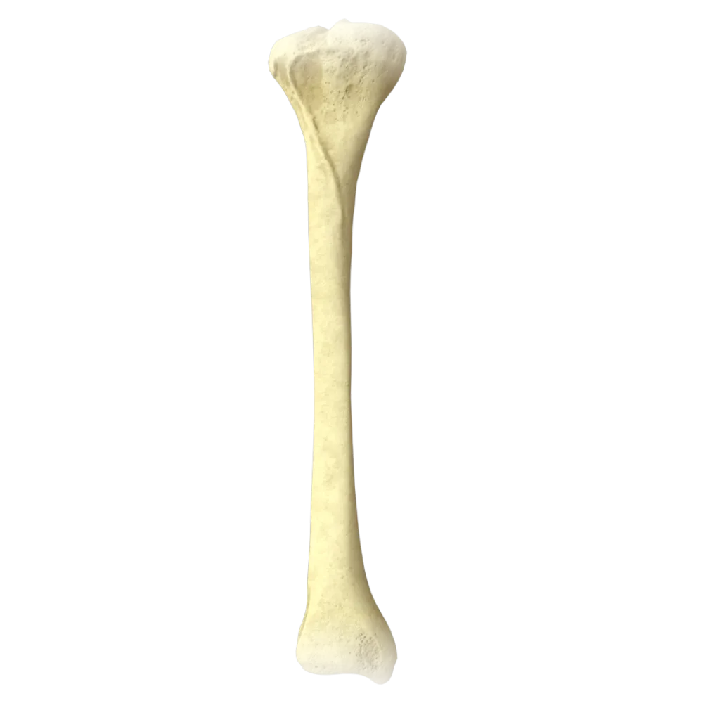
Bones of free part of lower limb
Tibia
0/20

Cookie Consent
We use cookies to enhance your browsing experience, analyze site traffic, and deliver content. Please choose whether you accept all cookies or wish to reject non-essential tracking.
Cookie Preferences
Manage your cookie preferences below:
Essential cookies enable basic functions and are necessary for the proper function of the website.
Name
Description
Duration
Geolocation Config
This cookie is used to store the consent settings based on the visitor's location.
30 days
Cookie Preferences
This cookie is used to store the user's cookie consent preferences.
30 days
Google reCAPTCHA helps protect websites from spam and abuse by verifying user interactions through challenges.
Name
Description
Duration
_GRECAPTCHA
Google reCAPTCHA sets a necessary cookie (_GRECAPTCHA) when executed for the purpose of providing its risk analysis.
179 days
Statistics cookies collect information anonymously. This information helps us understand how visitors use our website.
Google Analytics is a powerful tool that tracks and analyzes website traffic for informed marketing decisions.
Service URL: policies.google.com (opens in a new window)
Name
Description
Duration
__utma
ID used to identify users and sessions
2 years after last activity
__utmt
Used to monitor number of Google Analytics server requests
10 minutes
__utmb
Used to distinguish new sessions and visits. This cookie is set when the GA.js javascript library is loaded and there is no existing __utmb cookie. The cookie is updated every time data is sent to the Google Analytics server.
30 minutes after last activity
__utmc
Used only with old Urchin versions of Google Analytics and not with GA.js. Was used to distinguish between new sessions and visits at the end of a session.
End of session (browser)
__utmz
Contains information about the traffic source or campaign that directed user to the website. The cookie is set when the GA.js javascript is loaded and updated when data is sent to the Google Anaytics server
6 months after last activity
__utmv
Contains custom information set by the web developer via the _setCustomVar method in Google Analytics. This cookie is updated every time new data is sent to the Google Analytics server.
2 years after last activity
__utmx
Used to determine whether a user is included in an A / B or Multivariate test.
18 months
_ga
ID used to identify users
2 years
_gali
Used by Google Analytics to determine which links on a page are being clicked
30 seconds
_ga_
ID used to identify users
2 years
_gid
ID used to identify users for 24 hours after last activity
24 hours
_gat
Used to monitor number of Google Analytics server requests when using Google Tag Manager
1 minute
_gac_
Contains information related to marketing campaigns of the user. These are shared with Google AdWords / Google Ads when the Google Ads and Google Analytics accounts are linked together.
90 days
Clarity is a web analytics service that tracks and reports website traffic.
Service URL: clarity.microsoft.com (opens in a new window)
Name
Description
Duration
_clsk
Connects multiple page views by a user into a single Clarity session recording.
1 day
_clck
Stores a unique user identifier to analyze visitor behavior through heatmaps and session recordings.
1 year
ANONCHK
Indicates whether MUID is transferred to ANID, a cookie used for advertising. Clarity doesn't use ANID and so this is always set to 0.
Session
CLID
Identifies the first-time Clarity saw this user on any site using Clarity.
12 months
_clsk
Connects multiple page views by a user into a single Clarity session recording.
12 months
_clck
Persists the Clarity User ID and preferences, unique to that site is attributed to the same user ID.
12 months