
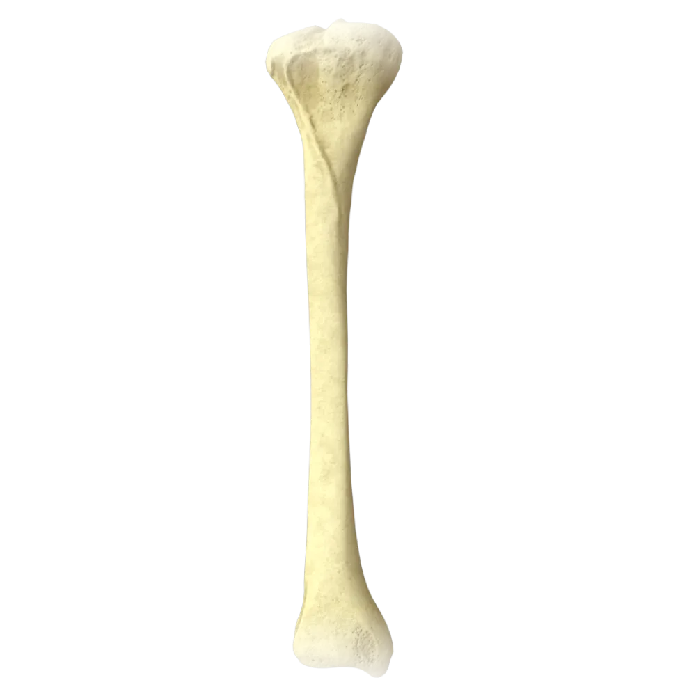
Fibula Anatomy Quiz
Normal anatomy quiz on the fibula: structure, processes, articular surfaces, connections, and functional landmarks.
1/20
bold
text
1. To which group of bones does the fibula belong according to the classification?
-
Short cancellous bone
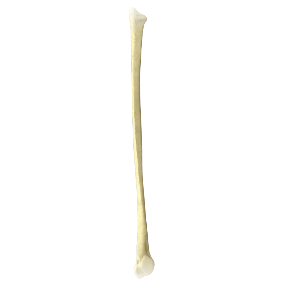
The fibula (fibula) is a long tubular bone comprising a shaft (diaphysis) and two epiphyses—proximal and distal.
-
Long tubular bone
The fibula (fibula) is a long tubular bone comprising a shaft (diaphysis) and two epiphyses—proximal and distal.
-
Flat bone
The fibula (fibula) is a long tubular bone comprising a shaft (diaphysis) and two epiphyses—proximal and distal.
-
Sesamoid bone
The fibula (fibula) is a long tubular bone comprising a shaft (diaphysis) and two epiphyses—proximal and distal.
-
I find it difficult to answer
The fibula (fibula) is a long tubular bone comprising a shaft (diaphysis) and two epiphyses—proximal and distal.
2. What is the proximal epiphysis of the fibula called?
-
Lateral malleolus
The proximal epiphysis of the fibula is called the head (caput fibulae). It bears an articular surface for articulation with the tibia.
-
Head of fibula
The proximal epiphysis of the fibula is called the head (caput fibulae). It bears an articular surface for articulation with the tibia.
-
Interosseous crest
The proximal epiphysis of the fibula is called the head (caput fibulae). It bears an articular surface for articulation with the tibia.
-
Medial condyle
The proximal epiphysis of the fibula is called the head (caput fibulae). It bears an articular surface for articulation with the tibia.
-
I find it difficult to answer
The proximal epiphysis of the fibula is called the head (caput fibulae). It bears an articular surface for articulation with the tibia.
3. Where on the head of the fibula is the articular surface for articulation with the tibia located?
-
On the anterior surface of the head
The articular surface of the head (facies articularis capitis fibulae) is located on the medial side of the head of the fibula and articulates with the fibular articular surface of the tibia.
-
On the medial surface of the head
The articular surface of the head (facies articularis capitis fibulae) is located on the medial side of the head of the fibula and articulates with the fibular articular surface of the tibia.
-
On the lateral surface of the head
The articular surface of the head (facies articularis capitis fibulae) is located on the medial side of the head of the fibula and articulates with the fibular articular surface of the tibia.
-
At the apex of the head
The articular surface of the head (facies articularis capitis fibulae) is located on the medial side of the head of the fibula and articulates with the fibular articular surface of the tibia.
-
I find it difficult to answer
The articular surface of the head (facies articularis capitis fibulae) is located on the medial side of the head of the fibula and articulates with the fibular articular surface of the tibia.
4. What is the pointed projection at the apex of the head of the fibula called?
-
Interosseous crest
At the apex of the head of the fibula, there is a pointed projection—the styloid process (apex capitis fibulae, processus styloideus).
-
Styloid process
At the apex of the head of the fibula, there is a pointed projection—the styloid process (apex capitis fibulae, processus styloideus).
-
Lateral malleolus
At the apex of the head of the fibula, there is a pointed projection—the styloid process (apex capitis fibulae, processus styloideus).
-
Anterior border
At the apex of the head of the fibula, there is a pointed projection—the styloid process (apex capitis fibulae, processus styloideus).
-
I find it difficult to answer
At the apex of the head of the fibula, there is a pointed projection—the styloid process (apex capitis fibulae, processus styloideus).
5. The distal epiphysis of the fibula forms the:
-
Medial malleolus
The distal epiphysis of the fibula forms the lateral malleolus (malleolus lateralis), which is part of the ankle joint.
-
Lateral malleolus
The distal epiphysis of the fibula forms the lateral malleolus (malleolus lateralis), which is part of the ankle joint.
-
Trochlea of talus
The distal epiphysis of the fibula forms the lateral malleolus (malleolus lateralis), which is part of the ankle joint.
-
Fibular notch
The distal epiphysis of the fibula forms the lateral malleolus (malleolus lateralis), which is part of the ankle joint.
-
I find it difficult to answer
The distal epiphysis of the fibula forms the lateral malleolus (malleolus lateralis), which is part of the ankle joint.
6. Which surface of the lateral malleolus bears the articular surface for the talus?
-
Posterior surface
The medial surface of the lateral malleolus (facies articularis malleoli lateralis) bears an articular surface that articulates with the lateral surface of the trochlea of the talus.
-
Lateral surface
The medial surface of the lateral malleolus (facies articularis malleoli lateralis) bears an articular surface that articulates with the lateral surface of the trochlea of the talus.
-
Medial surface
The medial surface of the lateral malleolus (facies articularis malleoli lateralis) bears an articular surface that articulates with the lateral surface of the trochlea of the talus.
-
Inferior surface
The medial surface of the lateral malleolus (facies articularis malleoli lateralis) bears an articular surface that articulates with the lateral surface of the trochlea of the talus.
-
I find it difficult to answer
The medial surface of the lateral malleolus (facies articularis malleoli lateralis) bears an articular surface that articulates with the lateral surface of the trochlea of the talus.
7. The posterior surface of the lateral malleolus has a groove through which the following pass:
-
Tendons of the anterior lower leg muscles
The posterior surface of the lateral malleolus bears the malleolar groove (sulcus malleolaris), through which the tendons of the fibularis longus and fibularis brevis pass.
-
Tendons of the fibular muscles
The posterior surface of the lateral malleolus bears the malleolar groove (sulcus malleolaris), through which the tendons of the fibularis longus and fibularis brevis pass.
-
Tendon of the flexor digitorum longus
The posterior surface of the lateral malleolus bears the malleolar groove (sulcus malleolaris), through which the tendons of the fibularis longus and fibularis brevis pass.
-
Tendon of the tibialis posterior
The posterior surface of the lateral malleolus bears the malleolar groove (sulcus malleolaris), through which the tendons of the fibularis longus and fibularis brevis pass.
-
I find it difficult to answer
The posterior surface of the lateral malleolus bears the malleolar groove (sulcus malleolaris), through which the tendons of the fibularis longus and fibularis brevis pass.
8. By which joint are the proximal epiphyses of the tibia and fibula connected?
-
By a syndesmosis
The proximal ends of the lower leg bones are connected by the proximal tibiofibular joint (articulatio tibiofibularis), which is classified as a plane joint.
-
By an interosseous synchondrosis
The proximal ends of the lower leg bones are connected by the proximal tibiofibular joint (articulatio tibiofibularis), which is classified as a plane joint.
-
By the proximal tibiofibular joint
The proximal ends of the lower leg bones are connected by the proximal tibiofibular joint (articulatio tibiofibularis), which is classified as a plane joint.
-
By the ankle joint
The proximal ends of the lower leg bones are connected by the proximal tibiofibular joint (articulatio tibiofibularis), which is classified as a plane joint.
-
I find it difficult to answer
The proximal ends of the lower leg bones are connected by the proximal tibiofibular joint (articulatio tibiofibularis), which is classified as a plane joint.
9. What type of connection joins the distal ends of the tibia and fibula?
-
Planar joint
The distal ends of the lower leg bones are connected by an interosseous syndesmosis—the distal tibiofibular syndesmosis (syndesmosis tibiofibularis)—forming the ankle mortise.
-
Synchondrosis
The distal ends of the lower leg bones are connected by an interosseous syndesmosis—the distal tibiofibular syndesmosis (syndesmosis tibiofibularis)—forming the ankle mortise.
-
Interosseous syndesmosis (ankle mortise)
The distal ends of the lower leg bones are connected by an interosseous syndesmosis—the distal tibiofibular syndesmosis (syndesmosis tibiofibularis)—forming the ankle mortise.
-
Sphenoidal joint
The distal ends of the lower leg bones are connected by an interosseous syndesmosis—the distal tibiofibular syndesmosis (syndesmosis tibiofibularis)—forming the ankle mortise.
-
I find it difficult to answer
The distal ends of the lower leg bones are connected by an interosseous syndesmosis—the distal tibiofibular syndesmosis (syndesmosis tibiofibularis)—forming the ankle mortise.
10. The interosseous membrane of the leg (membrana interossea cruris) is stretched between the:
-
Anterior borders of both lower leg bones
The interosseous membrane of the leg is stretched between the interosseous borders (margo interosseus) of the tibia and fibula, separating the anterior and posterior osteofascial compartments of the leg.
-
Interosseous borders of the tibia and fibula
The interosseous membrane of the leg is stretched between the interosseous borders (margo interosseus) of the tibia and fibula, separating the anterior and posterior osteofascial compartments of the leg.
-
Posterior surfaces of the lower leg bones
The interosseous membrane of the leg is stretched between the interosseous borders (margo interosseus) of the tibia and fibula, separating the anterior and posterior osteofascial compartments of the leg.
-
Medial surfaces of both lower leg bones
The interosseous membrane of the leg is stretched between the interosseous borders (margo interosseus) of the tibia and fibula, separating the anterior and posterior osteofascial compartments of the leg.
-
I find it difficult to answer
The interosseous membrane of the leg is stretched between the interosseous borders (margo interosseus) of the tibia and fibula, separating the anterior and posterior osteofascial compartments of the leg.
11. Which border of the shaft of the fibula faces the tibia and the interosseous membrane?
-
Anterior border
The interosseous border (margo interosseus) of the fibula faces medially toward the tibia and serves as the attachment site for the interosseous membrane of the leg.
-
Posterior border
The interosseous border (margo interosseus) of the fibula faces medially toward the tibia and serves as the attachment site for the interosseous membrane of the leg.
-
Interosseous border
The interosseous border (margo interosseus) of the fibula faces medially toward the tibia and serves as the attachment site for the interosseous membrane of the leg.
-
Lateral crest
The interosseous border (margo interosseus) of the fibula faces medially toward the tibia and serves as the attachment site for the interosseous membrane of the leg.
-
I find it difficult to answer
The interosseous border (margo interosseus) of the fibula faces medially toward the tibia and serves as the attachment site for the interosseous membrane of the leg.
12. How many surfaces are distinguished on the shaft of the fibula?
-
Two
Four surfaces are distinguished on the shaft of the fibula: anterior, posterior, medial, and lateral (according to the classical nomenclature—the anterolateral surface and the posterior part with a crest).
-
Three
Four surfaces are distinguished on the shaft of the fibula: anterior, posterior, medial, and lateral (according to the classical nomenclature—the anterolateral surface and the posterior part with a crest).
-
Four
Four surfaces are distinguished on the shaft of the fibula: anterior, posterior, medial, and lateral (according to the classical nomenclature—the anterolateral surface and the posterior part with a crest).
-
Five
Four surfaces are distinguished on the shaft of the fibula: anterior, posterior, medial, and lateral (according to the classical nomenclature—the anterolateral surface and the posterior part with a crest).
-
I find it difficult to answer
Four surfaces are distinguished on the shaft of the fibula: anterior, posterior, medial, and lateral (according to the classical nomenclature—the anterolateral surface and the posterior part with a crest).
13. On which surface of the shaft of the fibula is the nutrient foramen (foramen nutricium) located?
-
On the anterior surface
The nutrient foramen of the fibula, through which the nutrient artery passes, is generally located on the posterior surface of the shaft.
-
On the medial surface
The nutrient foramen of the fibula, through which the nutrient artery passes, is generally located on the posterior surface of the shaft.
-
On the posterior surface
The nutrient foramen of the fibula, through which the nutrient artery passes, is generally located on the posterior surface of the shaft.
-
On the lateral surface
The nutrient foramen of the fibula, through which the nutrient artery passes, is generally located on the posterior surface of the shaft.
-
I find it difficult to answer
The nutrient foramen of the fibula, through which the nutrient artery passes, is generally located on the posterior surface of the shaft.
14. The fibularis brevis muscle (m. peroneus brevis) originates from the:
-
Head of the fibula
The fibularis brevis originates from the lower two-thirds of the lateral surface of the fibula, whereas the fibularis longus originates from the upper two-thirds.
-
Lower two-thirds of the lateral surface of the fibula
The fibularis brevis originates from the lower two-thirds of the lateral surface of the fibula, whereas the fibularis longus originates from the upper two-thirds.
-
Upper two-thirds of the fibula and the interosseous membrane
The fibularis brevis originates from the lower two-thirds of the lateral surface of the fibula, whereas the fibularis longus originates from the upper two-thirds.
-
Medial surface of the fibula
The fibularis brevis originates from the lower two-thirds of the lateral surface of the fibula, whereas the fibularis longus originates from the upper two-thirds.
-
I find it difficult to answer
The fibularis brevis originates from the lower two-thirds of the lateral surface of the fibula, whereas the fibularis longus originates from the upper two-thirds.
15. The fibularis longus muscle (m. peroneus longus) originates from the:
-
Lower two-thirds of the lateral surface of the fibula
The fibularis longus originates from the head and upper two-thirds of the lateral surface of the fibula, as well as from the interosseous membrane.
-
Head and upper two-thirds of the lateral surface of the fibula
The fibularis longus originates from the head and upper two-thirds of the lateral surface of the fibula, as well as from the interosseous membrane.
-
Medial surface of the fibula and the interosseous membrane
The fibularis longus originates from the head and upper two-thirds of the lateral surface of the fibula, as well as from the interosseous membrane.
-
Posterior surface of the fibula
The fibularis longus originates from the head and upper two-thirds of the lateral surface of the fibula, as well as from the interosseous membrane.
-
I find it difficult to answer
The fibularis longus originates from the head and upper two-thirds of the lateral surface of the fibula, as well as from the interosseous membrane.
16. Does the tibialis anterior muscle (m. tibialis anterior) partially attach to the fibula?
-
Yes, to the head of the fibula
The tibialis anterior originates from the lateral condyle and lateral surface of the tibia, not from the fibula.
-
Yes, to the lateral surface of the fibula
The tibialis anterior originates from the lateral condyle and lateral surface of the tibia, not from the fibula.
-
No, this muscle does not originate from the fibula
The tibialis anterior originates from the lateral condyle and lateral surface of the tibia, not from the fibula.
-
Yes, to the interosseous border of the fibula
The tibialis anterior originates from the lateral condyle and lateral surface of the tibia, not from the fibula.
-
I find it difficult to answer
The tibialis anterior originates from the lateral condyle and lateral surface of the tibia, not from the fibula.
17. Which nerve winds around the fibular neck laterally?
-
Tibial nerve
The common fibular nerve (n. fibularis communis) winds around the fibular neck laterally and then divides into the superficial and deep fibular nerves.
-
Femoral nerve
The common fibular nerve (n. fibularis communis) winds around the fibular neck laterally and then divides into the superficial and deep fibular nerves.
-
Common fibular nerve
The common fibular nerve (n. fibularis communis) winds around the fibular neck laterally and then divides into the superficial and deep fibular nerves.
-
Sural nerve
The common fibular nerve (n. fibularis communis) winds around the fibular neck laterally and then divides into the superficial and deep fibular nerves.
-
I find it difficult to answer
The common fibular nerve (n. fibularis communis) winds around the fibular neck laterally and then divides into the superficial and deep fibular nerves.
18. The neck of the fibula (collum fibulae) is:
-
The part of the diaphysis immediately inferior to the distal epiphysis
The neck of the fibula (collum fibulae) is the narrowed portion connecting the head (proximal epiphysis) to the shaft of the bone.
-
The narrowed portion of the bone between the head and shaft
The neck of the fibula (collum fibulae) is the narrowed portion connecting the head (proximal epiphysis) to the shaft of the bone.
-
The interosseous crest in the middle third of the bone
The neck of the fibula (collum fibulae) is the narrowed portion connecting the head (proximal epiphysis) to the shaft of the bone.
-
The superior border of the interosseous membrane
The neck of the fibula (collum fibulae) is the narrowed portion connecting the head (proximal epiphysis) to the shaft of the bone.
-
I find it difficult to answer
The neck of the fibula (collum fibulae) is the narrowed portion connecting the head (proximal epiphysis) to the shaft of the bone.
19. Does the fibula support the body during standing?
-
Yes, it bears approximately 50% of the body-weight load
The fibula is not the primary weight-bearing bone—the tibia bears the axial load from body weight. The fibula serves as a site of muscle attachment and stabilizes the ankle joint.
-
Yes, it bears the primary load during walking
The fibula is not the primary weight-bearing bone—the tibia bears the axial load from body weight. The fibula serves as a site of muscle attachment and stabilizes the ankle joint.
-
No, it bears virtually no axial load from body weight
The fibula is not the primary weight-bearing bone—the tibia bears the axial load from body weight. The fibula serves as a site of muscle attachment and stabilizes the ankle joint.
-
Yes, it bears approximately 30% of the body-weight load
The fibula is not the primary weight-bearing bone—the tibia bears the axial load from body weight. The fibula serves as a site of muscle attachment and stabilizes the ankle joint.
-
I find it difficult to answer
The fibula is not the primary weight-bearing bone—the tibia bears the axial load from body weight. The fibula serves as a site of muscle attachment and stabilizes the ankle joint.
20. Which ligament connects the lateral malleolus to the calcaneus?
-
Anterior talofibular ligament
The calcaneofibular ligament (lig. calcaneofibulare) connects the lateral malleolus of the fibula to the lateral surface of the calcaneus and is one of the three ligaments of the lateral ligament complex of the ankle joint.
-
Posterior talofibular ligament
The calcaneofibular ligament (lig. calcaneofibulare) connects the lateral malleolus of the fibula to the lateral surface of the calcaneus and is one of the three ligaments of the lateral ligament complex of the ankle joint.
-
Calcaneofibular ligament
The calcaneofibular ligament (lig. calcaneofibulare) connects the lateral malleolus of the fibula to the lateral surface of the calcaneus and is one of the three ligaments of the lateral ligament complex of the ankle joint.
-
Talocalcaneal interosseous ligament
The calcaneofibular ligament (lig. calcaneofibulare) connects the lateral malleolus of the fibula to the lateral surface of the calcaneus and is one of the three ligaments of the lateral ligament complex of the ankle joint.
-
I find it difficult to answer
The calcaneofibular ligament (lig. calcaneofibulare) connects the lateral malleolus of the fibula to the lateral surface of the calcaneus and is one of the three ligaments of the lateral ligament complex of the ankle joint.
Retake this quiz?
Your current progress will be reset.
