
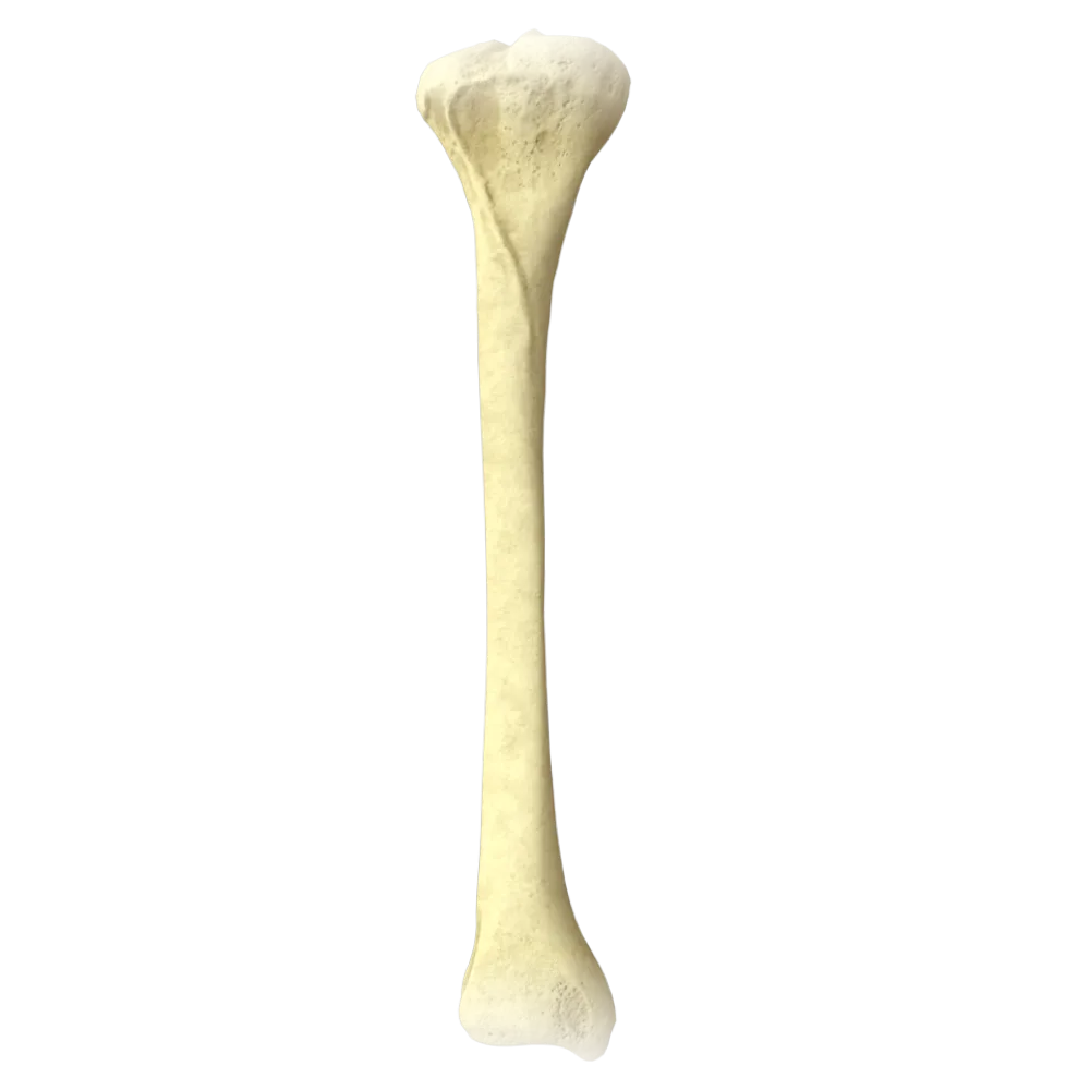
Patella anatomy quiz
Systematic anatomy quiz of the patella: structure, articular connections, blood supply, innervation, and functional relationships.
1/20
bold
text
1. To which type of bone does the patella belong according to the classification?
-
Short cancellous bone
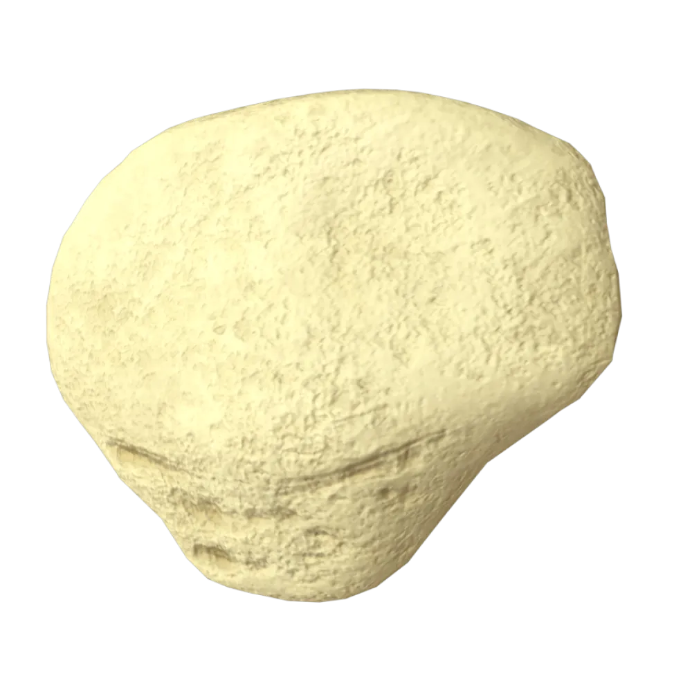
The patella is the largest sesamoid bone in the human body and develops within the tendon of the quadriceps femoris muscle.
-
Sesamoid bone
The patella is the largest sesamoid bone in the human body and develops within the tendon of the quadriceps femoris muscle.
-
Flat bone
The patella is the largest sesamoid bone in the human body and develops within the tendon of the quadriceps femoris muscle.
-
Long tubular bone
The patella is the largest sesamoid bone in the human body and develops within the tendon of the quadriceps femoris muscle.
-
I find it difficult to answer
The patella is the largest sesamoid bone in the human body and develops within the tendon of the quadriceps femoris muscle.
2. What shape does the patella normally comprise?
-
Cylindrical
The patella comprises a triangular shape with a broad base (basis patellae) directed superiorly and a rounded apex (apex patellae) directed inferiorly.
-
Triangular with a rounded apex
The patella comprises a triangular shape with a broad base (basis patellae) directed superiorly and a rounded apex (apex patellae) directed inferiorly.
-
Oval
The patella comprises a triangular shape with a broad base (basis patellae) directed superiorly and a rounded apex (apex patellae) directed inferiorly.
-
Rectangular
The patella comprises a triangular shape with a broad base (basis patellae) directed superiorly and a rounded apex (apex patellae) directed inferiorly.
-
I find it difficult to answer
The patella comprises a triangular shape with a broad base (basis patellae) directed superiorly and a rounded apex (apex patellae) directed inferiorly.
3. What is the inferior pole of the patella called?
-
Basis patellae
The inferior pole of the patella is designated as the apex patellae (apex of the patella). The base (basis patellae) is located superiorly.
-
Apex patellae
The inferior pole of the patella is designated as the apex patellae (apex of the patella). The base (basis patellae) is located superiorly.
-
Facies anterior
The inferior pole of the patella is designated as the apex patellae (apex of the patella). The base (basis patellae) is located superiorly.
-
Margo lateralis
The inferior pole of the patella is designated as the apex patellae (apex of the patella). The base (basis patellae) is located superiorly.
-
I find it difficult to answer
The inferior pole of the patella is designated as the apex patellae (apex of the patella). The base (basis patellae) is located superiorly.
4. Which surface of the patella articulates with the femur?
-
Facies anterior
The posterior surface of the patella—the facies articularis—is covered with hyaline cartilage and articulates with the facies patellaris of the femur.
-
Basis patellae
The posterior surface of the patella—the facies articularis—is covered with hyaline cartilage and articulates with the facies patellaris of the femur.
-
Facies articularis
The posterior surface of the patella—the facies articularis—is covered with hyaline cartilage and articulates with the facies patellaris of the femur.
-
Apex patellae
The posterior surface of the patella—the facies articularis—is covered with hyaline cartilage and articulates with the facies patellaris of the femur.
-
I find it difficult to answer
The posterior surface of the patella—the facies articularis—is covered with hyaline cartilage and articulates with the facies patellaris of the femur.
5. A vertical ridge on the posterior articular surface of the patella divides it into two facets. Which of them is larger?
-
Medial surface
The lateral part of the posterior articular surface of the patella is normally larger than the medial part, corresponding to the larger surface area of the lateral femoral condyle.
-
Lateral surface
The lateral part of the posterior articular surface of the patella is normally larger than the medial part, corresponding to the larger surface area of the lateral femoral condyle.
-
Both facets are equal in size
The lateral part of the posterior articular surface of the patella is normally larger than the medial part, corresponding to the larger surface area of the lateral femoral condyle.
-
Superior surface
The lateral part of the posterior articular surface of the patella is normally larger than the medial part, corresponding to the larger surface area of the lateral femoral condyle.
-
I find it difficult to answer
The lateral part of the posterior articular surface of the patella is normally larger than the medial part, corresponding to the larger surface area of the lateral femoral condyle.
6. Within the tendon of which muscle is the patella embedded?
-
Biceps femoris muscle
The patella is a sesamoid bone located within the tendon of the m. quadriceps femoris. All four heads of this muscle contribute to maintaining its position.
-
Quadriceps femoris muscle
The patella is a sesamoid bone located within the tendon of the m. quadriceps femoris. All four heads of this muscle contribute to maintaining its position.
-
Sartorius muscle
The patella is a sesamoid bone located within the tendon of the m. quadriceps femoris. All four heads of this muscle contribute to maintaining its position.
-
Semimembranosus muscle
The patella is a sesamoid bone located within the tendon of the m. quadriceps femoris. All four heads of this muscle contribute to maintaining its position.
-
I find it difficult to answer
The patella is a sesamoid bone located within the tendon of the m. quadriceps femoris. All four heads of this muscle contribute to maintaining its position.
7. Which ligament runs from the apex of the patella to the tibial tuberosity?
-
Lig. collaterale tibiale
Lig. patellae (patellar ligament) is a continuation of the quadriceps femoris tendon inferior to the patella and attaches to the tuberositas tibiae.
-
Lig. patellae
Lig. patellae (patellar ligament) is a continuation of the quadriceps femoris tendon inferior to the patella and attaches to the tuberositas tibiae.
-
Retinaculum patellae mediale
Lig. patellae (patellar ligament) is a continuation of the quadriceps femoris tendon inferior to the patella and attaches to the tuberositas tibiae.
-
Lig. popliteum obliquum
Lig. patellae (patellar ligament) is a continuation of the quadriceps femoris tendon inferior to the patella and attaches to the tuberositas tibiae.
-
I find it difficult to answer
Lig. patellae (patellar ligament) is a continuation of the quadriceps femoris tendon inferior to the patella and attaches to the tuberositas tibiae.
8. The medial patellar retinaculum (retinaculum patellae mediale) is formed by an expansion of the tendon of which muscle?
-
Vastus medialis muscle
The retinaculum patellae mediale is formed predominantly by the aponeurotic expansion of the m. vastus medialis and prevents lateral displacement of the patella.
-
Gracilis muscle
The retinaculum patellae mediale is formed predominantly by the aponeurotic expansion of the m. vastus medialis and prevents lateral displacement of the patella.
-
Semimembranosus muscle
The retinaculum patellae mediale is formed predominantly by the aponeurotic expansion of the m. vastus medialis and prevents lateral displacement of the patella.
-
Sartorius muscle
The retinaculum patellae mediale is formed predominantly by the aponeurotic expansion of the m. vastus medialis and prevents lateral displacement of the patella.
-
I find it difficult to answer
The retinaculum patellae mediale is formed predominantly by the aponeurotic expansion of the m. vastus medialis and prevents lateral displacement of the patella.
9. Which nerve provides innervation to the muscles that maintain the patella in the correct position (the quadriceps femoris muscle)?
-
Obturator nerve
The m. quadriceps femoris, within whose tendon the patella is embedded, is innervated by the femoral nerve (n. femoralis, L2–L4).
-
Femoral nerve
The m. quadriceps femoris, within whose tendon the patella is embedded, is innervated by the femoral nerve (n. femoralis, L2–L4).
-
Sciatic nerve
The m. quadriceps femoris, within whose tendon the patella is embedded, is innervated by the femoral nerve (n. femoralis, L2–L4).
-
Common fibular nerve
The m. quadriceps femoris, within whose tendon the patella is embedded, is innervated by the femoral nerve (n. femoralis, L2–L4).
-
I find it difficult to answer
The m. quadriceps femoris, within whose tendon the patella is embedded, is innervated by the femoral nerve (n. femoralis, L2–L4).
10. The patella participates in the formation of which joint?
-
Hip joint
The patella is part of the knee joint (articulatio genus), forming the patellofemoral articulation with the femur.
-
Knee joint
The patella is part of the knee joint (articulatio genus), forming the patellofemoral articulation with the femur.
-
Ankle joint
The patella is part of the knee joint (articulatio genus), forming the patellofemoral articulation with the femur.
-
Proximal tibiofibular joint
The patella is part of the knee joint (articulatio genus), forming the patellofemoral articulation with the femur.
-
I find it difficult to answer
The patella is part of the knee joint (articulatio genus), forming the patellofemoral articulation with the femur.
11. With which surface of the femur does the patella articulate?
-
Facies poplitea
The articular surface of the patella glides along the facies patellaris femoris—a grooved surface on the anterior part of the distal epiphysis of the femur.
-
Facies patellaris femoris
The articular surface of the patella glides along the facies patellaris femoris—a grooved surface on the anterior part of the distal epiphysis of the femur.
-
Epicondylus medialis
The articular surface of the patella glides along the facies patellaris femoris—a grooved surface on the anterior part of the distal epiphysis of the femur.
-
Condylus lateralis femoris
The articular surface of the patella glides along the facies patellaris femoris—a grooved surface on the anterior part of the distal epiphysis of the femur.
-
I find it difficult to answer
The articular surface of the patella glides along the facies patellaris femoris—a grooved surface on the anterior part of the distal epiphysis of the femur.
12. Which synovial bursa is located immediately superior to the patella?
-
Bursa infrapatellaris profunda
The bursa suprapatellaris is located superior to the patella between the quadriceps femoris tendon and the femur; it usually communicates with the cavity of the knee joint.
-
Bursa suprapatellaris
The bursa suprapatellaris is located superior to the patella between the quadriceps femoris tendon and the femur; it usually communicates with the cavity of the knee joint.
-
Bursa subcutanea infrapatellaris
The bursa suprapatellaris is located superior to the patella between the quadriceps femoris tendon and the femur; it usually communicates with the cavity of the knee joint.
-
Bursa anserina
The bursa suprapatellaris is located superior to the patella between the quadriceps femoris tendon and the femur; it usually communicates with the cavity of the knee joint.
-
I find it difficult to answer
The bursa suprapatellaris is located superior to the patella between the quadriceps femoris tendon and the femur; it usually communicates with the cavity of the knee joint.
13. The blood supply to the patella is provided predominantly by an arterial network formed by branches of which artery?
-
The anterior tibial artery
The patella receives its blood supply from the rete articulare genus—the arterial network of the knee joint—formed primarily by branches of the popliteal artery (a. poplitea): aa. genus superior medialis, superior lateralis, inferior medialis, and inferior lateralis.
-
The popliteal artery
The patella receives its blood supply from the rete articulare genus—the arterial network of the knee joint—formed primarily by branches of the popliteal artery (a. poplitea): aa. genus superior medialis, superior lateralis, inferior medialis, and inferior lateralis.
-
The posterior tibial artery
The patella receives its blood supply from the rete articulare genus—the arterial network of the knee joint—formed primarily by branches of the popliteal artery (a. poplitea): aa. genus superior medialis, superior lateralis, inferior medialis, and inferior lateralis.
-
The fibular artery
The patella receives its blood supply from the rete articulare genus—the arterial network of the knee joint—formed primarily by branches of the popliteal artery (a. poplitea): aa. genus superior medialis, superior lateralis, inferior medialis, and inferior lateralis.
-
I find it difficult to answer
The patella receives its blood supply from the rete articulare genus—the arterial network of the knee joint—formed primarily by branches of the popliteal artery (a. poplitea): aa. genus superior medialis, superior lateralis, inferior medialis, and inferior lateralis.
14. Through which surface of the patella do the nutrient vessels (vasa nutricia) predominantly enter?
-
Through the anterior (rough) surface
The nutrient vessels enter the patella primarily through its rough anterior surface (facies anterior), in the middle and inferior thirds of the bone.
-
Through the apex (apex patellae)
The nutrient vessels enter the patella primarily through its rough anterior surface (facies anterior), in the middle and inferior thirds of the bone.
-
Through the posterior hyaline cartilage
The nutrient vessels enter the patella primarily through its rough anterior surface (facies anterior), in the middle and inferior thirds of the bone.
-
Through the base (basis patellae)
The nutrient vessels enter the patella primarily through its rough anterior surface (facies anterior), in the middle and inferior thirds of the bone.
-
I find it difficult to answer
The nutrient vessels enter the patella primarily through its rough anterior surface (facies anterior), in the middle and inferior thirds of the bone.
15. What biomechanical function does the patella perform in the mechanism of leg extension?
-
Increases the lever arm of the quadriceps femoris muscle acting on the knee joint
The patella displaces the quadriceps femoris tendon anteriorly from the axis of rotation of the knee joint, thereby increasing the lever arm (moment) and improving the efficiency of leg extension by approximately 30–50%.
-
Decreases the torque of the quadriceps femoris muscle
The patella displaces the quadriceps femoris tendon anteriorly from the axis of rotation of the knee joint, thereby increasing the lever arm (moment) and improving the efficiency of leg extension by approximately 30–50%.
-
Transmits force from the biceps femoris muscle to the leg
The patella displaces the quadriceps femoris tendon anteriorly from the axis of rotation of the knee joint, thereby increasing the lever arm (moment) and improving the efficiency of leg extension by approximately 30–50%.
-
Stabilizes the head of the fibula
The patella displaces the quadriceps femoris tendon anteriorly from the axis of rotation of the knee joint, thereby increasing the lever arm (moment) and improving the efficiency of leg extension by approximately 30–50%.
-
I find it difficult to answer
The patella displaces the quadriceps femoris tendon anteriorly from the axis of rotation of the knee joint, thereby increasing the lever arm (moment) and improving the efficiency of leg extension by approximately 30–50%.
16. What is the fibrous continuation of the retinaculum patellae laterale that attaches to the lateral condyle of the tibia called?
-
Tractus iliotibialis
The retinaculum patellae laterale—the lateral patellar retinaculum—runs from the lateral border of the patella to the lateral condyle of the tibia and to the tractus iliotibialis, maintaining the position of the patella.
-
Retinaculum patellae laterale
The retinaculum patellae laterale—the lateral patellar retinaculum—runs from the lateral border of the patella to the lateral condyle of the tibia and to the tractus iliotibialis, maintaining the position of the patella.
-
Lig. collaterale fibulare
The retinaculum patellae laterale—the lateral patellar retinaculum—runs from the lateral border of the patella to the lateral condyle of the tibia and to the tractus iliotibialis, maintaining the position of the patella.
-
Lig. transversum genus
The retinaculum patellae laterale—the lateral patellar retinaculum—runs from the lateral border of the patella to the lateral condyle of the tibia and to the tractus iliotibialis, maintaining the position of the patella.
-
I find it difficult to answer
The retinaculum patellae laterale—the lateral patellar retinaculum—runs from the lateral border of the patella to the lateral condyle of the tibia and to the tractus iliotibialis, maintaining the position of the patella.
17. In which direction does the patella move during full extension of the knee joint?
-
Moves inferiorly and medially
During full extension of the knee joint, the patella moves superiorly (proximally) together with the quadriceps femoris tendon and slightly laterally, entering the lateral part of the facies patellaris femoris.
-
Moves superiorly and slightly laterally
During full extension of the knee joint, the patella moves superiorly (proximally) together with the quadriceps femoris tendon and slightly laterally, entering the lateral part of the facies patellaris femoris.
-
Remains stationary
During full extension of the knee joint, the patella moves superiorly (proximally) together with the quadriceps femoris tendon and slightly laterally, entering the lateral part of the facies patellaris femoris.
-
Moves directly inferiorly
During full extension of the knee joint, the patella moves superiorly (proximally) together with the quadriceps femoris tendon and slightly laterally, entering the lateral part of the facies patellaris femoris.
-
I find it difficult to answer
During full extension of the knee joint, the patella moves superiorly (proximally) together with the quadriceps femoris tendon and slightly laterally, entering the lateral part of the facies patellaris femoris.
18. Which synovial bursa is located immediately INFERIOR to the patella?
-
Bursa infrapatellaris profunda
The bursa infrapatellaris profunda (deep infrapatellar bursa) lies deeply, between the posterior surface of the patellar ligament (lig. patellae) and the anterior surface of the tibia (in the region of the tuberosity). Unlike the superficial infrapatellar bursa (bursa subcutanea infrapatellaris—option C), which lies beneath the skin over the tuberosity, the deep bursa does not communicate with the joint cavity (unlike the suprapatellar bursa).
-
Bursa suprapatellaris
The bursa infrapatellaris profunda (deep infrapatellar bursa) lies deeply, between the posterior surface of the patellar ligament (lig. patellae) and the anterior surface of the tibia (in the region of the tuberosity). Unlike the superficial infrapatellar bursa (bursa subcutanea infrapatellaris—option C), which lies beneath the skin over the tuberosity, the deep bursa does not communicate with the joint cavity (unlike the suprapatellar bursa).
-
Bursa subcutanea infrapatellaris
The bursa infrapatellaris profunda (deep infrapatellar bursa) lies deeply, between the posterior surface of the patellar ligament (lig. patellae) and the anterior surface of the tibia (in the region of the tuberosity). Unlike the superficial infrapatellar bursa (bursa subcutanea infrapatellaris—option C), which lies beneath the skin over the tuberosity, the deep bursa does not communicate with the joint cavity (unlike the suprapatellar bursa).
-
Bursa anserina
The bursa infrapatellaris profunda (deep infrapatellar bursa) lies deeply, between the posterior surface of the patellar ligament (lig. patellae) and the anterior surface of the tibia (in the region of the tuberosity). Unlike the superficial infrapatellar bursa (bursa subcutanea infrapatellaris—option C), which lies beneath the skin over the tuberosity, the deep bursa does not communicate with the joint cavity (unlike the suprapatellar bursa).
-
I find it difficult to answer
The bursa infrapatellaris profunda (deep infrapatellar bursa) lies deeply, between the posterior surface of the patellar ligament (lig. patellae) and the anterior surface of the tibia (in the region of the tuberosity). Unlike the superficial infrapatellar bursa (bursa subcutanea infrapatellaris—option C), which lies beneath the skin over the tuberosity, the deep bursa does not communicate with the joint cavity (unlike the suprapatellar bursa).
19. Of which tissue does the articular surface of the patella (facies articularis) consist?
-
Fibrocartilage
The articular surface of the patella is covered with hyaline cartilage, which is the thickest articular cartilage in the human body (up to 6–7 mm in the central part).
-
Hyaline cartilage
The articular surface of the patella is covered with hyaline cartilage, which is the thickest articular cartilage in the human body (up to 6–7 mm in the central part).
-
Elastic cartilage
The articular surface of the patella is covered with hyaline cartilage, which is the thickest articular cartilage in the human body (up to 6–7 mm in the central part).
-
Compact bone tissue
The articular surface of the patella is covered with hyaline cartilage, which is the thickest articular cartilage in the human body (up to 6–7 mm in the central part).
-
I find it difficult to answer
The articular surface of the patella is covered with hyaline cartilage, which is the thickest articular cartilage in the human body (up to 6–7 mm in the central part).
20. Which element of the knee joint passes along the anterior surface and surrounds the patella on both sides?
-
Anterior cruciate ligament
The fibrous capsule of the knee joint thickens on the anterior surface, forming the medial and lateral patellar retinacula (retinacula patellae mediale et laterale), which surround the patella on both sides and blend with the patellar ligament.
-
Transverse ligament of knee
The fibrous capsule of the knee joint thickens on the anterior surface, forming the medial and lateral patellar retinacula (retinacula patellae mediale et laterale), which surround the patella on both sides and blend with the patellar ligament.
-
Quadrangular fibrous capsule with retinacula patellae
The fibrous capsule of the knee joint thickens on the anterior surface, forming the medial and lateral patellar retinacula (retinacula patellae mediale et laterale), which surround the patella on both sides and blend with the patellar ligament.
-
Arcuate popliteal ligament
The fibrous capsule of the knee joint thickens on the anterior surface, forming the medial and lateral patellar retinacula (retinacula patellae mediale et laterale), which surround the patella on both sides and blend with the patellar ligament.
-
I find it difficult to answer
The fibrous capsule of the knee joint thickens on the anterior surface, forming the medial and lateral patellar retinacula (retinacula patellae mediale et laterale), which surround the patella on both sides and blend with the patellar ligament.
Retake this quiz?
Your current progress will be reset.
