Fractures of Clavicle: Classification, Symptoms, Treatment Options, and Rehabilitation
Aleksandr K.Orthopedic surgeon, MD
18 min read·January 29, 2026
This article is for informational purposes only
The content on this website, including text, graphics, and other materials, is provided for informational purposes only. It is not intended as advice or guidance. Regarding your specific medical condition or treatment, please consult your healthcare provider.
The clavicle is an S‑shaped bone that articulates with the sternum at the sternoclavicular joint and with the scapula at the acromioclavicular joint. Laterally, at its acromial end, it is flattened; the body is cylindrical, and the sternal end may be described as prismatic. It plays a key role in maintaining shoulder stability.
Etiology and Mechanism of Injury
Clavicle fractures occur primarily as a result of direct or indirect trauma.
Most common causes include:
Falling onto the shoulder or an outstretched hand.
Sports injuries (most often cycling or football).
Road traffic accidents.
The primary mechanism of injury is a fall directly onto the lateral aspect of the shoulder, producing a compressive force that leads to fracture (seen in approximately 85 % of cases). Less commonly, the fracture may result from a direct blow to the clavicle or from indirect force transmitted through the arm.
Displacement of clavicular fragments is determined by the pull of muscles attached to the bone:
Medial (central) fragments: displaced superiorly and posteriorly due to the pull of the sternocleidomastoid muscle.
Lateral fragments: displaced inferiorly and medially due to the action of the pectoralis major muscle and the weight of the upper limb.
Epidemiology
Clavicle fractures account for approximately 2.6–5 % of all fractures and up to 44 % of injuries to the pectoral girdle. They are most common in young, active individuals and children, with peak incidence in males aged 13–20 years.
Classification of Clavicle Fractures
The basic classification relies on the location of the fracture relative to the anatomical regions of the clavicle:
Midshaft (middle 1/3) fractures: 75–80 % of all clavicle fractures.
Acromial end (lateral 1/3) fractures: 15–25 %.
Sternal end (medial 1/3) fractures: 2–5 %.
The high frequency of midshaft (middle 1/3) fractures is due to this region being the thinnest and narrowest portion of the clavicle and lacking ligamentous and muscular support.
Neer Classification (for Acromial End Fractures)
The Neer classification is widely used in clinical practice to guide treatment decisions for fractures of the acromial (distal) end of the clavicle. It identifies five major fracture types and two subtypes.
Neer Type
Fracture Characteristics
Recommended Treatment
Type I
Extra‑articular fracture. The fracture line runs laterally to the attachment of the coracoclavicular ligaments. Ligaments intact. Minimal displacement. Stable fracture
Medical
Type II
Extra‑articular fracture. The fracture line runs medially to the attachment of the coracohumeral ligaments. Ligaments intact. Significant medial displacement. Unstable fracture
Surgical. Nonunion rate with medical therapy may amount to 56 %
Type II
Extra‑articular fracture. Two anatomical patterns: 1. The fracture line runs through the ligament attachment (the conoid ligament disrupted, the trapezoid ligament intact). 2. The fracture line runs laterally to the ligament attachment, but the ligaments are injured.
Surgical. Nonunion rate with medical therapy may amount to 30–45 %
Type III
Intra‑articular fracture. The fracture line runs laterally to the ligament attachment, extending into the acromioclavicular joint. Ligaments intact. Minimal displacement. Stable fracture
Medical. Risk of post‑traumatic acromioclavicular arthritis
Type IV
Extra‑articular fracture. In cases of open growth plates in the physeal region. Ligaments intact. Lateral clavicle fragment is displaced superiorly due to periosteal disruption. Stable fracture
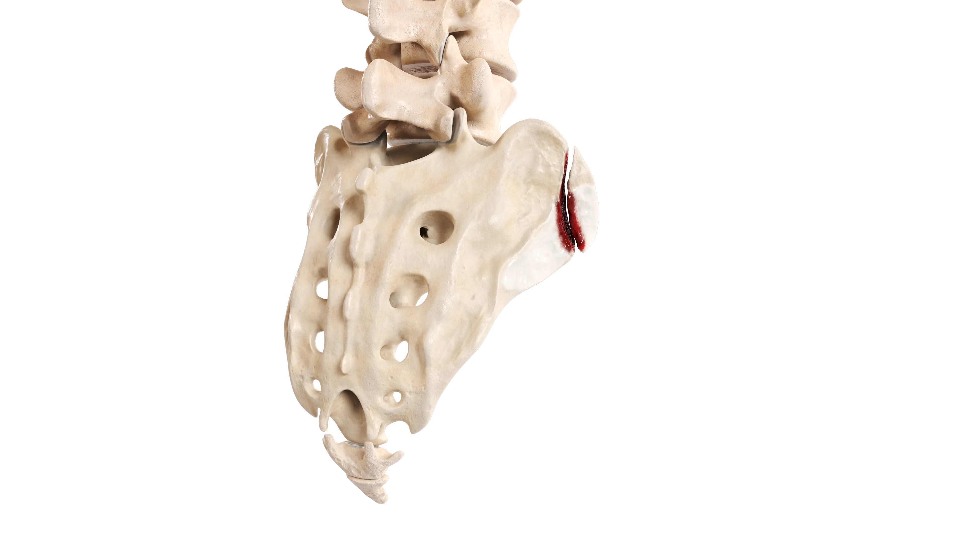
Extra‑Articular Fracture of Sternal End of Clavicle
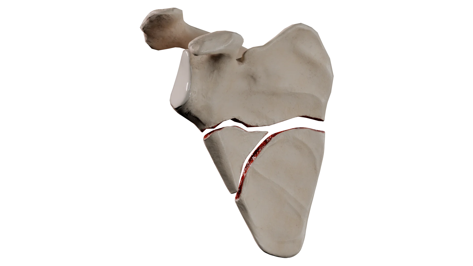
Comminuted Midshaft Fracture
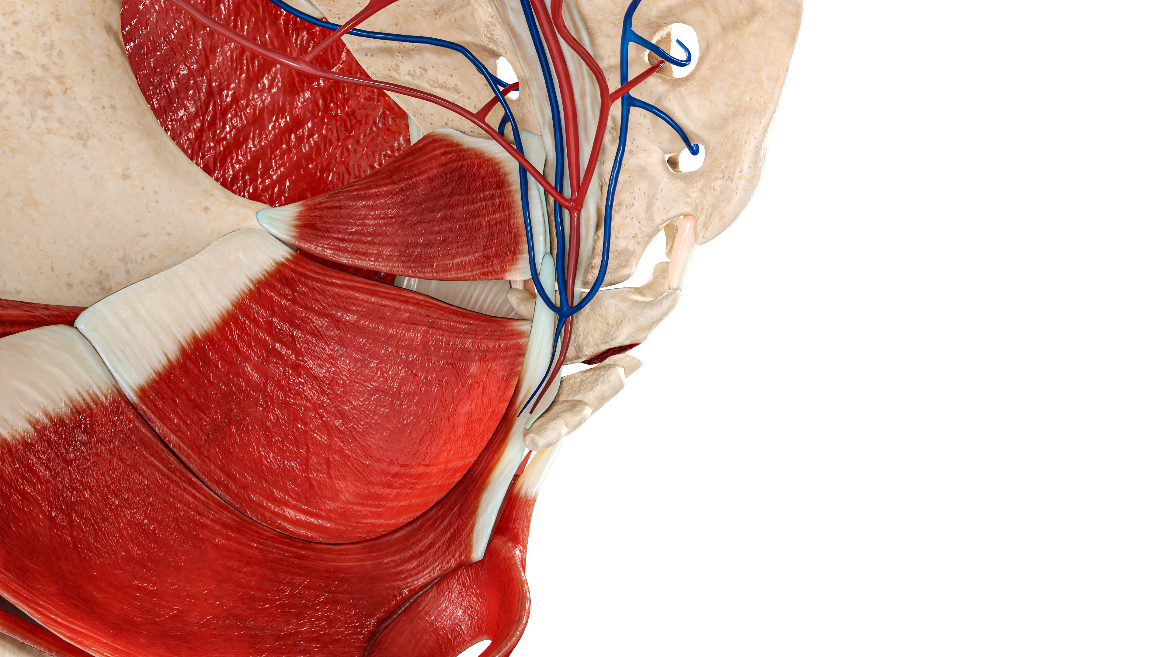
Extra‑Articular Comminuted Fracture of Acromial End of Clavicle (Neer Type V)
Clinical Manifestations and Symptoms
Typical clinical manifestations of clavicle fractures include:
Immediate shoulder pain and loss of function.
The arm is held close to the body and supported by the opposite hand.
Swelling, hematoma, and a palpable bony prominence beneath the skin.
Possible “skin tenting”, or, less commonly, an open fracture.
Possible injury to adjacent vessels and nerves.
Diagnosis
Diagnosis is based on clinical evaluation and radiologic imaging.
Clinical Evaluation
History of injury: The patient reports a traumatic event accompanied by a distinct cracking sound followed by pain.
Examination: Visible deformity in the clavicular region, soft‑tissue swelling, and a hematoma over the fracture site.
Palpation: Localized tenderness and crepitus.
Range of motion: Limited movement of the ipsilateral upper extremity due to pain.
Radiologic Methods
Radiography: Standard anteroposterior view and a 15° cephalic tilt (Zanca view) demonstrate the fracture location, displacement, and presence of fragments.
CT (computed tomography): Indicated for complex comminuted fractures and fractures of the sternal or acromial ends.
Treatment of Clavicle Fractures
Medical Therapy
Medical therapy is associated with a high rate of bone union in children and in minimally displaced fractures in adults.
Indications:
Midshaft fractures without displacement or with minimal displacement.
No associated vascular or nerve injury.
No skin compromise or significant shortening of the shoulder girdle (less than 2 cm).
Procedure:
Sling or figure‑of‑eight brace (Delbet rings) for immobilization.
Pain control and gradual return to activity over 6–12 weeks.
Physical therapy: Passive, pain‑free range of motion beginning at Week 2; resistance exercises starting at Week 6; return to full athletic activity by Month 4–6.
Surgical therapy
Surgery may provide earlier functional recovery and reduce the risk of nonunion or malunion in significantly displaced fractures.
Indications:
Diaphyseal fractures with significant shortening (> 2 cm) and displacement.
Open fractures.
Associated vascular or nerve injury.
Skin tenting around fragments with risk of skin breakdown.
Multifragmentary fractures; injury to the capsuloligamentous structures of the acromioclavicular or sternoclavicular joints.
Procedure:
Open reduction and internal fixation (ORIF) using plates and screws. Immobilization in a soft sling for 7–10 days, followed by active motion in a pain‑free range. Resistance and strengthening exercises beginning at Week 6. Return to athletic activity at approximately 3 months.
Intramedullary osteosynthesis (less frequently).
Rehabilitation: Principles and Phases
Rehabilitation follows a staged progression of orthopedic protection and gradually increasing load: immobilization (sling) → early passive motion → active motion → muscle strengthening → return to activity. The rate of progression depends on fracture type and treatment method.
Rehabilitation for Fractures of Sternal End of Clavicle
Timeline (Weeks)
Weight‑Bearing
Range of Motion
Notes
0-2
Contraindicated
Full range of motion is allowed for the elbow and wrist. Shoulder motion is contraindicated
Sling immobilization. Monitoring for vascular or visceral injury
2-6
Contraindicated
Pendular movements, passive shoulder flexion up to 90 ° (as tolerated)
–
6-8
Weight limit: up to 1 kg
Active shoulder motion > 90 ° (as tolerated)
Continued radiographic monitoring to assess fracture healing
8-12
Gradual increase in allowable load (as tolerated)
Full active shoulder range of motion. Isometric strengthening
–
12+
No restriction
No restriction
Continued radiographic monitoring to assess fracture healing. Sports activities may be resumed based on sport‑specific demands and radiographic evidence of union.
Rehabilitation for Midshaft Clavicle Fractures
Medical Therapy
Timeline (Weeks)
Weight‑Bearing
Range of Motion
0-2
Contraindicated
Full range of motion is allowed for the elbow and wrist. Shoulder motion contraindicated
2-6
Contraindicated
Pendular passive movements (shoulder flexion not exceeding 90 °), progressing to active motion as tolerated
6-8
Gradual increase in load up to 3 kg
Full active shoulder range of motion. Isometric strengthening
8-12
Gradual increase in allowable load (as tolerated)
Full active shoulder range of motion. Exercises focus on restoring strength in the limb
12+
Complete
Return to daily activities Return to sports depends on the specific sport
Surgical Therapy
Timeline (Weeks)
Weight‑Bearing
Range of Motion
0-2
Contraindicated. Sling immobilization
Full range of motion is allowed for the elbow and wrist. Pendular shoulder movements within a pain‑free range
2-4
Up to 1 kg
Abduction and flexion up to 90 ° assisted by the healthy arm as tolerated
4-6
Up to 2 kg
Progression to full active range of motion
6-8
Gradual increase in allowable load (as tolerated)
Progression to full active range of motion. Strengthening exercises
8+
No restriction
Gradual increase in exercise load. Return to sports no earlier than Week 12
Rehabilitation for Fractures of Acromial End of Clavicle
Medical Therapy
Timeline (Weeks)
Weight‑Bearing
Range of Motion
0-2
Contraindicated. Sling immobilization
Full range of motion is allowed for the elbow and wrist. Shoulder motion contraindicated
2-6
Up to 1 kg
Passive shoulder flexion and abduction up to 90 °
6-10
Gradual increase in allowable load (as tolerated)
Active shoulder motion > 90 ° (as tolerated) Strengthening exercises
10+
No restriction
Return to sports once radiographic union is confirmed
Surgical Therapy
Timeline (Weeks)
Weight‑Bearing
Range of Motion
0-2
Contraindicated. Immobilization in a soft sling
Full range of motion is allowed for the elbow and wrist. Shoulder motion contraindicated
2-4
Up to 1 kg
Passive pendular movements and passive flexion/abduction up to 90 °
4-6
Up to 2 kg
Active shoulder motion > 90 ° (as tolerated) Strengthening exercises
6-10
Gradual increase in allowable load (as tolerated)
Full range of motion. Progressive strengthening without restrictions
10+
No restriction
Return to sports once radiographic union is confirmed
Return‑to‑Sport Protocol
General Requirements:
No local tenderness at the fracture site.
Radiographic evidence of fracture union
Full, pain‑free active range of motion in the shoulder.
Ability to perform sport‑specific movements without limitation.
The process of returning to sport should be structured according to the timeline of the injury.
Load Progression by Phases:
Phase 1 (up to 2–3 weeks): Only isometric activation of the muscles of the shoulder girdle is permitted.
Phase 2 (2–6 weeks): Activities not involving the injured upper extremity are allowed (stationary cycling, lower‑extremity training without impact, aerobic activity without resistance). Resistance training for the injured arm, overhead exercises, and lifting weights are not permitted.
Phase 3 (6–9 weeks): Light weight training and body‑weight exercises (planks, push‑ups, burpees) are allowed as tolerated.
Phase 4 (9–12 weeks): Non‑contact sports is permitted, if entry criteria are met (radiographic evidence of union, pain‑free motion, etc.).
Phase 5 (beyond 12 weeks): Contact sports and competitive activities are allowed.
Return to contact sports is permitted no earlier than 12 weeks after injury and only when fracture union is radiographically confirmed. Healing and recovery take more time in older adults, patients with metabolic disorders, and those who sustained high‑energy trauma. Children typically heal and recover more quickly.
Find more scientifically accurate content on our social media
Subscribe and don’t miss out the latest resources
Prognosis
The prognosis for clavicle fractures is generally favorable, although outcomes depend significantly on fracture location and treatment strategy.
Fractures of sternal end of clavicle: These are relatively rare. Medical therapy is associated with a high union rate and minimal complications. Surgical treatment may allow earlier functional recovery of the injured limb.
Midshaft fractures: When treatment indications are followed, union occurs in approximately 85 % of cases with medical therapy and in more than 95 % of cases with surgical treatment. Surgery provides a faster return to full physical activity.
Fractures of acromial end of clavicle: Medical therapy is associated with nonunion in up to 30 % of cases. There is also a risk of impaired shoulder function due to reduced deltoid muscle strength. Surgical treatment tends to restore the limb function more quickly and shows high union rates.
FAQ
1. How long does a clavicle fracture take to heal?
In adults, a clavicle fracture heals in 6 to 12 weeks, depending on fracture complexity and management strategy. Complete bone remodeling may take up to one year.
2. Why can the arm become numb after a clavicle fracture?
Numbness or tingling in the fingers may indicate that displaced fracture fragments or swelling are compressing the brachial plexus (nerves) or the vessels beneath the clavicle. This is a concerning symptom that requires immediate medical evaluation.
3. Is surgery always necessary for displaced fractures?
No. If clavicle shortening is less than 2 cm and there is no skin compromise, conservative treatment (reduction and immobilization) may be appropriate. However, surgery allows faster restoration of arm function and reduces the risk of malunion.
4. How should you sleep with a clavicle fracture?
During the first weeks, it is recommended to sleep in a semi‑upright position (on elevated pillows) to reduce swelling and pain. Sleeping on the healthy side is acceptable if the immobilizing bandage is secure. Sleeping on the stomach or on the injured side is prohibited.
5. What complications can occur after a clavicle fracture?
With proper treatment, the prognosis is favorable. Possible complications include malunion (shortening, deformity), nonunion (pseudoarthrosis), post‑traumatic osteoarthritis, and numbness of the skin around the surgical scar after surgery.
References
1.
VOKA 3D Anatomy & Pathology – Complete Anatomy and Pathology 3D Atlas [Internet]. VOKA 3D Anatomy & Pathology.
Available from: https://catalog.voka.io/
2.
Moverley R, Little N, Gulihar A, Singh B. Current concepts in the management of clavicle fractures. J Clin Orthop Trauma. 2020 Feb;11(Suppl 1):S25-S30.
3.
Kang LX, Faulkner HJ, Howard WH, Low AK. Displaced medial clavicle fractures: a systematic review of outcomes after nonoperative and operative management. JSES Int. 2022 Oct 15;7(1):79-85.
4.
von Rüden C, Rehme-Röhrl J, Augat P, Friederichs J, Hackl S, Stuby F, Trapp O. Evidence on treatment of clavicle fractures. Injury. 2023 Oct;54 Suppl 5:110818.
5.
Yan MZ, Yuen WS, Yeung SC, Wing-Yin CW, Wong SC, Si-Qi WW, Tian E, Rashed S, Yung CSY, Fang CX. Operative management of midshaft clavicle fractures demonstrates better long-term outcomes: A systematic review and meta-analysis of randomised controlled trials. PLoS One. 2022 Apr 29;17(4):e0267861.
6.
Panagopoulos A, Solou K, Tatani I, Triantafyllopoulos IK, Lakoumentas J, Kouzelis A, Athanasiou V, Kokkalis ZT. What is the optimal surgical treatment for Neer type IIB (IIC) distal clavicle fractures? A systematic review and meta-analysis. J Orthop Surg Res. 2022 Apr 7;17(1):215.
7.
Wang C, Li X, Dong S, Xie W, Ling Z, Meng C, Stöckle U. Midshaft clavicle fractures with associated ipsilateral acromioclavicular joint injuries: a systematic review. BMC Surg. 2025 Feb 28;25(1):87.
8.
Yue L, Huang C, Zhang J, Wang Z, Wang S, Sun H. Treatment Strategies for Distal Clavicle Fractures: A Narrative Review. Orthop Res Rev. 2025 May 19;17:221-227.
9.
Kilkenny CJ, Daly GR, Whelehan SP, Vukanic D, Alrawashdeh M, Boland F, Quinlan JF, Molony DC. Return to play following clavicular fracture – A systematic review and meta analysis. JSES Rev Rep Tech. 2024 Dec 14;5(2):259-269.
405 Lexington Avenue, 26th Floor New York, NY 10174 United States
Thank you!
Your message is sent! Our experts will contact you shortly. If you have any additional questions, please contact us at info@voka.io
Cookie Consent
We use cookies to enhance your browsing experience, analyze site traffic, and deliver content. Please choose whether you accept all cookies or wish to reject non-essential tracking.
Cookie Preferences
Manage your cookie preferences below:
Essential cookies enable basic functions and are necessary for the proper function of the website.
Name
Description
Duration
Geolocation Config
This cookie is used to store the consent settings based on the visitor's location.
30 days
Cookie Preferences
This cookie is used to store the user's cookie consent preferences.
30 days
Google reCAPTCHA helps protect websites from spam and abuse by verifying user interactions through challenges.
Name
Description
Duration
_GRECAPTCHA
Google reCAPTCHA sets a necessary cookie (_GRECAPTCHA) when executed for the purpose of providing its risk analysis.
179 days
Statistics cookies collect information anonymously. This information helps us understand how visitors use our website.
Google Analytics is a powerful tool that tracks and analyzes website traffic for informed marketing decisions.
Used to monitor number of Google Analytics server requests
10 minutes
__utmb
Used to distinguish new sessions and visits. This cookie is set when the GA.js javascript library is loaded and there is no existing __utmb cookie. The cookie is updated every time data is sent to the Google Analytics server.
30 minutes after last activity
__utmc
Used only with old Urchin versions of Google Analytics and not with GA.js. Was used to distinguish between new sessions and visits at the end of a session.
End of session (browser)
__utmz
Contains information about the traffic source or campaign that directed user to the website. The cookie is set when the GA.js javascript is loaded and updated when data is sent to the Google Anaytics server
6 months after last activity
__utmv
Contains custom information set by the web developer via the _setCustomVar method in Google Analytics. This cookie is updated every time new data is sent to the Google Analytics server.
2 years after last activity
__utmx
Used to determine whether a user is included in an A / B or Multivariate test.
18 months
_ga
ID used to identify users
2 years
_gali
Used by Google Analytics to determine which links on a page are being clicked
30 seconds
_ga_
ID used to identify users
2 years
_gid
ID used to identify users for 24 hours after last activity
24 hours
_gat
Used to monitor number of Google Analytics server requests when using Google Tag Manager
1 minute
_gac_
Contains information related to marketing campaigns of the user. These are shared with Google AdWords / Google Ads when the Google Ads and Google Analytics accounts are linked together.
90 days
Clarity is a web analytics service that tracks and reports website traffic.